Posture and Movement Regulation
Posture and movement are regulated through a complex interplay of the central nervous system (CNS), peripheral nervous system (PNS), and various sensory systems. The primary components involved in this regulatory system include the brain, spinal cord, muscles, and sensory feedback mechanisms.
- Central Nervous System (CNS): The CNS, which includes the brain and spinal cord, plays a crucial role in coordinating posture and movement. Within the CNS, specific areas are responsible for different aspects of motor control.
- Peripheral Nervous System (PNS): The PNS consists of nerves that connect the CNS to limbs and organs. It transmits signals from the CNS to muscles to initiate movement and conveys sensory information back to the CNS regarding body position and movement.
- Sensory Feedback Mechanisms: Sensory receptors located in muscles, tendons, joints, and skin provide feedback about body position (proprioception) and external stimuli (exteroception). This feedback is essential for adjusting movements and maintaining balance.
- Motor Control Systems: These systems can be broadly categorized into:
- Voluntary Control: Involves conscious decision-making processes originating from higher brain centers.
- Involuntary Control: Includes reflexes that occur without conscious thought, primarily mediated by spinal circuits.
Function of Main Components of Regulatory Systems
- Brain: The brain integrates sensory information to plan and execute movements. Key areas include:
- Cerebral Cortex: Involved in voluntary motor control.
- Basal Ganglia: Regulates initiation of movement.
- Cerebellum: Coordinates timing and precision of movements.
- Spinal Cord: Acts as a conduit for signals between the brain and peripheral nerves. It also contains neural circuits that mediate reflex actions.
- Muscles: Effectors that carry out movements based on commands received from motor neurons.
- Sensory Receptors: Provide real-time feedback about body position, muscle tension, and external forces acting on the body.
Cortical Motor Area
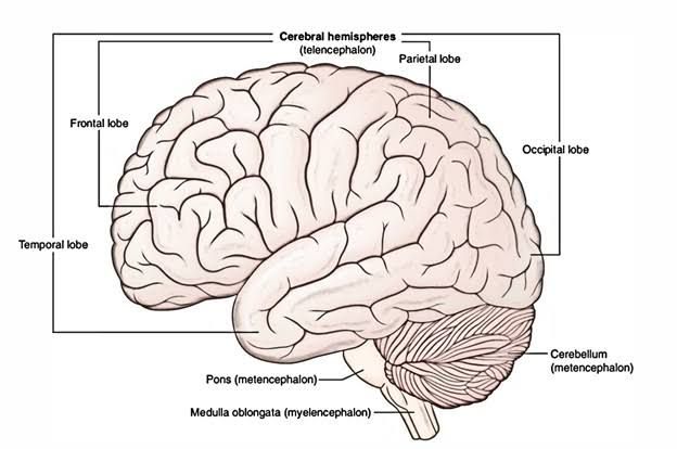
The cortical motor area primarily refers to regions within the frontal lobe of the brain responsible for planning, controlling, and executing voluntary movements. The primary motor cortex (M1), located along the precentral gyrus, is particularly significant as it sends signals directly to spinal motor neurons that innervate skeletal muscles. Other areas such as the premotor cortex contribute to planning movements based on sensory input or learned behaviors.
Pyramids
The pyramids are structures located in the medulla oblongata at the base of the brainstem where corticospinal tracts decussate (cross over) from one side of the brain to the opposite side of the body. This crossing is critical because it means that each hemisphere of the brain controls voluntary movements on the opposite side of the body. The pyramids contain bundles of axons from upper motor neurons descending from cortical areas toward lower motor neurons in the spinal cord.
Corticospinal Tracts
The corticospinal tracts are major pathways that convey motor commands from the cerebral cortex down to spinal motoneurons responsible for voluntary movement control. There are two main types:
- Lateral Corticospinal Tract: Comprises approximately 90% of corticospinal fibers; it decussates at the level of pyramids in medulla oblongata before descending into lateral columns of spinal cord where it synapses with lower motor neurons.
- Anterior Corticospinal Tract: Comprises about 10% of fibers; these do not decussate until they reach their target segment within the spinal cord.
Both tracts play essential roles in fine motor control, particularly for distal limb muscles involved in skilled tasks such as writing or playing an instrument.
In summary, posture and movement regulation involves a sophisticated network comprising various components including cortical areas for planning and executing movements, pyramidal structures facilitating crossover communication between hemispheres, and corticospinal tracts transmitting commands to effectors throughout the body.
Function of the Pyramidal System in Relation to Skilled Voluntary Movement
The pyramidal system, primarily composed of the corticospinal tract, plays a crucial role in the control of skilled voluntary movements. This system originates in the motor cortex of the brain and descends through the brainstem and spinal cord. It is responsible for transmitting motor commands from the brain to the muscles, facilitating precise and coordinated movements.
- Anatomy of the Pyramidal System: The pyramidal system consists mainly of upper motor neurons that originate in the primary motor cortex (M1) and project downwards through various pathways. The corticospinal tract is particularly significant as it innervates lower motor neurons located in the spinal cord that directly control skeletal muscles.
- Role in Skilled Movements: Skilled voluntary movements require fine motor control, which is heavily reliant on feedback mechanisms and precise timing. The pyramidal system enables this by allowing for direct cortical control over muscle activity. For instance, when a person performs tasks such as writing or playing a musical instrument, the pyramidal tract facilitates rapid adjustments based on sensory feedback.
- Integration with Other Systems: While the pyramidal system is essential for initiating and controlling voluntary movements, it works in conjunction with other neural systems, including extrapyramidal pathways (which regulate involuntary movements) and cerebellar circuits (which coordinate timing and precision). This integration ensures that skilled movements are not only initiated but also executed smoothly.
- Clinical Relevance: Damage to the pyramidal system can lead to various movement disorders characterized by weakness or loss of fine motor skills. Conditions such as stroke or multiple sclerosis can disrupt these pathways, highlighting their importance in maintaining skilled voluntary movement.
Decerebrate and Decorticate Rigidity
Decerebrate rigidity and decorticate rigidity are two types of postural abnormalities that arise from different levels of brain injury affecting motor control.
- Decerebrate Rigidity:
- Definition: Decerebrate rigidity is characterized by extension of all four limbs and often involves hyperextension of the back.
- Cause: This condition results from lesions at or below the level of the red nucleus (located in the midbrain), which disrupts normal inhibitory signals from higher brain centers. As a result, there is unopposed activity from extensor muscles due to loss of descending inhibition from structures like the cerebral cortex.
- Clinical Significance: Decerebrate rigidity indicates severe brain damage and often correlates with poor prognosis due to its association with dysfunction in vital autonomic functions.
- Decorticate Rigidity:
- Definition: Decorticate rigidity presents as flexion of the arms at the elbows with extension of legs; it reflects a more preserved level of function compared to decerebrate rigidity.
- Cause: This condition arises from lesions above the red nucleus but below cortical areas, leading to disinhibition of flexor muscle activity while extensor activity remains inhibited due to intact cortical influences.
- Clinical Significance: While decorticate rigidity suggests significant brain injury, it may indicate better preservation of some cortical functions compared to decerebrate rigidity.
In summary, both types of rigidity reflect different levels and locations of neurological impairment affecting voluntary movement control but indicate severe underlying pathology.
Postural Reflexes Integrated in the Medulla Oblongata, Pons, Midbrain, and Cerebral Cortex
Postural reflexes are essential for maintaining balance and posture. These reflexes are integrated at various levels of the central nervous system, including the medulla oblongata, pons, midbrain, and cerebral cortex.
- Medulla Oblongata: The medulla is responsible for several autonomic functions and plays a crucial role in postural reflexes. It integrates sensory information from the vestibular system (which helps with balance) and proprioceptive inputs from muscles and joints. The medulla coordinates reflexive adjustments to maintain posture when there are changes in body position or external forces acting on the body. For example, if a person begins to lean to one side, the medulla will trigger muscle contractions on the opposite side to restore balance.
- Pons: The pons acts as a relay station between different parts of the brain and is involved in regulating motor control and sensory analysis. It contributes to postural reflexes by integrating information from higher brain centers (like the cerebellum) with signals coming from the spinal cord. The pons helps coordinate movements that require precise timing and smooth transitions, such as walking or running.
- Midbrain: The midbrain contains structures like the superior colliculus and substantia nigra that are involved in visual reflexes and motor control respectively. In terms of postural reflexes, it integrates visual stimuli with motor responses to help maintain posture while moving through space. For instance, if an object suddenly appears in a person’s peripheral vision, the midbrain can initiate a reflexive response to adjust posture accordingly.
- Cerebral Cortex: While most postural reflexes are automatic and occur at lower levels of the CNS, higher-level processing occurs in the cerebral cortex. The primary motor cortex is involved in planning voluntary movements that require postural adjustments. It sends signals down through descending pathways to execute these movements while maintaining stability.
Motor Functions of the Brainstem
The brainstem comprises three main parts: the midbrain, pons, and medulla oblongata. It serves as a critical hub for motor functions:
- Regulation of Muscle Tone: The brainstem helps regulate muscle tone through descending pathways that influence spinal cord activity.
- Coordination of Reflexes: Many basic reflex actions (like swallowing or blinking) are coordinated here.
- Integration of Sensory Information: It processes sensory inputs related to balance (vestibular), proprioception (muscle/joint position), and visual cues necessary for coordinated movement.
Major Functions of Descending Motor Pathways Originating in the Brainstem
Descending motor pathways originating from the brainstem include:
- Corticobulbar Tract: This pathway originates in the motor cortex but descends through the brainstem to innervate cranial nerve nuclei responsible for facial expressions, mastication, swallowing, etc.
- Reticulospinal Tract: This tract originates from neurons in both the pons and medulla oblongata; it modulates voluntary movements by influencing muscle tone and posture through connections with spinal cord interneurons.
- Vestibulospinal Tract: This pathway arises from vestibular nuclei located in the pons/medulla; it plays a significant role in maintaining balance by facilitating extensor muscle activity while inhibiting flexor muscles during changes in head position.
- Tectospinal Tract: Originating from the superior colliculus within the midbrain, this tract coordinates head movements towards visual stimuli by influencing neck muscles based on visual input.
- Rubrospinal Tract: This pathway originates from the red nucleus in the midbrain; it primarily facilitates flexor muscle activity while inhibiting extensors during voluntary movement execution.
In summary, postural reflexes integrated across various regions of the CNS ensure stability during movement while descending motor pathways originating from these areas facilitate coordinated muscular responses necessary for complex activities.


