Development of the Endocrine Glands
1. Thyroid Gland Development
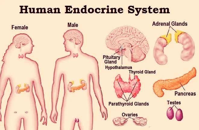
The thyroid gland originates from the endodermal layer of the embryo. It begins to develop around the third week of gestation when a small outpouching, known as the thyroid diverticulum, forms at the base of the tongue. This diverticulum descends into the neck and eventually becomes the thyroid gland. By the seventh week of gestation, it has migrated to its final position in front of the trachea. The gland is fully formed by approximately 10-12 weeks, and it begins producing thyroid hormones (thyroxine and triiodothyronine) by the end of the first trimester.
2. Parathyroid Gland Development
The parathyroid glands develop from the third and fourth pharyngeal pouches during embryogenesis. The inferior parathyroids arise from the third pouch, while the superior parathyroids originate from the fourth pouch. As these pouches develop, they migrate downwards to their final positions on the posterior aspect of the thyroid gland. The parathyroid glands begin to produce parathyroid hormone (PTH) around week 8 of gestation.
3. Pituitary Gland Development
The pituitary gland develops from two distinct embryonic origins: the anterior pituitary (adenohypophysis) arises from an ectodermal structure called Rathke’s pouch, which forms in the roof of the mouth during early development; this process begins around week 3-4 of gestation. The posterior pituitary (neurohypophysis) develops from an extension of neural tissue from the hypothalamus. By approximately week 8, both parts are well established, and by week 12, they start producing hormones such as growth hormone and prolactin.
4. Adrenal Gland Development
The adrenal glands consist of two parts: the adrenal cortex and adrenal medulla. The adrenal cortex develops from mesodermal tissue surrounding each kidney during weeks 6-7 of gestation. The adrenal medulla arises from neural crest cells that migrate into each adrenal gland around week 8. By mid-gestation, both components are functional; however, full maturation occurs postnatally.
4. Pancreas Development
The pancreas develops from two separate buds: dorsal and ventral pancreatic buds that emerge from endodermal tissue in weeks 4-5 of gestation. These buds eventually fuse together to form a single organ by about week 7. The pancreas starts producing insulin by approximately week 10-12 but does not reach full functionality until after birth when it can effectively regulate blood glucose levels.
In summary, each endocrine gland has a unique developmental pathway influenced by various embryonic tissues and structures that ultimately lead to their formation and functionality within the body.
Microscopic Structure and Cells of the Pituitary Gland
The pituitary gland, often referred to as the “master gland,” is a small but crucial endocrine organ located at the base of the brain. Its microscopic structure can be divided into two main lobes: the anterior pituitary (adenohypophysis) and the posterior pituitary (neurohypophysis). Each lobe has distinct cellular compositions and functions.
Anterior Pituitary (Adenohypophysis)
The anterior pituitary is primarily composed of hormone-secreting epithelial cells. It is larger than the posterior lobe and accounts for about 80% of the total weight of the pituitary gland. The cells in this region are derived from an outpouching of the roof of the pharynx known as Rathke’s pouch during embryonic development. Under a light microscope, these cells appear relatively homogeneous; however, they can be classified into at least five distinct types based on their hormonal secretions:
- Somatotrophs: These cells secrete growth hormone (GH), which plays a vital role in growth, metabolism, and muscle development.
- Lactotrophs: These cells produce prolactin (PRL), which is essential for milk production in lactating females.
- Thyrotrophs: Responsible for synthesizing and secreting thyroid-stimulating hormone (TSH), these cells regulate thyroid function.
- Gonadotrophs: These cells secrete luteinizing hormone (LH) and follicle-stimulating hormone (FSH), both critical for reproductive function.
- Corticotrophs: These cells produce adrenocorticotropic hormone (ACTH), which stimulates cortisol production in the adrenal glands.
The anterior pituitary’s cellular arrangement allows for efficient hormone synthesis and release into the bloodstream.
Posterior Pituitary (Neurohypophysis)
In contrast to the anterior lobe, the posterior pituitary does not synthesize hormones but instead stores and releases hormones produced by the hypothalamus. This lobe consists largely of unmyelinated secretory neurons that extend from the hypothalamus through a nerve tract known as the infundibulum or pituitary stalk.
The primary hormones stored and released by the posterior pituitary include:
- Oxytocin: Involved in childbirth and lactation, oxytocin stimulates uterine contractions during labor and promotes milk ejection during breastfeeding.
- Antidiuretic Hormone (ADH): Also known as vasopressin, ADH regulates water balance in the body by promoting water reabsorption in kidney tubules.
The posterior pituitary’s structure facilitates rapid release of these hormones directly into circulation when signaled by nerve impulses from the hypothalamus.
Conclusion
Overall, both lobes of the pituitary gland exhibit specialized microscopic structures that enable them to perform their respective functions effectively—hormone synthesis in the anterior lobe and hormone storage/release in the posterior lobe.
Microscopic Structure of Thyroid Follicle, Follicular and Parafollicular Cells
1. Thyroid Follicle Structure
The thyroid gland is composed of numerous spherical structures known as thyroid follicles. Each follicle is lined by a single layer of cuboidal epithelial cells known as follicular cells (or thyrocytes). The interior of the follicle contains a colloid-filled lumen, which is rich in thyroglobulin, a precursor to thyroid hormones. The follicles are surrounded by a basement membrane that provides structural support.
The arrangement of these follicles allows for efficient synthesis and storage of thyroid hormones. The colloid serves as a reservoir for iodinated thyroglobulin, which is essential for hormone production. The overall architecture facilitates the rapid release of hormones into the bloodstream when needed.
2. Follicular Cells (Thyrocytes)
Follicular cells are the primary cell type within the thyroid follicles and play a crucial role in hormone production. These cells have a cuboidal shape and are characterized by their basolateral membrane, which contains receptors for thyroid-stimulating hormone (TSH). This interaction is vital for regulating the activity of these cells.
Follicular cells actively transport iodide from the bloodstream into their cytoplasm via sodium-iodide symporters located on their basolateral membranes. Once inside, they synthesize thyroglobulin and thyroperoxidase from amino acids and secrete these proteins into the colloid along with iodide. After iodination occurs within the colloid, follicular cells endocytose iodinated thyroglobulin back into their cytoplasm where proteases cleave it to release active thyroid hormones—triiodothyronine (T3) and thyroxine (T4)—which are then secreted into circulation.
3. Parafollicular Cells (C Cells)
Interspersed among the follicular cells are parafollicular cells, also known as C cells. These cells have a distinct appearance compared to follicular cells; they typically have lighter staining cytoplasm due to their different functional roles. Parafollicular cells produce calcitonin, a hormone involved in calcium homeostasis.
Calcitonin secretion helps lower blood calcium levels by inhibiting osteoclast activity in bones and promoting calcium excretion in kidneys. While parafollicular cells do not participate directly in thyroid hormone synthesis, they play an essential role in maintaining mineral balance within the body.
In summary, the microscopic structure of the thyroid gland consists of well-organized follicles lined with follicular cells responsible for synthesizing T3 and T4 hormones, while parafollicular cells contribute to calcium regulation through calcitonin production.
Microscopic Structure and Cells of the Parathyroid Gland
The parathyroid glands are small, oval-shaped endocrine glands typically located on the posterior surface of the thyroid gland. They play a crucial role in regulating calcium levels in the blood through the secretion of parathyroid hormone (PTH). The microscopic structure of the parathyroid glands is characterized by specific types of cells and their arrangement within the gland.
- Histological Composition: The parathyroid gland is encapsulated by a connective tissue capsule that separates it from surrounding tissues. Upon microscopic examination, two primary types of cells can be identified within the parathyroid glands: chief cells and oxyphil cells.
- Chief Cells:
- Description: Chief cells are the predominant cell type found in the parathyroid glands. They are smaller than oxyphil cells and are responsible for synthesizing and secreting PTH.
- Characteristics: These cells contain a well-developed rough endoplasmic reticulum and Golgi apparatus, which facilitate the production and secretion of PTH. The cytoplasm of chief cells appears basophilic due to the presence of ribosomes involved in protein synthesis.
- Function: Chief cells respond to low blood calcium levels by releasing PTH, which acts to increase calcium concentrations in the bloodstream through various mechanisms, including stimulating osteoclast activity in bones, enhancing renal reabsorption of calcium, and promoting vitamin D synthesis.
- Oxyphil Cells:
- Description: Oxyphil cells are larger than chief cells but less abundant within the parathyroid gland. Their exact function remains unclear; however, they tend to increase in number with age.
- Characteristics: These cells have a more eosinophilic (pink-staining) cytoplasm compared to chief cells due to their higher mitochondrial content.
- Function: While their precise role is not fully understood, oxyphil cells may have some involvement in regulating calcium metabolism or may serve as a reserve population for chief cell function.
- Adipose Tissue: In addition to these two main cell types, adipose (fat) tissue can also be observed within the parathyroid gland, particularly as individuals age. This fat infiltration may vary among individuals and could influence gland function.
- Vascularization: The parathyroid glands are richly vascularized, which is essential for delivering hormones into circulation efficiently and responding rapidly to changes in blood calcium levels.
In summary, the microscopic structure of the parathyroid gland consists primarily of chief cells responsible for PTH secretion and oxyphil cells whose function remains uncertain. The organization and characteristics of these cell types enable effective regulation of calcium homeostasis in conjunction with other hormonal signals.
Zones and Cells of the Adrenal Gland
The adrenal glands, located atop each kidney, are composed of two main parts: the adrenal cortex and the adrenal medulla. Each part has distinct zones and cell types that produce specific hormones essential for various bodily functions.
Adrenal Cortex
The adrenal cortex is the outer region of the adrenal gland and is divided into three distinct zones, each responsible for producing different classes of hormones:
- Zona Glomerulosa
- This is the outermost layer of the adrenal cortex.
- The primary cells in this zone are called glomerulosa cells.
- These cells primarily produce mineralocorticoids, with aldosterone being the most significant hormone. Aldosterone plays a crucial role in regulating blood pressure and electrolyte balance by promoting sodium retention and potassium excretion in the kidneys.
- Zona Fasciculata
- The middle layer of the adrenal cortex is known as the zona fasciculata.
- It contains fasciculata cells, which are organized in long columns or fascicles.
- This zone primarily produces glucocorticoids, with cortisol being the most important hormone. Cortisol helps regulate metabolism, suppress inflammation, control blood sugar levels, and manage stress responses.
- Zona Reticularis
- The innermost layer of the adrenal cortex is called the zona reticularis.
- This zone consists of reticularis cells, which produce weak androgens such as dehydroepiandrosterone (DHEA) and androstenedione. These hormones serve as precursors to more potent sex hormones like testosterone and estrogen.
Adrenal Medulla
The adrenal medulla is located at the center of the adrenal gland and functions differently from the cortex:
- It consists mainly of chromaffin cells, which are modified postganglionic sympathetic neurons.
- The primary hormones produced by these cells are catecholamines, specifically epinephrine (adrenaline) and norepinephrine (noradrenaline). These hormones are critical for initiating the body’s fight-or-flight response during stressful situations by increasing heart rate, blood flow to muscles, and energy availability.
In summary, each zone of the adrenal gland plays a vital role in hormone production that affects various physiological processes such as metabolism, stress response, blood pressure regulation, and electrolyte balance.
Microscopic Structure of the Pancreas
The pancreas is a complex organ with both exocrine and endocrine components, each having distinct microscopic structures.
Exocrine Pancreas
The exocrine portion of the pancreas constitutes more than 95% of its mass and is primarily responsible for producing digestive enzymes. This part is made up of acinar cells and ductal cells:
- Acinar Cells: These are the primary functional units of the exocrine pancreas. Acinar cells are pyramidal in shape and are organized into clusters called acini. Each acinus contains zymogen granules that store inactive forms of digestive enzymes such as amylase, lipase, and proteases. The cytoplasm of these cells is basophilic due to the presence of rough endoplasmic reticulum, which synthesizes proteins.
- Ductal Cells: The ducts that transport the digestive enzymes from the acini to the duodenum consist of ductal epithelial cells. These cells are cuboidal to columnar in shape and form a branching network that leads to larger ducts. The main pancreatic duct (Wirsung’s duct) collects secretions from smaller ducts and transports them to the duodenum. Ductal cells also secrete bicarbonate ions, which help neutralize gastric acid in the small intestine.
- Connective Tissue: Surrounding these acini and ducts is a stroma composed of connective tissue, blood vessels, lymphatics, and nerves that support the pancreatic structure.
Endocrine Pancreas
The endocrine component comprises about 1-2% of pancreatic mass and consists mainly of clusters known as islets of Langerhans:
- Islets of Langerhans: These are small clusters scattered throughout the pancreas, containing several types of hormone-secreting cells:
- Alpha Cells: Produce glucagon, which raises blood glucose levels.
- Beta Cells: Produce insulin, which lowers blood glucose levels.
- Delta Cells: Secrete somatostatin, which regulates other hormones.
- PP Cells (Pancreatic Polypeptide Cells): Secrete pancreatic polypeptide involved in regulating both endocrine and exocrine functions.
- Vascularization: Islets are highly vascularized compared to exocrine tissue, allowing for rapid release of hormones into the bloodstream.
- Cell Arrangement: The arrangement within islets allows for efficient interaction between different cell types, facilitating coordinated hormonal responses to changes in blood glucose levels.
In summary, the pancreas exhibits a dual function with distinct microscopic structures tailored for its roles in digestion (exocrine) and metabolic regulation (endocrine). The organization into acini for enzyme production and islets for hormone secretion reflects its complex physiological functions.


