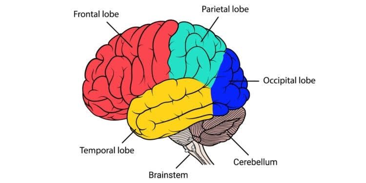
Major Lobes and Regions of the Cerebellum
The cerebellum can be divided into several anatomical lobes and functional regions, each with distinct roles in motor control and coordination.
1. Anatomical Lobes:
- Anterior Lobe: This lobe is located at the front of the cerebellum and is primarily involved in regulating posture and voluntary movement.
- Posterior Lobe: Situated behind the anterior lobe, this lobe plays a crucial role in coordinating fine motor movements and is involved in motor learning.
- Flocculonodular Lobe: This small lobe is located at the bottom of the cerebellum and is essential for balance and eye movements. It receives input from the vestibular system.
2. Zones:
- Vermis: The vermis is the narrow midline region of the cerebellum that connects the two hemispheres. It is involved in controlling posture and locomotion.
- Intermediate Zone: Located on either side of the vermis, this zone contributes to limb coordination and muscle tone.
- Lateral Hemispheres: These are situated laterally to the intermediate zone and are responsible for planning movements, motor learning, and visually guided actions.
3. Functional Divisions:
- Cerebrocerebellum: This division includes the lateral hemispheres and is involved in planning movements, motor learning, and coordination of muscle activation.
- Spinocerebellum: Comprising the vermis and intermediate zones, this area integrates sensory input with motor commands to regulate body posture and movement.
- Vestibulocerebellum: This region corresponds to the flocculonodular lobe and plays a critical role in maintaining balance by processing information from the vestibular system.
In summary, the major lobes of the cerebellum include the anterior lobe, posterior lobe, and flocculonodular lobe. The regions consist of the vermis, intermediate zone, lateral hemispheres, while its functional divisions include cerebrocerebellum, spinocerebellum, and vestibulocerebellum.
Structure of the Cerebellar Cortex
The cerebellar cortex is a highly convoluted layer that encases the deep cerebellar nuclei and is integral to the overall function of the cerebellum. It is composed of three primary layers:
- Molecular Layer: This outermost layer contains few neurons but is rich in synaptic connections. It consists primarily of parallel fibers, which are the axons of granule cells, and various types of inhibitory interneurons, including basket and stellate cells. The molecular layer plays a crucial role in integrating sensory information and modulating motor commands.
- Purkinje Cell Layer: Situated beneath the molecular layer, this middle layer contains large Purkinje cells, which are characterized by their extensive dendritic trees that extend into the molecular layer. Purkinje cells are the sole output neurons of the cerebellar cortex, sending inhibitory signals to the deep cerebellar nuclei. Their activity is critical for coordinating motor control and balance.
- Granule Cell Layer: The innermost layer is densely packed with granule cells, which are small excitatory neurons that receive input from mossy fibers (afferent inputs from various brain regions). Granule cells send their axons into the molecular layer where they bifurcate to form parallel fibers that synapse with Purkinje cells.
The intricate organization of these layers allows for complex processing and integration of sensory and motor information within the cerebellum.
Deep Cerebellar Nuclei and Their Connections
The deep cerebellar nuclei are essential for relaying processed information from the cerebellar cortex to other parts of the central nervous system. There are four primary deep nuclei:
- Fastigial Nucleus: Located medially, it receives input primarily from the vermis region of the cerebellum as well as vestibular and proprioceptive information. It projects mainly to vestibular nuclei and reticular formation, playing a significant role in balance and posture.
- Interposed Nuclei: Comprising two components—the emboliform nucleus and globose nucleus—these nuclei are situated laterally to the fastigial nucleus. They receive inputs from the intermediate zone of the cerebellum and project to contralateral red nucleus, influencing voluntary movement coordination.
- Dentate Nucleus: The largest of all deep nuclei, located laterally, it receives afferent inputs from lateral zones of the cerebellum via corticopontocerebellar tracts. The dentate nucleus projects to various thalamic nuclei (such as ventrolateral thalamus) and then onto motor areas in the cerebral cortex, playing a crucial role in planning and initiating movements.
- Cerebellar Outputs: All outputs from these nuclei ultimately influence motor control pathways through descending projections that affect spinal cord motor neurons or relay signals back to higher brain centers involved in movement regulation.
In summary, both the structure of the cerebellar cortex with its layered arrangement and its associated deep nuclei with specific connections facilitate complex functions related to motor control, balance, coordination, and even cognitive processes.
Afferent and Efferent Connections of the Cerebellum
The cerebellum, often referred to as the “little brain,” plays a crucial role in motor control and coordination. Its connections can be categorized into afferent (incoming) and efferent (outgoing) pathways, which are organized through three pairs of cerebellar peduncles: the superior, middle, and inferior cerebellar peduncles.
Afferent Connections
Afferent connections to the cerebellum primarily originate from various regions of the central nervous system. These inputs provide sensory information that is essential for the cerebellum’s functions in motor control, balance, and coordination. The main sources of afferent fibers include:
- Mossy Fibers: These fibers arise from several sources:
- Pontine Nuclei: They relay information from the cerebral cortex regarding planned movements.
- Vestibular Nuclei: They provide input related to balance and spatial orientation.
- Spinal Cord: They convey proprioceptive information about body position and movement.
Mossy fibers synapse with granule cells in the cerebellar cortex, which then project parallel fibers that interact with Purkinje cells.
- Climbing Fibers: These fibers originate from the inferior olivary nucleus. Climbing fibers have a direct excitatory effect on Purkinje cells by synapsing on their proximal dendrites and cell bodies. They play a critical role in error signaling during motor tasks.
- Other Inputs: Additional afferent pathways include inputs from the reticular formation and other brainstem nuclei that contribute to the overall sensory integration within the cerebellum.
The arrangement of these afferent connections is primarily through the middle and inferior cerebellar peduncles:
- The middle cerebellar peduncle carries predominantly mossy fibers from the pontine nuclei.
- The inferior cerebellar peduncle transmits both mossy fibers (from spinal cord and vestibular nuclei) and climbing fibers (from the inferior olivary nucleus).
Efferent Connections
Efferent connections from the cerebellum are mainly directed towards various motor control centers in the brain. The primary output structures are located in four deep cerebellar nuclei:
- Dentate Nucleus
- Emboliform Nucleus
- Globose Nucleus
- Fastigial Nucleus
These nuclei send projections to different parts of the central nervous system, including:
- The thalamus (specifically ventrolateral nucleus), which relays signals to motor areas of the cerebral cortex.
- Brainstem nuclei involved in motor control, such as those regulating posture and balance.
The efferent pathways exit through:
- The superior cerebellar peduncle, which primarily carries outputs from the dentate nucleus to thalamic targets.
- The fastigial nucleus sends projections via both superior and inferior peduncles to influence autonomic functions related to balance.
In summary, afferent connections predominantly enter through middle and inferior peduncles while efferent signals exit mainly via the superior peduncle, establishing a complex network for integrating sensory input with motor output.
Major Functions of the Cerebellum
The cerebellum, often referred to as the “little brain,” plays a crucial role in various functions that are essential for motor control and cognitive processes. Its major functions can be categorized as follows:
1. Maintenance of Balance and Posture
The cerebellum is integral to maintaining balance and posture. It receives input from vestibular receptors (which detect changes in head position) and proprioceptors (which provide information about body position). By processing this sensory information, the cerebellum modulates motor commands sent to motor neurons, allowing for adjustments that help maintain stability during movement. Individuals with cerebellar damage often experience balance disorders, leading them to adopt compensatory postural strategies.
2. Coordination of Voluntary Movements
Another primary function of the cerebellum is the coordination of voluntary movements. It ensures that different muscle groups work together in a temporally coordinated manner, allowing for smooth and fluid motions. The cerebellum fine-tunes the timing and force of muscle contractions, which is vital for activities such as reaching for an object or playing a musical instrument.
3. Motor Learning
The cerebellum plays a significant role in motor learning, which involves adapting and refining motor programs through practice and experience. This process often occurs via trial-and-error mechanisms, enabling individuals to improve their performance over time. For example, when learning to hit a baseball or ride a bicycle, the cerebellum helps adjust movements based on feedback from previous attempts.
4. Cognitive Functions
While traditionally associated with motor control, recent research has shown that the cerebellum also contributes to certain cognitive functions, including aspects of language processing and executive functioning. Although these roles are not yet fully understood, they indicate that the cerebellum’s influence extends beyond purely motor tasks.
Control Mechanism: Ipsilateral Control
Each side of the cerebellum primarily controls the ipsilateral side of the body due to its unique neural pathways. The mechanism can be explained as follows:
- Cerebellar Inputs: The cerebellum receives sensory input from various parts of the body through multiple pathways.
- Cerebral Cortex Connection: The information processed by one hemisphere of the cerebellum is relayed back to corresponding areas in the cerebral cortex via deep nuclei (e.g., dentate nucleus).
- Decussation: While many pathways cross over (decussate) at different levels within the central nervous system, particularly at brainstem levels before reaching spinal cord targets, it is important to note that outputs from each side of the cerebellum predominantly influence muscles on the same side (ipsilateral) due to how these connections are organized.
In summary, while there are complex interactions involving contralateral pathways in some instances (especially concerning higher-order processing), each hemisphere’s primary influence remains on its respective side of the body.
Effects of Lesions of the Cerebellum
Lesions in the cerebellum can lead to a variety of motor disorders due to its critical role in coordinating voluntary movements, maintaining balance, and regulating muscle tone. The cerebellum is divided into several regions, each responsible for different aspects of motor control. Damage to these areas can result in specific deficits.
- Coordination Impairments: One of the most prominent effects of cerebellar lesions is ataxia, which refers to a lack of coordination during voluntary movements. This can manifest as:
- Dysmetria: The inability to judge distances accurately, leading to overshooting (hypermetria) or undershooting (hypometria) targets.
- Decomposition of Movement: Movements may be broken down into smaller parts rather than being smooth and fluid.
- Dysdiadochokinesia: Difficulty performing rapid alternating movements, such as pronation and supination of the hands.
- Gait Abnormalities: Individuals with cerebellar lesions often exhibit an unsteady gait characterized by:
- Broad-based Stance: A wider stance to maintain balance.
- Irregular Steps: Steps may be irregular and staggering, with frequent corrections or falls.
- Reduced Speed and Rhythm Disturbances: Gait may be slower with an altered rhythm.
- Speech Deficits: Ataxic dysarthria is common in patients with cerebellar damage. This condition is characterized by:
- Scanning Speech: Speech may have a staccato rhythm and can be explosive or slurred.
- Unintelligibility: Words may become difficult to understand due to poor articulation.
- Oculomotor Disturbances: Lesions can also affect eye movements leading to:
- Nystagmus: Involuntary eye movements that can disrupt visual stability.
- Gaze-Evoked Nystagmus: A common form where the eyes drift away from a target and then quickly snap back.
- Tremors and Muscle Tone Changes: Patients may experience:
- Cerebellar Tremor: Low-frequency oscillations that occur during movement (kinetic tremor) or when maintaining posture (postural tremor).
- Hypotonia or Hypertonia: Altered muscle tone can lead to either decreased muscle tension (hypotonia) or increased tension (hypertonia).
- Balance Issues: The inability to maintain equilibrium is a significant consequence of cerebellar lesions, leading to:
- Increased body sway and instability while standing.
- Difficulty in performing tasks that require fine motor skills due to compromised proprioception.
In summary, lesions in the cerebellum result in a constellation of motor disorders characterized by impaired coordination, abnormal gait patterns, speech difficulties, oculomotor disturbances, tremors, and balance issues.


