Types of Brain Hemorrhages
Brain hemorrhages, also known as intracranial hemorrhages, can be classified based on their location within the skull and brain. Understanding these types is crucial for diagnosis and treatment. Here are the different types of brain hemorrhages:
1. Intracerebral Hemorrhage (ICH)
This type of bleeding occurs within the brain tissue itself. It can happen in various regions of the brain but is most commonly found in areas such as the brainstem, cerebellum, and cerebral lobes. The bleeding leads to increased pressure on surrounding brain structures and can disrupt normal brain function.
2. Intraventricular Hemorrhage (IVH)
Intraventricular hemorrhage refers to bleeding that occurs in the ventricles, which are the fluid-filled cavities within the brain. This type of hemorrhage can complicate other forms of bleeding and may lead to serious complications due to its impact on cerebrospinal fluid circulation.
3. Epidural Hemorrhage
Epidural hemorrhage occurs between the skull and the dura mater, which is the outermost protective layer covering the brain. This type of bleed is often associated with traumatic head injuries and can lead to rapid increases in intracranial pressure.
4. Subdural Hemorrhage
Subdural hemorrhage happens just beneath the dura mater. It typically results from tearing of veins due to head trauma or sudden movement of the head. Symptoms may develop slowly over time as blood accumulates.
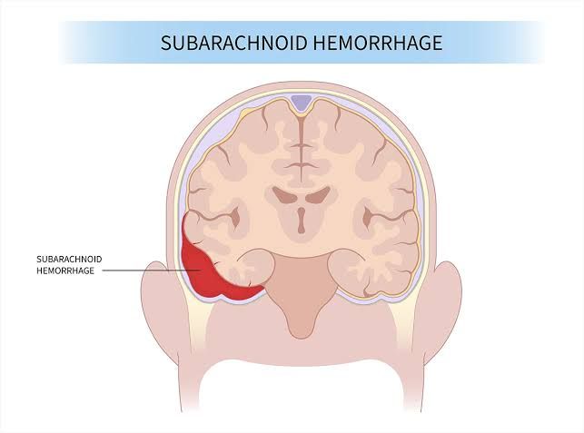
5. Subarachnoid Hemorrhage (SAH)
This type of hemorrhage occurs between the brain and the tissues that cover it, specifically in the subarachnoid space where cerebrospinal fluid circulates. Subarachnoid hemorrhages are often caused by ruptured aneurysms or head injuries and are considered a medical emergency.
Each type of brain hemorrhage has distinct causes, symptoms, and implications for treatment, making accurate identification essential for effective management.
Differentiating Between Subarachnoid and Intracerebral Hemorrhage
Definition
A subarachnoid hemorrhage (SAH) is defined as bleeding that occurs in the space between the brain and the tissues covering it, specifically in the subarachnoid space. This type of hemorrhage often results from the rupture of an aneurysm or arteriovenous malformation.
In contrast, an intracerebral hemorrhage (ICH) refers to bleeding that occurs within the brain tissue itself. This can be caused by various factors, including hypertension, trauma, or vascular malformations.
Causes
The causes of subarachnoid hemorrhage primarily include:
- Ruptured cerebral aneurysms.
- Arteriovenous malformations.
- Trauma to the head.
- Blood disorders that affect clotting.
On the other hand, intracerebral hemorrhage can result from:
- Chronic hypertension leading to small vessel disease.
- Trauma or head injury.
- Use of anticoagulant medications.
- Tumors that invade blood vessels.
Symptoms
The symptoms associated with subarachnoid hemorrhage typically include:
- Sudden severe headache often described as a “thunderclap” headache.
- Nausea and vomiting.
- Stiff neck due to irritation of the meninges.
- Sensitivity to light (photophobia).
- Loss of consciousness or altered mental status.
For intracerebral hemorrhage, symptoms may vary but commonly include:
- Sudden onset of headache (less intense than SAH).
- Neurological deficits such as weakness on one side of the body.
- Changes in consciousness or alertness.
- Nausea and vomiting.
- Seizures in some cases.
Diagnosis
To diagnose a subarachnoid hemorrhage, a computed tomography (CT) scan is typically performed first, followed by a cerebral angiogram if SAH is confirmed to assess for any underlying aneurysms.
For diagnosing an intracerebral hemorrhage, CT scans are also used to identify areas of bleeding within the brain tissue, along with magnetic resonance imaging (MRI) when necessary for further evaluation.
Treatment Options
Treatment for a subarachnoid hemorrhage may involve surgical intervention such as craniotomy to clip an aneurysm or neuroendovascular therapy using coils to occlude the aneurysm without open surgery.
In contrast, treatment for an intracerebral hemorrhage depends on its cause and severity; it may require surgical evacuation of hematomas or medical management aimed at controlling blood pressure and preventing further bleeding.
In summary, while both types of hemorrhages involve bleeding related to cerebrovascular issues, they differ significantly in their locations, causes, symptoms, diagnostic approaches, and treatment strategies.
Understanding the Presentation of Subarachnoid Haemorrhage
The presentation of SAH can vary, but common symptoms include:
- Sudden Severe Headache: Often described as a “thunderclap” headache or the worst headache ever experienced.
- Nausea and Vomiting: This may occur due to increased intracranial pressure or irritation of the meninges.
- Photophobia: Sensitivity to light is common due to meningeal irritation.
- Neck Stiffness: This can be a sign of meningeal irritation.
- Altered Consciousness: Patients may experience confusion, drowsiness, or loss of consciousness.
- Seizures: Some patients may present with seizures due to irritation of the cerebral cortex.
Diagnosis typically involves imaging studies such as a CT scan or MRI, which can reveal blood in the subarachnoid space.
Different Causes of SAH
The causes of SAH can be classified into several categories:
- Aneurysmal Rupture: The most common cause, where a cerebral aneurysm bursts, leading to bleeding.
- Arteriovenous Malformations (AVMs): Abnormal connections between arteries and veins can rupture and cause SAH.
- Trauma: Head injuries can lead to SAH through direct damage to blood vessels.
- Coagulopathy: Disorders that affect blood clotting can increase the risk of spontaneous bleeding.
- Vascular Disease: Conditions such as hypertension or vasculitis can weaken blood vessel walls and lead to hemorrhage.
- Idiopathic Cases: In some instances, no clear cause for SAH can be identified.
Surgical Management of Different Causes of SAH
The management of SAH often requires surgical intervention depending on its underlying cause:
- Aneurysmal SAH:
- Clipping: A neurosurgeon places a metal clip at the base of an aneurysm to prevent further bleeding.
- Endovascular Coiling: A less invasive procedure where coils are inserted into the aneurysm via catheterization to promote clotting and seal off the aneurysm from circulation.
- AVM-related SAH:
- Surgical Resection: The AVM may be surgically removed if accessible and safe to do so.
- Embolization: Endovascular techniques are used to block blood flow to the AVM before surgical resection.
- Traumatic SAH:
- Management depends on associated injuries; surgical intervention may involve decompression or evacuation if there is significant mass effect or ongoing bleeding.
- Coagulopathy-related SAH:
- Treatment focuses on correcting coagulopathy (e.g., administering clotting factors) rather than direct surgical intervention unless there is significant hemorrhage requiring evacuation.
- Supportive Care and Monitoring:
- Regardless of cause, patients with SAH require intensive monitoring in a critical care setting for complications such as rebleeding, vasospasm, and hydrocephalus.
In summary, timely diagnosis and appropriate surgical management are crucial in improving outcomes for patients with subarachnoid hemorrhage.
Diagnosis of Subarachnoid Hemorrhage (SAH)
To diagnose subarachnoid hemorrhage (SAH), it is crucial to consider the clinical presentation and then proceed with appropriate investigations.
- Clinical Presentation:
- Patients typically present with a sudden onset of a severe headache, often described as a “thunderclap” headache or the “worst headache of their life.”
- Accompanying symptoms may include nausea, vomiting, photophobia, neck stiffness, and altered consciousness.
- A thorough history should be taken to assess for risk factors such as family history of aneurysms, personal history of connective tissue disorders (like Ehlers-Danlos syndrome), hypertension, smoking, and substance abuse.
- Initial Investigations:
- CT Scan: The first-line investigation for suspected SAH is a non-contrast computed tomography (CT) scan of the head. This imaging modality is highly sensitive in detecting acute hemorrhage within the subarachnoid space.
- If the CT scan is negative but suspicion remains high, a lumbar puncture may be performed to analyze cerebrospinal fluid (CSF) for xanthochromia or red blood cells.
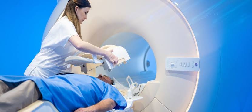
- MRI: Magnetic resonance imaging (MRI) can also be utilized if there are concerns about other intracranial pathologies or if CT findings are inconclusive.
- Angiography: Once SAH is confirmed, cerebral angiography (either CT or digital subtraction angiography) should be performed to identify any ruptured aneurysms or vascular malformations.
- CT Scan: The first-line investigation for suspected SAH is a non-contrast computed tomography (CT) scan of the head. This imaging modality is highly sensitive in detecting acute hemorrhage within the subarachnoid space.
Medical Management of SAH
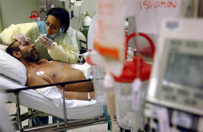
The management of SAH involves several critical steps aimed at stabilizing the patient and preventing complications:
- Initial Stabilization:
- Ensure airway protection and provide supplemental oxygen as needed.
- Establish intravenous access for fluid resuscitation and medication administration.
- Blood Pressure Management:
- Maintain systolic blood pressure within a target range (typically 140-160 mmHg) to prevent rebleeding while avoiding hypotension which could compromise cerebral perfusion.
- Pain Management:
- Administer analgesics for headache relief while being cautious with medications that could affect coagulation.
- Nimodipine Administration:
- Start oral nimodipine within 96 hours of SAH diagnosis to reduce the risk of delayed cerebral ischemia due to vasospasm.
- Monitoring for Complications:
- Continuous neurological assessment using standardized scales like the Glasgow Coma Scale (GCS).
- Monitor vital signs closely for signs of increased intracranial pressure or cardiovascular instability.
- Preventing Secondary Complications:
- Implement measures to prevent deep vein thrombosis (DVT) through early mobilization when feasible and use of compression devices.
- Manage electrolyte imbalances and monitor renal function closely.
- Surgical Intervention:
- Depending on the patient’s condition and findings from angiography, surgical options such as clipping or endovascular coiling may be indicated to secure any identified aneurysms.
- Multidisciplinary Approach:
- Involve neurosurgery, neurocritical care specialists, nursing staff trained in neurocritical care protocols, and rehabilitation services early in management to optimize outcomes.
By following these steps systematically, healthcare providers can effectively diagnose and manage patients with SAH while minimizing morbidity and mortality associated with this serious condition.
Understanding CT Scans of Subarachnoid Hemorrhage (SAH) and Intracerebral Hemorrhage (ICH)
1. CT Scan Evaluation of SAH
Computed tomography (CT) is the preferred initial imaging modality for diagnosing SAH due to its rapid acquisition time and high sensitivity for detecting acute hemorrhages. On a non-contrast CT scan, SAH typically appears as hyperdense areas in the subarachnoid space. The blood accumulates in the cerebral sulci, particularly overlying the convexities of the brain. In cases of significant trauma or severe hemorrhage, it may also be distributed diffusely throughout the subarachnoid space.
The typical appearance of SAH on CT includes:
- Hyperdensity: Blood appears brighter than surrounding cerebrospinal fluid (CSF).
- Distribution: Blood often collects in the sulci near the vertex of the skull while sparing basal cisterns unless there is extensive bleeding.
- Timing: The detection of SAH can be influenced by timing; small amounts may not be visible if imaging occurs several days after onset.
In addition to standard CT scans, advanced techniques such as CT angiography can help identify potential sources of bleeding like ruptured aneurysms or vascular malformations.
2. CT Scan Evaluation of ICH
Intracerebral hemorrhage is identified on CT scans as hyperdense areas within brain parenchyma. The appearance varies depending on factors such as age of bleed and location:
- Acute ICH: Appears hyperdense relative to surrounding brain tissue due to fresh blood.
- Chronic ICH: Over time, blood products undergo changes that may alter their density; older clots may appear hypodense compared to acute bleeds.
Common types of ICH identifiable on CT include:
- Epidural Hematoma: Characterized by a lens-shaped hyperdensity between skull and dura mater.
- Subdural Hematoma: Appears as crescent-shaped hyperdensity along the inner surface of the skull.
- Hemorrhagic Contusions: These are localized areas where blood accumulates due to trauma and appear as irregular hyperdense regions within brain tissue.
The evaluation process involves assessing not only for presence but also for volume and midline shift, which indicates increased intracranial pressure or mass effect from hematomas.
Conclusion
In summary, both SAH and ICH can be effectively evaluated using CT imaging techniques. Understanding their appearances on CT scans is crucial for timely diagnosis and management. Immediate recognition allows healthcare providers to initiate appropriate interventions that could significantly impact patient outcomes.
Presentation of Hypertensive Intracerebral Hemorrhage
Hypertensive intracerebral hemorrhage (ICH) typically presents with a sudden onset of neurological symptoms, which can vary based on the location and extent of the hemorrhage. The most common symptoms include:
- Headache: Patients often report a severe headache that may be described as a “thunderclap” headache.
- Altered Consciousness: This can range from confusion to complete loss of consciousness, depending on the severity of the hemorrhage.
- Neurological Deficits: Weakness or numbness is usually noted on one side of the body (hemiparesis), affecting the face, arm, or leg. This is due to damage in specific areas of the brain responsible for motor control.
- Nausea and Vomiting: These symptoms may accompany other neurological signs due to increased intracranial pressure.
- Seizures: Some patients may experience seizures as a result of irritation to the brain tissue caused by blood accumulation.
The presentation can occur progressively over minutes to hours, making it crucial for patients experiencing these symptoms to seek immediate medical attention.
Management of Intracerebral Hemorrhage
The management of intracerebral hemorrhage focuses on several key objectives: stopping the bleeding, relieving pressure on the brain, and addressing any underlying causes. The treatment approach includes:
- Immediate Medical Care: Patients should be stabilized in an emergency setting. This includes monitoring vital signs and performing a rapid assessment using imaging studies such as non-contrast computed tomography (CT) scans to confirm ICH and assess its size and location.
- Control Blood Pressure: Since hypertension is a primary cause of ICH, controlling blood pressure is critical. Medications such as intravenous antihypertensives may be administered to lower blood pressure safely.
- Surgical Intervention:
- Craniotomy: In cases where there is significant hematoma volume or mass effect causing increased intracranial pressure, surgical evacuation may be necessary.
- Minimally Invasive Techniques: Newer approaches involve minimally invasive surgery techniques that allow for clot removal with less trauma compared to traditional craniotomy.
- Supportive Care: This includes managing complications such as seizures, hydrocephalus (accumulation of cerebrospinal fluid), and maintaining adequate oxygenation and nutrition.
- Rehabilitation Services: Post-stabilization, rehabilitation services are essential for recovery, focusing on physical therapy, occupational therapy, and speech therapy depending on the deficits experienced by the patient.
- Monitoring for Complications: Continuous monitoring for potential complications such as rebleeding or delayed deterioration due to swelling is important during hospitalization.
In summary, hypertensive intracerebral hemorrhage presents acutely with neurological deficits requiring urgent medical intervention focused on stabilization and management strategies tailored to individual patient needs.


