Definition of Joints
Joints, also known as articulations, are connections between two or more bones in the skeletal system. They play a crucial role in providing stability to the skeleton while allowing for various types of movement. The structure and function of joints can vary significantly, depending on their classification.
Classification of Joints Based on Uniting Material
Joints can be classified based on the type of connective tissue that unites the bones. This classification includes three primary categories:
- Fibrous Joints: In fibrous joints, the bones are connected by dense connective tissue, primarily composed of collagen fibers. These joints typically allow little to no movement and are further sub-classified into:
- Sutures: Immovable joints found between the flat bones of the skull.
- Gomphoses: Immovable joints where teeth articulate with their sockets in the maxilla and mandible.
- Syndesmoses: Slightly movable joints where bones are connected by an interosseous membrane (e.g., middle radioulnar joint).
- Cartilaginous Joints: These joints unite bones through cartilage, either hyaline cartilage or fibrocartilage. They allow more movement than fibrous joints but less than synovial joints and are divided into:
- Synchondroses: Immovable joints connected by hyaline cartilage (e.g., growth plates).
- Symphyses: Slightly movable joints united by fibrocartilage (e.g., pubic symphysis).
- Synovial Joints: Characterized by a fluid-filled joint cavity surrounded by a fibrous capsule, synovial joints permit free movement and are classified into several types based on their shape and movement capabilities:
- Hinge (e.g., elbow joint)
- Saddle (e.g., carpometacarpal joint)
- Plane (e.g., acromioclavicular joint)
- Pivot (e.g., atlantoaxial joint)
- Condyloid (e.g., wrist joint)
- Ball and Socket (e.g., hip joint)
These classifications highlight how different types of connective tissue contribute to the functional characteristics of each joint type.
Overview of a Synovial Joint
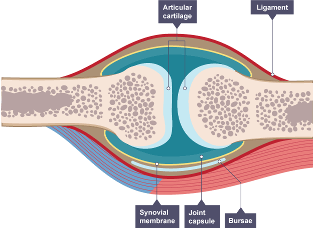
A synovial joint is characterized by the presence of a fluid-filled joint cavity contained within a fibrous capsule. It is the most common type of joint found in the human body and allows for a wide range of motion due to its unique structure. Synovial joints are classified as diarthroses, meaning they are freely mobile.
Features of Synovial Joints
- Articular Capsule: The articular capsule surrounds the joint and is continuous with the periosteum of the articulating bones. It consists of two layers:
- Fibrous Layer (Outer): Made up of white fibrous tissue known as capsular ligament, which holds together the articulating bones and supports the underlying synovium.
- Synovial Layer (Inner): A highly vascularized layer of serous connective tissue that absorbs and secretes synovial fluid, mediating nutrient exchange between blood and the joint.
- Articular Cartilage: The articulating surfaces of a synovial joint are covered by a thin layer of hyaline cartilage. This cartilage minimizes friction during movement and absorbs shock.
- Synovial Fluid: Located within the joint cavity, synovial fluid serves three primary functions:
- Lubrication: Reduces friction between articular cartilages during movement.
- Nutrient Distribution: Provides nutrients to avascular articular cartilage through passive diffusion.
- Shock Absorption: Helps absorb shocks during activities such as walking or running.
- Accessory Structures:
- Accessory Ligaments: These ligaments can be separate or part of the joint capsule, consisting of dense regular connective tissue that resists strain.
- Bursae: Small sacs lined by synovial membrane filled with synovial fluid that reduce friction at key points in joints.
- Innervation: Synovial joints have a rich supply from articular nerves, which provide sensory feedback regarding pain and proprioception.
- Additional Features:
- Fat pads that cushion between bones.
- Intrinsic and intracapsular ligaments that provide stability.
These features collectively enable synovial joints to facilitate smooth movements while protecting against injury.
Classification of Synovial Joints
1. Shape of Articulating Surfaces
Synovial joints can be classified based on the shape of their articulating surfaces into six main types:
- Hinge Joints: These joints allow movement primarily in one plane, facilitating flexion and extension. An example is the elbow joint.
- Saddle Joints: Characterized by opposing articular surfaces that have a concave-convex shape, allowing for a greater range of motion than hinge joints. The carpometacarpal joint at the base of the thumb is an example.
- Plane Joints: These joints feature relatively flat articular surfaces, permitting gliding movements between bones. An example is the acromioclavicular joint.
- Pivot Joints: These allow for rotational movement around a single axis. A classic example is the atlantoaxial joint, which enables side-to-side rotation of the head.
- Condyloid Joints: These joints permit movement in two planes (flexion/extension and abduction/adduction). The radiocarpal joint of the wrist serves as an example.
- Ball and Socket Joints: This type allows for movement in multiple directions and rotation around an axis. The hip joint is a prime example of a ball and socket joint.
2. Degree of Mobility
Synovial joints are also classified according to their degree of mobility:
- Diarthrosis: This term describes freely movable joints, which include all synovial joints. They allow for a wide range of motion due to their structure and design.
In summary, synovial joints can be classified based on both the shape of their articulating surfaces (hinge, saddle, plane, pivot, condyloid, ball and socket) and their degree of mobility (diarthrosis).
Innervation of Synovial Joints
The innervation of synovial joints is characterized by a rich supply of articular nerves. These nerves are responsible for transmitting sensory information from the joint to the central nervous system. The principles governing the innervation can be summarized as follows:
- Hilton’s Law: This principle states that the nerves supplying a joint also supply the muscles moving the joint and the skin covering their distal attachments. This means that sensory information related to joint position (proprioception) and pain (nociception) is conveyed through these nerves.
- Afferent Impulses: Articular nerves transmit afferent impulses, which include proprioceptive information that helps in understanding the position and movement of the joint, as well as nociceptive signals that indicate pain or discomfort within the joint.
- Distribution: The distribution of these nerves typically follows a pattern where they branch out from nearby structures, ensuring comprehensive coverage of the joint area.
Blood Supply of Synovial Joints
The blood supply to synovial joints is primarily provided by articular arteries, which arise from surrounding vessels. The key aspects of this blood supply include:
- Articular Arteries: These arteries are located within the joint capsule, predominantly in the synovial membrane. They ensure an adequate blood supply to both the synovium and other structures within the joint.
- Anastomoses: A common feature among articular arteries is frequent anastomoses, which are connections between adjacent arteries. This anatomical arrangement ensures that blood flow to and across the joint remains consistent regardless of its position during movement.
- Accompanying Veins: The venous drainage of synovial joints is facilitated by articular veins that accompany their respective arteries within the synovial membrane, helping to return deoxygenated blood back to circulation.
In summary, synovial joints receive a rich nerve supply for sensory feedback and motor control through articular nerves following Hilton’s Law, while their vascularization is ensured by a network of articular arteries with anastomoses for consistent blood flow.


