Specialized Excitatory and Conductive System of the Heart
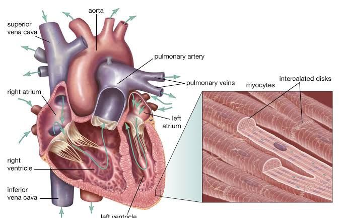
The heart’s specialized excitatory and conductive system is a complex network that plays a crucial role in maintaining the rhythm and coordination of heartbeats. This system consists of specialized cells that generate and conduct electrical impulses, ensuring that the heart contracts in a synchronized manner to effectively pump blood throughout the body.
1. Sinoatrial (SA) Node
The SA node, often referred to as the natural pacemaker of the heart, is located in the upper wall of the right atrium near the opening of the superior vena cava. It is composed of specialized pacemaker cells that generate electrical impulses spontaneously. These impulses initiate each heartbeat by causing the atria to contract, pushing blood into the ventricles. The rate at which the SA node fires is influenced by autonomic nervous system inputs, allowing it to increase or decrease heart rate based on physiological demands such as exercise or rest.
2. Atrioventricular (AV) Node
After originating from the SA node, electrical signals travel to the AV node, situated at the junction between the atria and ventricles. The AV node serves as a critical relay point for these signals; it introduces a slight delay (approximately 0.1 seconds) before transmitting them to ensure that the atria have fully contracted and emptied their blood into the ventricles before ventricular contraction begins. This delay is essential for effective cardiac function.
3. Bundle of His
From the AV node, electrical impulses are transmitted through a collection of fibers known as the Bundle of His (or atrioventricular bundle). This structure extends down through the interventricular septum and branches into two pathways: one for each ventricle (the right bundle branch and left bundle branch). The Bundle of His conducts impulses rapidly from the AV node toward both ventricles.
4. Purkinje Fibers
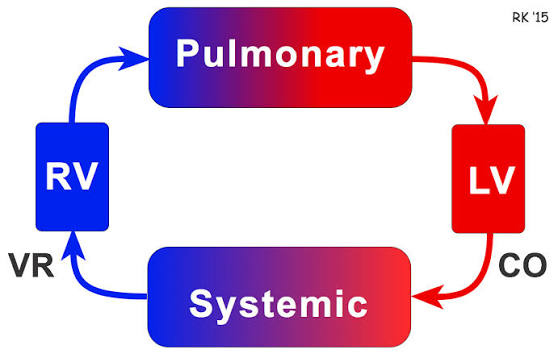
The final component of this conduction system consists of Purkinje fibers, which are specialized nerve-like fibers that spread throughout the ventricular walls. These fibers receive impulses from both branches of the Bundle of His and distribute them quickly across both ventricles, leading to their simultaneous contraction. This coordinated contraction is vital for efficiently pumping blood out of both ventricles—into pulmonary arteries from the right ventricle and into systemic circulation via the aorta from the left ventricle.
5. Summary of Electrical Conduction Pathway
To summarize, an electrical impulse starts at the SA node, travels to the AV node where it is delayed briefly, then moves down through the Bundle of His and its branches before reaching Purkinje fibers in both ventricles. This sequence ensures that blood flows efficiently through both chambers during each heartbeat.
In conclusion, this intricate network allows for precise control over heart rhythm and timing, adapting to various physiological needs while maintaining overall cardiovascular health.
Overview of Primary Pacemaker
The primary pacemaker of the heart is the sinoatrial (SA) node, which is located in the upper part of the right atrium near the entrance of the superior vena cava. The SA node consists of specialized cardiac muscle cells known as pacemaker cells that have the unique ability to spontaneously generate electrical impulses. These impulses initiate the heart’s rhythm and are responsible for setting the pace of contraction for the entire heart. The intrinsic firing rate of these pacemaker cells is approximately 100 beats per minute under normal physiological conditions, but this rate can be modulated by autonomic nervous system influences, resulting in an average resting heart rate of about 70 beats per minute in adults.
The SA node’s role as the primary pacemaker is crucial because it ensures that electrical impulses spread throughout the atria, leading to coordinated contraction and effective pumping of blood into the ventricles. If there is any dysfunction or damage to the SA node, other structures in the heart can take over as secondary pacemakers, such as the atrioventricular (AV) node or Bundle of His, but these typically operate at a slower intrinsic rate.
Function of Internodal Pathways
Internodal pathways are specialized conduction pathways that facilitate rapid transmission of electrical impulses from the SA node to the AV node. There are three main internodal pathways: Bachmann’s bundle, Wenckebach’s pathway, and Thorel’s pathway. Each pathway serves a specific route for impulse conduction:
- Bachmann’s Bundle: This pathway extends from the anterior part of the SA node and bifurcates to deliver impulses directly to the left atrium. It plays a critical role in ensuring that both atria contract simultaneously.
- Wenckebach’s Pathway: This middle pathway runs posteriorly from the superior part of the SA node and descends within the interatrial septum before connecting with other pathways leading to the AV node. It helps coordinate impulse transmission between both atria and facilitates communication with subsequent nodes.
- Thorel’s Pathway: This posterior pathway extends from the inferior part of the SA node through structures like crista terminalis and Eustachian ridge before reaching areas around coronary sinus and entering into AV node territory.
These internodal pathways ensure that electrical signals travel efficiently across both atria before reaching the AV node, allowing for synchronized contraction and optimal cardiac function. They also help maintain a clear distinction between fast conduction routes (which allow rapid impulse transmission) and slower routes (which may be utilized during higher heart rates).
In summary, while the primary pacemaker is defined as the sinoatrial (SA) node, which generates spontaneous electrical impulses to regulate heart rhythm, the internodal pathways serve as specialized conduits that facilitate rapid transmission of these impulses from the SA node to the atrioventricular (AV) node, ensuring coordinated contraction across both atria.
What are Purkinje Fibers?
Purkinje fibers are specialized cardiac muscle fibers that play a crucial role in the heart’s electrical conduction system. Named after the Czech scientist Jan Evangelista Purkyně, who discovered them in 1839, these fibers are located in the inner ventricular walls of the heart, specifically beneath the endocardium within a region known as the subendocardium.
Structure and Composition
Purkinje fibers are distinct from regular cardiomyocytes (heart muscle cells) in several ways. They are larger than typical myocardial cells and contain fewer myofibrils, which are the contractile units of muscle tissue. Instead, they have a higher concentration of mitochondria and glycogen. The presence of glycogen gives Purkinje fibers a lighter appearance under microscopic examination compared to surrounding muscle cells. Additionally, they often appear binucleated (having two nuclei) and are arranged longitudinally along the cardiac vector.
Function in Cardiac Conduction
The primary function of Purkinje fibers is to conduct electrical impulses rapidly throughout the ventricles during the cardiac cycle. They receive signals from both left and right bundle branches of the heart’s conduction system and transmit these impulses to the myocardium (the muscular layer of the heart). This rapid conduction is essential for ensuring synchronized contractions of the ventricles, allowing for effective pumping of blood either into pulmonary circulation from the right ventricle or systemic circulation from the left ventricle.
Role as Pacemaker Cells
While Purkinje fibers primarily serve as conduits for electrical impulses, they also possess intrinsic pacemaking abilities. In normal circumstances, their activity is suppressed by faster pacemaker activity from the sinoatrial (SA) node, which typically fires at a rate of 60-100 beats per minute. However, if there is compromise in upstream conduction or pacemaking ability (such as failure of the SA node), Purkinje fibers can act as backup pacemakers, firing at a slower rate of 20-40 beats per minute. When this occurs, it may result in premature ventricular contractions (PVCs) or other forms of escape rhythms.
Conclusion
In summary, Purkinje fibers are specialized conducting fibers within the heart that facilitate rapid impulse transmission necessary for coordinated ventricular contraction. Their unique structural characteristics enable them to perform this function efficiently while also providing an alternative pacemaking capability when needed.
Definition of a Cardiac Myocyte
A cardiac myocyte, also known as a cardiomyocyte, is defined as a muscle cell of the heart. These cells are specialized for the contraction and relaxation processes that enable the heart to pump blood throughout the body. Cardiac myocytes are unique in their structure and function compared to other muscle cells, such as skeletal or smooth muscle cells.
Characteristics of Cardiac Myocytes
- Structure: Cardiac myocytes are striated muscle cells that contain organized bundles of actin and myosin filaments, which are essential for muscle contraction. They have a single central nucleus and are interconnected by intercalated discs, which facilitate synchronized contractions by allowing electrical impulses to pass rapidly between cells.
- Function: The primary role of cardiac myocytes is to contract in response to electrical signals generated by pacemaker cells in the heart. This contraction is rhythmic and involuntary, allowing the heart to maintain a consistent heartbeat necessary for effective blood circulation.
- Regeneration: Unlike skeletal muscle, cardiac myocytes have limited regenerative capacity. Damage to these cells due to injury or disease can lead to heart failure since they cannot easily proliferate or replace themselves.
- Metabolism: Cardiac myocytes primarily rely on aerobic metabolism for energy production, utilizing fatty acids and glucose as fuel sources. This metabolic preference supports their continuous activity and endurance required for sustained heart function.
- Clinical Relevance: A deficiency of cardiac myocytes underlies many cases of heart failure, prompting research into methods for regenerating these cells through techniques such as stem cell therapy and cardiac reprogramming.
In summary, cardiac myocytes play a crucial role in maintaining heart function through their unique structural characteristics and specialized functions that allow them to contract rhythmically and efficiently.
Excitation and Contraction of Cardiac Myocyte
The process of excitation and contraction in cardiac myocytes (heart muscle cells) is a complex sequence of events that involves electrical signals leading to mechanical contraction. This process can be broken down into several key steps:
- Initiation of Action Potential: The excitation begins in the sinoatrial (SA) node, which acts as the heart’s natural pacemaker. The SA node generates an action potential due to the influx of sodium ions (Na+) through voltage-gated ion channels. This depolarization spreads through the atria, causing them to contract.
- Propagation Through the Heart: The action potential travels from the SA node to the atrioventricular (AV) node, where it is briefly delayed to allow for adequate ventricular filling. From there, it moves rapidly through the His bundle, bundle branches, and Purkinje fibers, ensuring that the ventricles contract almost simultaneously.
- Calcium Release and Contraction: As the action potential reaches the cardiac myocytes, it causes voltage-gated calcium channels in the sarcolemma (cell membrane) to open. Calcium ions (Ca2+) enter the cell from the extracellular space and also trigger further release of Ca2+ from the sarcoplasmic reticulum (SR), a specialized organelle that stores calcium.
- Cross-Bridge Cycling: The increase in intracellular calcium concentration leads to binding of calcium to troponin, a regulatory protein associated with actin filaments in muscle cells. This binding causes a conformational change that allows myosin heads to attach to actin filaments, initiating cross-bridge cycling and resulting in muscle contraction.
- Relaxation: After contraction, calcium ions are pumped back into the SR and out of the cell by various transport mechanisms, leading to a decrease in intracellular calcium levels. This reduction allows troponin to return to its original shape, blocking myosin binding sites on actin and resulting in muscle relaxation.
Role of Gap Junctions
Gap junctions play a critical role in facilitating communication between adjacent cardiac myocytes during excitation-contraction coupling. These specialized intercellular connections consist of protein channels called connexins that form low-resistance pathways allowing ions and small molecules to pass directly between cells.
- Electrical Coupling: Gap junctions enable rapid transmission of action potentials from one myocyte to another, ensuring coordinated contraction across large areas of cardiac tissue. This electrical coupling is essential for maintaining synchronized heartbeats.
- Propagation Velocity: The presence of gap junctions influences conduction velocity within different regions of the heart. For instance, they allow for faster conduction in regions like the atria compared to slower conduction through structures like the AV node.
- Response Coordination: By allowing direct communication between cells, gap junctions ensure that when one cell depolarizes due to an action potential, neighboring cells can quickly follow suit, leading to effective contraction patterns necessary for efficient pumping action.
In summary, gap junctions are vital for maintaining electrical continuity among cardiac myocytes and ensuring that contractions occur in a coordinated manner throughout the heart.


