Components of the Female Reproductive System
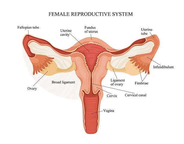
The female reproductive system is a complex network of organs and structures that play crucial roles in sexual reproduction, hormone production, and menstrual cycles. It consists of both external and internal components.
- External Genitalia (Vulva):
- The vulva encompasses all external female genital structures. Key parts include:
- Mons Pubis: The fleshy area above the vaginal opening.
- Labia Majora and Labia Minora: Two pairs of skin folds that protect the vaginal opening.
- Clitoris: A small, sensitive organ located at the front of the vulva, important for sexual arousal.
- Urethral Opening: The opening through which urine exits the body.
- Vaginal Opening: The entrance to the vagina.
- The vulva encompasses all external female genital structures. Key parts include:
- Vagina:
- A muscular, hollow tube extending from the vulva to the uterus. It serves multiple functions including:
- Receiving the penis during intercourse.
- Acting as a birth canal during childbirth.
- Providing an exit route for menstrual blood.
- A muscular, hollow tube extending from the vulva to the uterus. It serves multiple functions including:
- Cervix:
- The lower part of the uterus that connects to the vagina. It has a small opening that allows menstrual blood to flow out and sperm to enter.
- Uterus (Womb):
- A pear-shaped muscular organ where a fertilized egg can implant and develop into a fetus. It has three main layers:
- Fallopian Tubes:
- Two tubes that extend from either side of the uterus to the ovaries. They are responsible for transporting eggs from the ovaries to the uterus and are typically where fertilization occurs when sperm meets an egg.
- Ovaries:
- Two almond-shaped organs located on either side of the uterus. They produce eggs (ova) and secrete hormones such as estrogen and progesterone, which regulate various functions within the reproductive system.
General Organization of the Ovaries
The ovaries are essential female reproductive organs that are primarily responsible for producing eggs and hormones. Their organization can be understood through several key components:
1. Location and Structure
The ovaries are two small, oval-shaped glands located on either side of the uterus in the pelvic cavity. They are typically about the size of an almond, measuring approximately 3.5 cm in length, 2 cm in width, and 1 cm in thickness. Each ovary is held in place by peritoneal ligaments and is situated within shallow depressions known as ovarian fossae.
2. Layers of the Ovaries
The ovaries consist of three distinct layers:
- Germinal Epithelium: This outer layer is made up of simple cuboidal epithelium, which is actually a part of the visceral peritoneum enveloping the ovaries.
- Tunica Albuginea: Beneath the germinal epithelium lies a dense connective tissue capsule known as the tunica albuginea, which provides structural support.
- Cortex and Medulla: The substance of the ovary is divided into two main regions:
- Cortex: The outer region appears granular due to numerous ovarian follicles at various stages of development. Each follicle contains an oocyte (immature egg).
- Medulla: The inner region consists of loose connective tissue that houses blood vessels, lymphatic vessels, and nerve fibers.
3. Ovarian Follicles
Within the cortex, thousands of ovarian follicles exist. These follicles are small sacs that contain immature eggs (oocytes). During each menstrual cycle, several primary oocytes begin to mature under hormonal influence; however, typically only one follicle fully matures and releases its egg during ovulation.
4. Hormonal Function
The ovaries also play a crucial role in hormone production. They secrete key hormones such as estrogen and progesterone, which regulate various aspects of the menstrual cycle and prepare the body for potential pregnancy.
5. Oogenesis
Oogenesis is the process by which female gametes develop within the ovaries. It begins with primordial germ cells differentiating into oogonia during fetal development. These oogonia undergo mitosis to form primary oocytes that enter meiosis but remain arrested until puberty when they resume development under hormonal stimulation.
In summary, the organization of the ovaries includes their anatomical location, layered structure comprising germinal epithelium, tunica albuginea, cortex, and medulla; their role in housing ovarian follicles; their function in hormone production; and their involvement in oogenesis.
Identifying Ovary and Ovarian Follicles Under Microscope
1. Overview of the Ovary Structure
The ovary is a vital component of the female reproductive system, responsible for producing oocytes (eggs) and hormones such as estrogen and progesterone. When viewed under a microscope, the ovary exhibits a distinct cortex and medulla organization. The outer layer, known as the tunica albuginea, is a thick connective tissue capsule that encases the ovary. The surface of this capsule is covered by germinal epithelium, which consists of cuboidal cells.
2. Primordial Follicles
Primordial follicles are the earliest stage of ovarian follicle development and can be identified by their unique structure. Each primordial follicle contains a primary oocyte surrounded by a single layer of flattened follicular cells (also referred to as granulosa cells). Under microscopic examination, these follicles appear small and are typically located near the outer edge of the ovarian cortex.
3. Primary Follicles
As primordial follicles develop into primary follicles, they undergo significant changes. The primary oocyte enlarges, and the surrounding follicular cells transition from a single layer to multiple layers of cuboidal cells. This multilayered structure is characteristic of primary follicles. The zona pellucida, a glycoprotein layer that forms between the oocyte and granulosa cells, becomes more prominent during this stage.
4. Secondary Follicles
Secondary follicles are larger than primary follicles and can be recognized by the presence of an antrum—a fluid-filled space that develops among the granulosa cells. At this stage, there are more layers of granulosa cells surrounding the oocyte compared to primary follicles. The surrounding stroma differentiates into two layers: theca interna (which secretes hormones) and theca externa (a fibrous layer).
5. Graafian Follicles
The Graafian follicle represents a mature ovarian follicle ready for ovulation. It is characterized by a large antrum filled with follicular fluid and an oocyte that has completed its first meiotic division to become a secondary oocyte. The cumulus oophorus—granulosa cells that surround and support the oocyte—can also be observed at this stage.
6. Summary of Identification Techniques
To identify these structures under a microscope:
- Look for primordial follicles as small clusters with flattened epithelial cells.
- Identify primary follicles by observing enlarged oocytes surrounded by multiple layers of cuboidal granulosa cells.
- Recognize secondary follicles through their larger size and presence of an antrum.
- Finally, locate Graafian follicles, which will have a prominent antrum and well-defined cumulus oophorus.
By following these steps systematically while examining ovarian tissue slides under a microscope, one can accurately identify various stages of ovarian follicle development.
Layers and Cells of Fallopian Tubes Under Microscope
When examining the fallopian tubes (also known as uterine tubes or oviducts) under a microscope, several distinct layers and cell types can be identified. The structure of the fallopian tubes is composed of three main layers:
1. Mucosa
- The mucosa is the innermost layer and features longitudinal folds that are more pronounced in the infundibulum region. It is lined by a single layer of tall, columnar epithelium.
- Within this epithelial lining, there are three types of cells:
- Ciliated Cells: These cells have cilia on their apical surface and are more prevalent in the distal portion of the tube. They play a crucial role in moving the ovum toward the uterus through coordinated wave-like movements.
- Non-Ciliated Secretory Cells (Peg Cells): These cells are primarily found in the proximal part of the tube and secrete a nutrient-rich fluid that supports the ovum and aids sperm capacitation. They are particularly active around ovulation.
- Intercalated Cells: These may represent a morphological variant of secretory cells, contributing to the overall function of the mucosal lining.
2. Muscularis
- The muscularis layer consists of two sub-layers: an inner circular layer and an outer longitudinal layer. This arrangement allows for peristaltic contractions that assist in propelling the fertilized ovum through the tube toward the uterus.
3. Serosa
- The serosa is the outermost layer, which provides structural support to the fallopian tubes. It consists of loose connective tissue that connects to surrounding structures via mesosalpinx, part of the broad ligament.
In summary, when viewed under a microscope, one can identify these three layers—mucosa, muscularis, and serosa—along with their respective cell types: ciliated cells, non-ciliated secretory (peg) cells, and intercalated cells within the mucosal layer.


