General Principles of the Development of the Different Parts of the Female Genital Tract
1. Timeline of Development
The development of the female reproductive organs begins early in gestation, specifically between weeks 4 and 5 of pregnancy. This process continues until approximately the 20th week. The early stages are crucial as they set the foundation for the formation of various structures within the female genital tract.
2. Genetic Factors
Genetic factors play a significant role in the development of the female genital tract. Missing or damaged genes can lead to developmental differences. For instance, congenital adrenal hyperplasia is a condition caused by genetic variants that affect hormone production, leading to atypical development of external genitalia while preserving internal structures like the uterus and ovaries.
3. Hormonal Influence
Hormones are critical during fetal development, influencing how reproductive organs form and differentiate. The presence or absence of certain hormones can dictate whether an embryo develops male or female characteristics. For example, exposure to androgens can lead to masculinization, while a lack thereof typically results in female genitalia.
4. Environmental Factors
Environmental influences, such as maternal exposure to specific medications during pregnancy, can disrupt normal development. A notable example is diethylstilbestrol (DES), which was prescribed to prevent miscarriage but later found to cause abnormalities in the uterine structure and increased cancer risk in offspring.
5. Structural Development
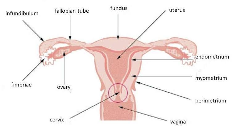
The female reproductive tract consists of several key components:
- Vagina: Develops from a combination of mesodermal and endodermal tissues.
- Uterus: Forms from the fusion of two Müllerian ducts; any disruption in this process can lead to uterine anomalies.
- Ovaries: Develop from gonadal ridges and migrate into their final position within the pelvis.
- Cervix: Forms from both uterine tissue and vaginal tissue; abnormalities may arise if there are issues during its formation.
6. Associated Anomalies
Developmental issues may not be isolated to just one part of the reproductive system; they often coincide with problems in adjacent systems such as urinary or gastrointestinal tracts due to their close anatomical relationships during embryonic development.
7. Clinical Implications
Understanding these principles is essential for diagnosing and managing congenital anomalies that may arise from disruptions during any stage of development. Conditions like cloacal abnormalities, imperforate hymen, or Mayer-Rokitansky-Küster-Hauser syndrome illustrate how variations in normal development can manifest clinically.
In summary, the development of different parts of the female genital tract is governed by a complex interplay between genetic factors, hormonal influences, environmental exposures, and structural formation processes that occur primarily during early gestation.
Types of Müllerian Anomalies and Their Clinical Presentation
1. Septate Uterus
- Description: The external shape of the uterus appears normal, but the cavity is divided by an extra wall of tissue called a septum. If the septum completely divides the cavity, it is termed a complete septate uterus; if it partially divides the cavity, it is known as a sub-septate uterus.
- Clinical Presentation: Patients may experience recurrent miscarriages due to the abnormal shape of the uterine cavity. Some may have no symptoms if menstrual flow is not obstructed.
2. Bicornuate Uterus
- Description: This anomaly features an abnormal external shape with a large indentation at the fundus (the top of the uterus), resulting in two cavities within one uterus due to incomplete fusion of the Müllerian ducts.
- Clinical Presentation: Women may present with complications during pregnancy, such as preterm labor or malpresentation of the fetus. Some may remain asymptomatic until pregnancy.
3. Unicornuate Uterus
- Description: In this condition, only one Müllerian duct develops, leading to a uterus that is smaller than normal and has only one horn.
- Clinical Presentation: Patients might experience infertility or complications during pregnancy, including ectopic pregnancies in the rudimentary horn if present.
4. Uterine Didelphys
- Description: This anomaly results in complete duplication of both the uterus and cervix, creating two separate uteri and two cervices while maintaining two fallopian tubes and ovaries.
- Clinical Presentation: Many women are asymptomatic; however, some may face obstetric complications such as preterm birth or difficulties during labor.
In addition to these specific anomalies, patients with Müllerian anomalies can also experience associated symptoms related to menstrual disorders or infertility. Often, these conditions are discovered incidentally during imaging studies conducted for other reasons or when complications arise during pregnancy.
Development and Congenital Anomalies of the Vulva
The vulva is the external part of the female genitalia, which includes structures such as the labia majora, labia minora, clitoris, and vaginal opening. The development of these structures occurs during embryonic growth and can be influenced by genetic and environmental factors. Congenital anomalies of the vulva are rare but can have significant implications for affected individuals.
Embryonic Development of the Vulva
The vulva develops from the urogenital sinus and the paired genital tubercles during fetal development. This process begins around the 6th week of gestation and continues until birth. Hormonal influences, particularly from estrogen and testosterone, play a crucial role in shaping these structures. Any disruption in this developmental process can lead to congenital anomalies.
Types of Congenital Anomalies
Congenital anomalies affecting the vulva can manifest in various forms:
- Childhood Asymmetry Labium Majus Enlargement (CALME): This condition involves an enlargement or swelling of one side of the labia majora due to excess tissue growth, leading to an asymmetrical appearance.
- Labial Hypoplasia: In this anomaly, one side of the labia is underdeveloped or absent entirely, which may affect both appearance and function.
- Other Rare Conditions: While less common, other conditions such as labial fusion or abnormal pigmentation may also occur.
Signs and Symptoms
Symptoms associated with congenital anomalies of the vulva can vary widely depending on the specific condition:
- Asymmetrical Appearance: Visible differences in size or shape between the two sides of the labia.
- Discomfort or Pain: Some individuals may experience discomfort due to enlarged tissues or structural abnormalities.
- Psychosocial Impact: The appearance of congenital anomalies can lead to psychological distress or body image issues for some individuals.
Diagnosis
Congenital anomalies are often diagnosed through physical examination by a healthcare provider. In some cases, imaging studies such as ultrasound may be utilized to assess internal structures if there are concerns about associated conditions.
Treatment Options
Treatment for congenital anomalies of the vulva depends on their nature and severity:
- Surgical Intervention: For conditions like CALME or labial hypoplasia that cause discomfort or psychosocial distress, surgical correction may be considered.
- Observation: In cases where no functional impairment exists, observation may be sufficient without any need for intervention.
In summary, congenital anomalies of the vulva are rare but can significantly impact individuals’ health and well-being. Early diagnosis and appropriate management are essential for addressing any complications that arise from these conditions.
Recognizing Different Müllerian Anomalies in Hysterosalpingogram (HSG)
Müllerian duct anomalies (MDAs) are a spectrum of congenital abnormalities resulting from improper development of the Müllerian ducts during embryogenesis. Hysterosalpingography (HSG) is a radiologic procedure that can help identify these anomalies by providing images of the uterine cavity and fallopian tubes. Here’s a detailed breakdown of how to recognize different types of Müllerian anomalies on HSG.
1. Normal Uterus (Class U0)
In a normal HSG, the uterine cavity appears as a single, smooth, and centrally located structure with no irregularities or indentations. The fallopian tubes should be patent, allowing for the contrast medium to spill into the peritoneal cavity.
2. Dysmorphic Uterus (Class U1)
A dysmorphic uterus may present as a T-shaped cavity on HSG due to abnormal thickening of the uterine walls or an abnormal outer contour. The internal contour may appear irregular but does not show significant indentations or septa.
3. Septate Uterus (Class U2)
On HSG, a septate uterus is characterized by an internal indentation in the uterine cavity while maintaining a normal external contour. The contrast will fill both sides of the cavity but will not extend through the central septum, which divides it into two compartments.
4. Bicornuate Uterus (Class U3)
A bicornuate uterus appears on HSG as two separate cavities with an external cleft at the fundus. The contrast medium will fill both horns distinctly, demonstrating that there are two distinct endometrial cavities separated by an external indentation.
5. Didelphys Uterus (Class U3)
Similar to a bicornuate uterus, a didelphys uterus shows two completely separate uterine cavities and cervices on HSG. Each cavity will be filled with contrast material, and there will be no communication between them.
6. Unicornuate Uterus (Class U4)
In cases of unicornuate uterus, only one side of the uterine cavity is visualized on HSG while the other side may be absent or rudimentary. The single horn will appear elongated and may have an associated renal anomaly on imaging.
7. Aplastic Uterus (Class U5)
An aplastic uterus may not be well visualized on HSG since it results from complete failure of Müllerian duct formation; hence, there might be little to no filling defect seen where the uterus would normally be located.
8. Unclassified Cases (Class U6)
Some anomalies may not fit neatly into any classification category based solely on HSG findings and require further imaging modalities like ultrasound or MRI for accurate diagnosis.
Recognizing these anomalies requires careful interpretation of HSG images in conjunction with clinical history and additional imaging when necessary to ensure accurate diagnosis and appropriate management strategies for affected individuals.
Development and Congenital Anomalies of the Hymen
The hymen is a thin membrane located at the vaginal opening, which is present at birth. Its development occurs during fetal growth, where it typically forms as a ring-like structure surrounding the vaginal opening. The hymen’s primary purpose is not definitively understood, but it is thought to play a role in protecting the vagina from bacteria during early life.
Congenital Anomalies of the Hymen
Congenital anomalies of the hymen can occur when there are developmental issues during fetal formation. The most notable congenital anomalies include:
- Imperforate Hymen: This condition arises when the hymenal tissue completely covers the vaginal opening, resulting in no passage for menstrual blood or other fluids. It is a rare condition, occurring in approximately 0.5% of individuals. Symptoms may not be evident until puberty when menstruation begins; trapped blood can lead to pain and complications such as endometriosis or infections.
- Microperforate Hymen: In this case, there is a small opening in the hymen that may allow some menstrual blood or mucus to escape but not enough for normal menstrual flow. This can cause prolonged periods and discomfort during tampon use or sexual intercourse.
- Septate Hymen: A septate hymen features an extra band of tissue that divides the hymenal opening into two parts. This anomaly can hinder tampon insertion and may cause discomfort or tearing during sexual activity.
These conditions are typically diagnosed through physical examination and imaging techniques like ultrasound if necessary. Treatment usually involves surgical intervention to correct the anomaly, allowing for normal menstrual function and sexual health.
Surgical Correction
Surgical procedures such as hymenectomy are performed to correct these anomalies. For an imperforate hymen, surgery involves creating an opening in the hymenal tissue under general anesthesia, allowing trapped blood to drain and securing the new opening with absorbable stitches to prevent reblockage.
After surgical correction, individuals with previously diagnosed congenital anomalies of the hymen generally experience normal menstrual cycles and have no long-term reproductive issues, including fertility.
In summary, congenital anomalies of the hymen primarily include imperforate hymens, microperforate hymens, and septate hymens, each presenting unique challenges regarding menstruation and sexual health. Surgical interventions provide effective solutions for these conditions.
Development and Congenital Anomalies of the Vagina
The vagina develops during embryonic growth through a process where two paired structures, known as the Müllerian ducts, fuse to form a single canal. This fusion typically occurs around the 12th week of gestation. However, various congenital anomalies can arise from disruptions in this developmental process, leading to conditions that may affect the structure and function of the vagina.
Types of Vaginal Anomalies
- Vaginal Agenesis/Atresia: This condition is characterized by the incomplete development or absence of the vagina. It affects approximately 1 in every 5,000 female infants and is often associated with other reproductive tract anomalies, such as absent or underdeveloped uterus and kidney abnormalities. In cases where there is complete vaginal agenesis, women may experience retrograde menstruation if they have a uterus but no cervix.
- Transverse Vaginal Septum: A transverse vaginal septum is a horizontal wall of fibrous tissue that forms during embryologic development, creating a blockage within the vagina. This septum can occur at various levels within the vaginal canal. Some women may have a small opening (fenestration) in this septum, allowing for menstrual flow; however, if there is no opening, menstrual blood can accumulate above the septum, leading to complications such as hematometra (blood accumulation in the uterus). Surgical resection of the septum is typically required to restore normal anatomy and function.
- Longitudinal Vaginal Septum: This anomaly involves a vertical division of the vagina into two separate canals due to incomplete resorption of the Müllerian ducts. While menstrual blood can exit normally in these cases, individuals may face challenges with tampon insertion or sexual intercourse due to anatomical differences.
- Imperforate Hymen: This condition occurs when the hymen fails to perforate properly during fetal development, resulting in an obstruction at the vaginal opening. Adolescent girls with an imperforate hymen often present with pelvic pain and may not experience menstruation until surgical intervention is performed to create an adequate opening.
- Complete Vaginal Septum: Similar to longitudinal septa but more severe, this condition results in two distinct vaginas and typically presents with duplication of reproductive structures such as uteri and cervices. Women may notice symptoms during tampon use or sexual activity due to anatomical discrepancies.
Diagnosis and Treatment
Diagnosis of these anomalies often occurs during adolescence or early adulthood when symptoms manifest—such as pelvic pain or difficulties with menstruation or sexual activity. Imaging studies like ultrasound or MRI can help visualize structural abnormalities.
Treatment options vary depending on the specific anomaly:
- For vaginal agenesis, surgical creation of a neovagina (vaginoplasty) may be performed.
- Transverse and longitudinal septa are usually addressed surgically through resection.
- An imperforate hymen requires surgical intervention to create an appropriate vaginal opening.
Post-surgical care often includes monitoring for complications such as scarring or stenosis (narrowing) that could affect future sexual function or reproductive capabilities.
In summary, congenital anomalies of the vagina encompass a range of conditions arising from developmental disruptions during embryogenesis. These anomalies can significantly impact reproductive health but are generally manageable through appropriate medical interventions.
Comprehensive overview of Development and Congenital Anomalies of the Uterus
The uterus develops during embryonic life from two paired structures known as Müllerian ducts. These ducts typically fuse together to form a single, normal uterus by the end of the first trimester of pregnancy. However, if there are disruptions in this process, congenital uterine anomalies can occur. These anomalies can range from minor variations in shape to complete absence of the uterus.
Types of Congenital Uterine Anomalies
Congenital uterine anomalies can be classified into several categories based on their structural characteristics:
- Septate Uterus: This anomaly features a normal external uterine contour but has two separate endometrial cavities due to a fibrous or muscular septum dividing the uterus.
- Bicornuate Uterus: In this case, the uterus has an indented external surface and contains two endometrial cavities, resembling a heart shape.
- Unicornuate Uterus: This anomaly occurs when only one half of the uterus develops, resulting in a smaller, banana-shaped organ.
- Didelphys Uterus: This condition is characterized by two completely separate uteri, each with its own cervix and possibly a vaginal septum.
- Arcuate Uterus: Considered a variation of normal development, this type features a slight indentation (1 cm or less) at the top of the endometrial cavity while maintaining a normal external contour.
- Absent Uterus (Mayer-Rokitansky-Küster-Hauser Syndrome): In rare cases, women may be born without a uterus due to developmental failure of the Müllerian ducts.
Causes of Congenital Uterine Anomalies
The exact causes of congenital uterine anomalies remain largely unknown; however, most affected women have a normal chromosomal pattern (46 XX). Historical exposure to diethylstilbestrol (DES), a synthetic estrogen given to pregnant women between 1938 and 1971 to prevent miscarriages, has been linked to an increased risk for certain uterine anomalies in daughters exposed in utero.
Currently, there are no established risk factors for developing these anomalies nor any known preventive measures that can be taken during pregnancy.
Symptoms and Diagnosis
Congenital uterine anomalies are often asymptomatic and may not be detected until evaluations for infertility or pregnancy complications arise. Some women may experience painful menstrual periods due to associated conditions like endometriosis or fibroids rather than directly from the anomaly itself.
Diagnosis typically involves imaging studies such as:
- Hysterosalpingogram (HSG): An X-ray procedure where dye is injected into the uterine cavity to visualize its shape.
- Ultrasound: Both conventional and 3D ultrasound can provide detailed images of uterine structure.
- Magnetic Resonance Imaging (MRI): Used for complex cases where detailed anatomical information is needed.
A comprehensive medical history and physical examination may also lead healthcare providers to suspect an anomaly.
Treatment Options
There are no non-surgical treatments available for congenital uterine anomalies. Surgical intervention is generally reserved for specific cases:
- For women with a septate uterus who have experienced recurrent miscarriages or infertility issues, surgical resection of the septum is often recommended as it can improve reproductive outcomes.
- Other types of anomalies like bicornuate or unicornuate uteri rarely require surgery unless complications arise.
It’s important to note that while surgical correction may enhance pregnancy outcomes for some women with septate uteri, it does not eliminate all risks associated with congenital anomalies—such as preterm birth or fetal growth restriction—especially if other factors contribute to these risks.
In conclusion, congenital uterine anomalies represent significant variations in female reproductive anatomy that can impact fertility and pregnancy outcomes. Understanding their development, classification, diagnosis, and treatment options is crucial for effective management and care.
Development and Congenital Anomalies of the Fallopian Tubes
The fallopian tubes, also known as uterine tubes or salpinges, are essential components of the female reproductive system. They play a critical role in the transport of ova from the ovaries to the uterus and are typically about 10 cm long. The development of these tubes occurs during embryogenesis, specifically from the paramesonephric (Müllerian) ducts.
Embryonic Development of Fallopian Tubes
During early fetal development, around the sixth week of gestation, the Müllerian ducts begin to form. These ducts develop into various structures within the female reproductive tract, including the fallopian tubes, uterus, cervix, and upper two-thirds of the vagina. The proper fusion and differentiation of these ducts are crucial for normal anatomical development.
As development progresses, each Müllerian duct differentiates into distinct segments that will become parts of the fallopian tube:
- Infundibulum – The funnel-shaped end near the ovary.
- Ampulla – The widest section where fertilization typically occurs.
- Isthmus – A narrower segment connecting to the uterus.
- Intramural Portion – The part embedded within the uterine wall.
Any disruption in this developmental process can lead to congenital anomalies.
Congenital Anomalies of Fallopian Tubes
Congenital anomalies affecting the fallopian tubes are relatively rare but can have significant implications for fertility and reproductive health. These anomalies may include:
- Agenesis or Hypoplasia: This condition refers to either complete absence (agenesis) or underdevelopment (hypoplasia) of one or both fallopian tubes. Women with this anomaly may not experience any menstrual irregularities but may face challenges with fertility.
- Accessory Uterine Tubes: Some individuals may have additional fallopian tubes alongside normal ones, which is known as accessory uterine tubes or duplication.
- Paratubal Cysts: These are fluid-filled sacs that can develop near the fallopian tubes and may not cause symptoms unless they grow large enough to exert pressure on surrounding structures.
- Abnormal Length or Shape: Variations in length or structural abnormalities can occur but are less common.
- Other Müllerian Duct Anomalies (MDAs): Since fallopian tube anomalies often coexist with other MDAs affecting the uterus and vagina, comprehensive evaluation is necessary when diagnosing such conditions.
Diagnosis and Treatment
Most congenital anomalies of the fallopian tubes remain undiagnosed until puberty when a girl experiences menstrual irregularities or infertility issues later in life. Diagnosis typically involves:
- Pelvic examinations by gynecologists.
- Imaging techniques such as pelvic ultrasound.
- Laparoscopy for direct visualization.
- Biopsy if abnormal tissue is present.
Treatment options depend on individual circumstances and symptoms:
- For agenesis without associated complications, no treatment may be necessary.
- Surgical intervention might be required for accessory tubes or cysts if they cause discomfort or complications.
- In cases where there is significant impact on fertility, assisted reproductive technologies like in vitro fertilization (IVF) may be considered.
In summary, while congenital anomalies of the fallopian tubes are rare and often asymptomatic initially, they can significantly affect reproductive health and require careful diagnosis and management based on individual needs.
Development and Congenital Anomalies of the Ovary
The ovaries are critical components of the female reproductive system, responsible for producing eggs (ova) and hormones such as estrogen and progesterone. Their development occurs during embryogenesis, specifically from the mesodermal tissue, which is distinct from the tissues that form the uterus and other parts of the reproductive tract. The formation of ovaries typically begins around the sixth week of gestation and continues through fetal development.
Ovarian Development
- Embryonic Origin: Ovaries develop from the gonadal ridges, which are formed from mesodermal tissue. The presence of specific genes, such as those on the Y chromosome (in males), influences whether these gonads will develop into testes or ovaries. In females, the absence of these male-determining factors leads to ovarian development.
- Hormonal Influence: During fetal development, hormones play a crucial role in ovarian differentiation. The absence of testosterone allows for the development of female characteristics, including ovaries.
- Follicle Formation: By approximately 16 weeks gestation, primordial follicles begin to form within the ovaries. Each follicle contains an immature egg surrounded by granulosa cells. This process continues until birth when a female is born with a finite number of follicles.
- Puberty Changes: At puberty, hormonal changes trigger further maturation of these follicles, leading to ovulation cycles throughout a woman’s reproductive years.
Congenital Anomalies of the Ovary
Congenital anomalies affecting the ovaries can arise due to various developmental disruptions during embryogenesis:
- Ovarian Agenesis or Dysgenesis: This condition refers to either absent ovaries (agenesis) or poorly developed ovaries (dysgenesis). Ovarian agenesis is often associated with Turner syndrome, where one X chromosome is missing or partially missing in females.
- Ectopic Ovaries: In some cases, an ovary may be located outside its normal anatomical position within the pelvic cavity due to abnormal migration during development.
- Polycystic Ovarian Syndrome (PCOS): While not strictly a congenital anomaly since it typically manifests later in life, PCOS has been linked to genetic factors that may influence ovarian development and function.
- Ovarian Tumors: Some congenital anomalies may predispose individuals to tumors in ovarian tissue; however, these tumors can also arise independently due to genetic mutations or environmental factors.
- Associated Anomalies: Congenital anomalies of the ovaries are often associated with other developmental disorders affecting adjacent structures like the uterus or urinary tract due to their shared embryonic origins.
- Symptoms and Diagnosis: Many congenital anomalies may not present symptoms until puberty or later when menstrual irregularities occur or fertility issues arise. Diagnosis can involve imaging techniques such as ultrasound or MRI and may require surgical exploration for definitive assessment.
- Management Options: Treatment for congenital anomalies varies depending on their nature and severity but may include hormonal therapy for dysgenesis cases or surgical intervention for ectopic locations or associated complications.
In summary, while congenital anomalies affecting ovarian development are relatively rare compared to other reproductive tract abnormalities, they can have significant implications for reproductive health and overall well-being in affected individuals.


