Normal Coronary Blood Flow During Systole and Diastole
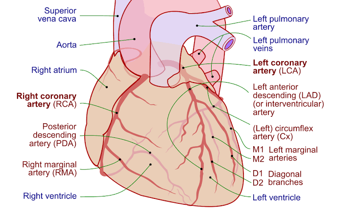
Coronary blood flow is essential for supplying oxygen and nutrients to the myocardium (heart muscle). This flow occurs through the coronary arteries, which branch off from the aorta. Understanding how this flow changes during different phases of the cardiac cycle—systole and diastole—is crucial for comprehending cardiac physiology.
Coronary Blood Flow During Diastole
During diastole, the heart muscle relaxes, allowing the chambers of the heart to fill with blood. This phase is characterized by:
- Myocardial Relaxation: As the left ventricle relaxes, there is a decrease in intraventricular pressure. The relaxation of myocardial fibers reduces extravascular compression on coronary vessels.
- Increased Coronary Perfusion Pressure: The aortic valve closes at the end of systole, leading to a rise in aortic pressure that drives blood into the coronary arteries. This increased pressure allows for enhanced perfusion of the myocardium.
- Peak Blood Flow: Coronary blood flow reaches its peak during diastole as there are no compressive forces acting on the coronary vessels. The left coronary artery experiences significant flow due to its anatomical position and relationship with the left ventricle.
- Distribution of Blood Flow: The distribution of blood flow during diastole is not uniform; it tends to be greater in subendocardial regions (inner layer) compared to subepicardial regions (outer layer). This is because subendocardial areas are more susceptible to ischemia due to their location within the heart wall.
Coronary Blood Flow During Systole
During systole, when the heart contracts to pump blood out of the ventricles, coronary blood flow experiences distinct changes:
- Ventricular Contraction: As myocytes contract, they exert compressive forces on nearby coronary vessels, particularly those supplying the left ventricle. This phenomenon is known as “extravascular compression.”
- Decreased Coronary Perfusion Pressure: The contraction leads to an increase in intraventricular pressure that exceeds aortic pressure temporarily, causing a reduction in perfusion through coronary arteries.
- Flow Interruption and Reversal: In some cases, particularly during isovolumetric contraction (the initial phase of systole), blood flow through certain segments of coronary arteries can be halted or even reversed due to high pressures within contracting myocardial fibers.
- Restoration of Flow During Ejection Phase: Once ventricular pressures exceed aortic pressures and the aortic valve opens (during ejection), coronary blood flow can resume as perfusion pressure increases again. However, this flow may still be lower than during diastolic periods due to ongoing extravascular compression.
- Regional Variability: Similar to diastolic conditions, there can be variability in how different regions receive blood during systole; however, overall myocardial oxygen demand increases significantly during this phase due to heightened metabolic activity associated with contraction.
Conclusion
In summary, normal coronary blood flow varies significantly between systole and diastole due to mechanical factors related to myocardial contraction and relaxation. During diastole, there is maximal perfusion as compressive forces are absent; conversely, during systole, especially at peak contraction phases like isovolumetric contraction and ejection, there can be reduced or reversed flow due to extravascular compression.
Local Factors for Control of Coronary Blood Flow
The regulation of coronary blood flow is a complex process that is influenced by various local factors, with local metabolism playing a crucial role. Understanding these mechanisms is essential for appreciating how the heart meets its oxygen and nutrient demands during different physiological states.
Local Metabolism as a Primary Factor
Local metabolism refers to the metabolic processes occurring within the myocardial tissue that influence coronary blood flow. The heart has a high metabolic demand due to its continuous activity and need for oxygen. As such, it relies heavily on local metabolic signals to regulate blood flow effectively.
When myocardial tissue experiences an increase in metabolic activity—such as during exercise or increased heart rate—it requires more oxygen and substrates (like glucose and fatty acids) to sustain this heightened activity. This increased demand leads to several biochemical changes that promote vasodilation, thereby enhancing coronary blood flow.
Key metabolites produced during increased myocardial metabolism include adenosine, carbon dioxide (CO2), hydrogen ions (H+), and lactate. These metabolites act as potent vasodilators:
- Adenosine: Released from cardiac myocytes during periods of low oxygen availability or high energy demand, adenosine promotes vasodilation by acting on specific receptors (A2 receptors) on vascular smooth muscle cells.
- Carbon Dioxide: Increased levels of CO2 in the myocardium can lead to a decrease in pH (acidosis), which also contributes to vasodilation.
- Hydrogen Ions: Elevated H+ concentrations resulting from anaerobic metabolism further stimulate vasodilation.
- Lactate: Produced during anaerobic glycolysis, lactate can also induce vasodilation and improve blood flow.
These local metabolic factors ensure that coronary blood flow matches the oxygen demand of the myocardium, maintaining an optimal balance between supply and demand.
Oxygen Demand
The heart’s oxygen demand is primarily determined by its workload, which can be influenced by several factors including heart rate, contractility, and afterload. As the workload increases—such as during physical exertion—the heart’s requirement for oxygen rises significantly.
To meet this increased oxygen demand, several compensatory mechanisms are activated:
- Increased Heart Rate: A higher heart rate increases cardiac output but also raises myocardial oxygen consumption.
- Enhanced Contractility: Increased force of contraction requires more energy and thus more oxygen.
- Afterload Considerations: Higher systemic vascular resistance can increase the workload on the heart, necessitating greater oxygen delivery.
As these demands rise, local metabolic signals trigger vasodilation in coronary vessels to increase blood flow accordingly. This dynamic adjustment allows the coronary circulation to respond rapidly to changes in myocardial oxygen requirements.
In summary, local metabolism serves as a primary factor in controlling coronary blood flow through the release of various metabolites that induce vasodilation in response to increased myocardial oxygen demand. This intricate interplay ensures that the heart receives adequate blood supply relative to its functional needs at any given moment.
Effect of Autonomic Nervous System on Coronary Arteries
The autonomic nervous system (ANS) plays a crucial role in regulating coronary blood flow and the function of coronary arteries. It is divided into two main branches: the sympathetic and parasympathetic nervous systems, each having distinct effects on the heart and blood vessels.
Sympathetic Nervous System
The sympathetic nervous system primarily influences coronary arteries through adrenergic receptors, which are classified into alpha (α) and beta (β) receptors.
- Alpha-Adrenergic Receptors: Activation of α-adrenergic receptors generally leads to vasoconstriction of coronary arteries. When catecholamines like norepinephrine bind to these receptors, they cause the smooth muscle in the vascular walls to contract, reducing the cross-sectional area of large coronary arteries. This response can decrease coronary blood flow despite an increase in coronary distending pressure.
- Beta-Adrenergic Receptors: There are two main types of β-adrenergic receptors involved in coronary artery regulation—β1 and β2 receptors.
- Beta-1 Receptors: These receptors are primarily located in the heart and play a significant role in increasing heart rate and contractility. Their activation during sympathetic stimulation enhances myocardial oxygen demand.
- Beta-2 Receptors: These receptors are found in both cardiac tissue and vascular smooth muscle. Activation of β2-receptors leads to vasodilation, which increases blood flow through the coronary arteries. This effect is particularly important during periods of increased myocardial oxygen demand, such as during exercise.
The balance between α-adrenergic-mediated constriction and β-adrenergic-mediated dilation determines overall coronary vascular tone. In healthy individuals, sympathetic activation typically results in an increase in coronary blood flow due to the predominance of β-receptor-mediated vasodilation over α-receptor-mediated constriction.
Parasympathetic Nervous System
The role of the parasympathetic nervous system in regulating coronary circulation is more complex and somewhat controversial. While it is well-established that parasympathetic stimulation can lead to vasodilation in certain species (like dogs), its effects on humans can vary significantly:
- In some studies involving primates, parasympathetic activation has been shown to induce vasoconstriction rather than dilation.
- The mechanisms underlying these responses may involve various factors such as species differences, local metabolic demands, and interactions with other signaling pathways.
Overall, while the sympathetic nervous system predominantly facilitates increased blood flow through adrenergic receptor activation during stress or exercise, the parasympathetic system’s role appears more nuanced and context-dependent.
In summary, the autonomic nervous system regulates coronary arteries through a complex interplay between alpha-adrenergic receptor-mediated vasoconstriction and beta-adrenergic receptor-mediated vasodilation, with additional modulation by parasympathetic influences that can vary among species.
Ischemic Heart Disease
Ischemic heart disease (IHD), also known as coronary artery disease or coronary heart disease, is a condition characterized by damage to the heart muscle due to reduced blood flow caused by narrowed or blocked coronary arteries. This reduction in blood flow leads to decreased oxygen supply to the heart muscle, which can result in chest pain (angina) and potentially lead to a heart attack if the blood supply is severely compromised.
Cause of Cardiac Pain
The primary cause of cardiac pain associated with ischemic heart disease is myocardial ischemia, which occurs when the demand for oxygen by the heart muscle exceeds the supply delivered through the coronary arteries. Factors that can increase this demand include physical exertion, emotional stress, cold exposure, or any situation that requires increased cardiac output. When the heart muscle does not receive enough oxygen-rich blood, it can result in symptoms such as:
- Chest pain or discomfort (angina pectoris)
- Pain radiating to other areas such as the arms, neck, jaw, back, or stomach
- Shortness of breath
- Sweating
- Nausea or vomiting
These symptoms arise because ischemia triggers a complex interplay of biochemical processes within the heart muscle cells. The lack of oxygen leads to anaerobic metabolism and accumulation of metabolic waste products like lactic acid, which stimulate nerve endings and result in pain sensations.
Mechanism of Collateral Circulation
Collateral circulation refers to the network of small blood vessels that can develop over time to provide alternative routes for blood flow when primary pathways are obstructed due to conditions like ischemic heart disease. The mechanism behind collateral circulation involves several key processes:
- Vascular Remodeling: When there is chronic ischemia due to narrowed arteries, existing small vessels may undergo remodeling and enlargement. This process allows these vessels to accommodate increased blood flow.
- Angiogenesis: In response to low oxygen levels (hypoxia), new blood vessels can form from pre-existing ones through a process called angiogenesis. Growth factors such as vascular endothelial growth factor (VEGF) play a crucial role in stimulating this process.
- Hemodynamic Changes: As larger coronary arteries become narrowed or blocked, changes in pressure gradients can stimulate collateral vessels to open up and increase their diameter, allowing more blood flow through these alternate routes.
- Functional Adaptation: Over time, collateral vessels may adapt functionally by improving their ability to dilate and respond effectively during periods of increased demand for blood flow.
The development of collateral circulation can help mitigate some effects of ischemia by providing additional pathways for oxygenated blood delivery to areas of the myocardium that are at risk but not yet infarcted.
Diagnosis of Coronary Artery Disease, Angina Pectoris, and Myocardial Infarction
1. Understanding Coronary Artery Disease (CAD)
Coronary artery disease is characterized by the narrowing or blockage of coronary arteries due to atherosclerosis, which is the buildup of cholesterol plaques. This condition can lead to reduced blood flow to the heart muscle, resulting in ischemia. Diagnosis typically involves a combination of clinical evaluation, risk factor assessment, and diagnostic testing.
Clinical Evaluation
The initial step in diagnosing CAD involves a thorough clinical history and physical examination. Key symptoms to assess include chest pain or discomfort (angina), shortness of breath, fatigue during exertion, and any history of heart disease in the family. The nature of angina can be classified as typical, atypical, or non-anginal based on specific criteria:
- Typical Angina: Constricting discomfort in the chest or other areas (neck, shoulders, jaw) that occurs with exertion and is relieved by rest or nitroglycerin.
- Atypical Angina: Presence of two out of three features of typical angina.
- Non-Anginal Chest Pain: Presence of one or none of the features.
Diagnostic Testing for CAD
Several tests are utilized to confirm a diagnosis of CAD:
- Electrocardiogram (ECG): A resting ECG can reveal signs of ischemia or previous myocardial infarctions through ST-segment changes.
- Stress Testing: This may involve exercise or pharmacological stress testing to evaluate how the heart responds under stress conditions.
- Coronary Angiography: This invasive procedure allows visualization of coronary arteries and helps identify blockages. It remains a gold standard for diagnosing obstructive coronary artery disease.
2. Diagnosis of Angina Pectoris
Angina pectoris is diagnosed based on symptomatology and response to treatment:
- Patients presenting with chest pain consistent with angina should undergo further evaluation using the aforementioned tests.
- The presence of obstructive coronary artery disease on angiography confirms angina; however, it is important to note that many patients may have angina without obstructive lesions (INOCA – Ischemia with No Obstructive Coronary Artery Disease).
3. Myocardial Infarction Diagnosis
Myocardial infarction (MI) occurs when there is prolonged ischemia leading to heart muscle damage. The diagnosis is made based on:
- Clinical Presentation: Symptoms such as severe chest pain that may radiate to arms, neck, back; sweating; nausea; and shortness of breath.
- Biomarkers: Elevated levels of cardiac troponins (I or T) in blood tests indicate myocardial injury.
- ECG Changes: ST-segment elevation indicates ST-Elevation Myocardial Infarction (STEMI), while non-ST elevation indicates Non-ST-Elevation Myocardial Infarction (NSTEMI).
- Imaging Studies: Echocardiography may be used to assess heart function post-MI.
In summary, diagnosing CAD involves evaluating symptoms through clinical history and physical examination followed by appropriate diagnostic testing including ECGs, stress tests, and angiography. For angina pectoris specifically, symptom classification plays a crucial role alongside diagnostic imaging. Myocardial infarction diagnosis hinges on clinical presentation combined with biomarker analysis and ECG findings.


