Neoplasms of the Anterior Pituitary and Their Clinical Syndromes
Overview of Anterior Pituitary Neoplasms
The anterior pituitary gland, also known as the adenohypophysis, is responsible for producing several key hormones that regulate various bodily functions. Neoplasms, or tumors, of the anterior pituitary can be classified primarily into two categories: functional and non-functional adenomas. Functional adenomas secrete excess hormones, leading to specific clinical syndromes, while non-functional adenomas do not produce hormones but can cause symptoms due to mass effects.
Types of Anterior Pituitary Adenomas
- Growth Hormone-Secreting Adenomas (Somatotropinomas)
- These tumors produce excess growth hormone (GH), leading to conditions such as acromegaly in adults and gigantism in children. Acromegaly is characterized by abnormal growth of bones and tissues, particularly in the hands, feet, and face.
- Symptoms may include joint pain, thickened skin, and increased sweating. Patients are also at higher risk for cardiovascular disease and diabetes.
- Prolactin-Secreting Adenomas (Lactotropinomas)
- Prolactinomas are the most common type of functional pituitary adenoma. They lead to hyperprolactinemia, which can cause galactorrhea (milk production), amenorrhea (absence of menstruation), infertility in women, and erectile dysfunction in men.
- In women, elevated prolactin levels can disrupt menstrual cycles and lead to decreased libido.
- Adrenocorticotropic Hormone-Secreting Adenomas (Corticotropinomas)
- These tumors produce excess adrenocorticotropic hormone (ACTH), resulting in Cushing’s disease. This condition is characterized by excessive cortisol production from the adrenal glands.
- Symptoms include rapid weight gain, particularly around the abdomen and face (moon facies), high blood pressure, diabetes mellitus, and skin changes such as easy bruising.
- Thyroid-Stimulating Hormone-Secreting Adenomas (Thyrotropinomas)
- Thyrotropinomas are rare tumors that secrete thyroid-stimulating hormone (TSH). They can lead to hyperthyroidism with symptoms such as weight loss, increased heart rate, anxiety, and tremors.
- Diagnosis often involves measuring TSH levels alongside thyroid hormone levels.
- Gonadotropin-Secreting Adenomas
- These tumors produce luteinizing hormone (LH) and follicle-stimulating hormone (FSH). They are less common than other types of adenomas.
- Clinical manifestations may include hypogonadism or other reproductive issues due to disrupted hormonal balance.
Non-Functional Adenomas
- Non-functional adenomas do not secrete active hormones but can grow large enough to exert pressure on surrounding structures within the sella turcica or brain. This can lead to headaches, visual disturbances due to optic nerve compression, and hypopituitarism due to compression of normal pituitary tissue.
- These tumors may be discovered incidentally during imaging studies conducted for other reasons.
Clinical Syndromes Associated with Anterior Pituitary Neoplasms
Each type of anterior pituitary neoplasm is associated with distinct clinical syndromes based on the hormones they secrete:
- Acromegaly/Gigantism: Caused by GH-secreting adenomas; leads to abnormal growth patterns.
- Hyperprolactinemia: Resulting from prolactin-secreting adenomas; affects reproductive health.
- Cushing’s Disease: Due to ACTH-secreting adenomas; presents with metabolic syndrome features.
- Hyperthyroidism: From TSH-secreting adenomas; causes systemic symptoms related to increased metabolism.
- Hypogonadism: Associated with gonadotropin-secreting adenomas; impacts fertility and sexual function.
In summary, anterior pituitary neoplasms present a range of clinical syndromes primarily determined by their hormonal activity. Understanding these conditions is crucial for diagnosis and management.
Causes of Hypopituitarism
Hypopituitarism can arise from a variety of causes that affect the pituitary gland or hypothalamus, leading to a deficiency in one or more hormones produced by the pituitary gland. The primary causes include:
- Tumors: Tumors in the pituitary gland (pituitary adenomas) or hypothalamus can exert pressure on these structures, disrupting their function and hormone production.
- Trauma: Head injuries, particularly those that result in traumatic brain injury, can damage the pituitary gland or hypothalamus.
- Surgery: Surgical procedures involving the brain, especially those targeting tumors or other conditions affecting the pituitary gland, can inadvertently damage it.
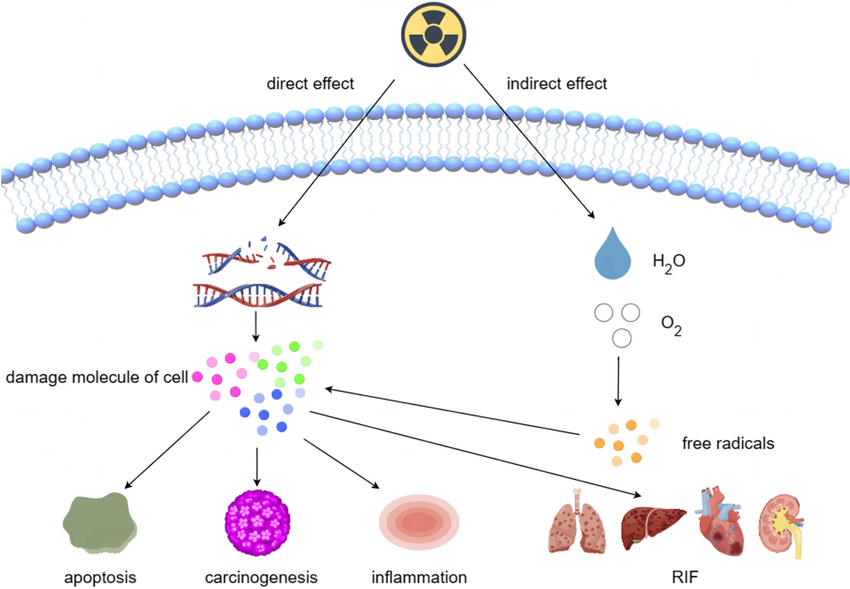
- Radiation Therapy: Treatment for brain tumors or other cancers may involve radiation that affects the pituitary gland’s ability to produce hormones.
- Infections and Inflammation: Conditions such as meningitis or encephalitis can lead to inflammation of the tissues surrounding the brain and pituitary gland, resulting in hypopituitarism.
- Pituitary Apoplexy: This is a sudden hemorrhage into the pituitary gland, which can cause rapid hormone deficiencies and is often associated with pre-existing tumors.
- Vascular Events: Rarely, strokes or subarachnoid hemorrhages (bleeding around the brain) can impair blood flow to the pituitary gland, causing tissue death and loss of hormone production.
- Autoimmune Conditions: Disorders like lymphocytic hypophysitis involve an autoimmune attack on the pituitary tissue, leading to hormonal deficiencies.
- Metabolic Disorders: Conditions such as hemochromatosis (excess iron accumulation) and histiocytoses (abnormal proliferation of immune cells) can also affect pituitary function.
- Sheehan Syndrome: This rare condition occurs due to severe blood loss during or after childbirth, leading to tissue death in the pituitary gland.
- Medications: Certain medications, particularly glucocorticoids used for inflammatory conditions and some treatments for prostate cancer, may suppress normal pituitary function.
Clinical Entities Related to Hypopituitarism
Hypopituitarism is associated with various clinical entities depending on which hormones are deficient:
- Growth Hormone Deficiency (GHD):
- In children, it leads to stunted growth and development issues.
- In adults, it may cause decreased muscle mass and increased body fat.
- Thyroid-Stimulating Hormone (TSH) Deficiency:
- Results in hypothyroidism symptoms such as fatigue, weight gain, cold intolerance, and depression.
- Adrenocorticotropic Hormone (ACTH) Deficiency:
- Leads to adrenal insufficiency characterized by fatigue, weakness, low blood pressure, and potential life-threatening adrenal crisis if untreated.
- Gonadotropin Deficiency (FSH/LH):
- In males: Low testosterone levels result in reduced libido and infertility.
- In females: Can lead to menstrual irregularities or amenorrhea and infertility.
- Prolactin Deficiency:
- Primarily affects lactation; women may experience insufficient milk production post-delivery.
- Antidiuretic Hormone (ADH) Deficiency:
- Causes diabetes insipidus characterized by excessive thirst and urination due to inability of kidneys to concentrate urine effectively.
- Oxytocin Deficiency:
- May impact labor contractions during childbirth and milk ejection during breastfeeding but is less commonly discussed clinically compared to other deficiencies.
The management of hypopituitarism involves identifying specific hormone deficiencies through diagnostic testing and implementing appropriate hormone replacement therapies tailored to each patient’s needs based on their clinical presentation.
Diabetes Insipidus and Syndrome of Inappropriate Antidiuretic Hormone Secretion
Overview of Diabetes Insipidus (DI)
Diabetes insipidus is a condition characterized by excessive urination and persistent thirst. It results from a deficiency in the hormone vasopressin, also known as antidiuretic hormone (ADH), which is produced by the hypothalamus and secreted by the posterior pituitary gland. This hormone plays a crucial role in regulating water balance in the body by signaling the kidneys to retain water. When there is insufficient vasopressin, the kidneys fail to reabsorb water effectively, leading to increased urine output and potential dehydration.
There are several types of diabetes insipidus:
- Central Diabetes Insipidus: This form occurs due to inadequate production of vasopressin, often resulting from damage to the hypothalamus or pituitary gland due to surgery, infections, tumors, or head injuries.
- Nephrogenic Diabetes Insipidus: In this type, the kidneys do not respond appropriately to vasopressin. Causes can include certain medications (like lithium), electrolyte imbalances (such as high calcium or low potassium levels), genetic factors, or chronic kidney disease.
- Dipsogenic Diabetes Insipidus: This variant arises from excessive fluid intake driven by an abnormal thirst mechanism, often linked to psychological conditions or lesions in the hypothalamus.
- Gestational Diabetes Insipidus: A temporary condition that can occur during pregnancy due to placental enzymes that break down vasopressin.
The symptoms of diabetes insipidus primarily include frequent urination (polyuria), intense thirst (polydipsia), and pale urine output. These symptoms can lead to complications such as dehydration and electrolyte imbalances.
Overview of Syndrome of Inappropriate Antidiuretic Hormone Secretion (SIADH)
In contrast, the syndrome of inappropriate antidiuretic hormone secretion leads to excessive retention of water due to overproduction of vasopressin. This condition results in dilutional hyponatremia—low sodium levels in the blood—because excess water dilutes sodium concentrations.
SIADH can be caused by various factors including:
- Neurological conditions such as brain tumors or infections
- Certain cancers (notably small cell lung cancer)
- Medications like carbamazepine and oxcarbazepine
- Lung diseases such as pneumonia
- Post-surgical states where pain sensors may stimulate ADH release
Symptoms associated with SIADH primarily stem from hyponatremia and may include nausea, vomiting, headaches, fatigue, confusion, irritability, seizures, and even coma in severe cases.
Diagnosis and Management
Diagnosing these conditions involves careful evaluation of clinical symptoms alongside laboratory tests measuring serum sodium levels and urine osmolality. For diabetes insipidus, low urine osmolality despite high plasma osmolality indicates a lack of ADH action. Conversely, SIADH typically shows low serum sodium with high urine osmolality.
Management strategies differ significantly between these two disorders:
- Diabetes Insipidus Treatment: The primary treatment involves administering desmopressin—a synthetic form of vasopressin—which can be given orally or via injection. Additional measures may include ensuring adequate hydration.
- SIADH Treatment: Management focuses on fluid restriction to prevent further dilution of serum sodium levels. In some cases, medications such as vaptans (vasopressin receptor antagonists) may be used to counteract the effects of excess vasopressin.
Both conditions highlight critical aspects of fluid balance regulation within the body and underscore how disruptions in hormonal control can lead to significant clinical challenges.
Craniopharyngioma
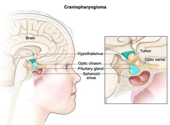
A craniopharyngioma is a rare, benign tumor that typically develops near the pituitary gland in the brain. These tumors are slow-growing and can significantly impact the endocrine system and cranial nerves, particularly those responsible for vision. Craniopharyngiomas are most commonly diagnosed in children aged 5 to 14 years and adults aged 50 to 74 years.
Types of Craniopharyngioma
There are two main types of craniopharyngiomas:
- Adamantinomatous Craniopharyngioma: This is the most common type and primarily affects children and younger adults.
- Papillary Craniopharyngioma: This type is less common and predominantly occurs in adults.
Symptoms
The symptoms of craniopharyngiomas can vary widely depending on their size and location. Common symptoms include:
- Changes in mood or behavior
- Headaches
- Hearing loss
- Hormonal changes
- Loss of balance
- Nausea and vomiting
As the tumor grows, it may exert pressure on surrounding structures, leading to more severe symptoms such as:
- Adrenal failure (causing dizziness, sleepiness, muscle weakness)
- Changes in sexual function (impotence, menstrual irregularities)
- Growth and developmental issues in children (delayed puberty, stunted growth)
- Excessive thirst and urination (diabetes insipidus)
- Hypothyroidism (weight gain, fatigue)
- Decreased vision or peripheral vision problems
Diagnosis
Diagnosing a craniopharyngioma typically involves a thorough medical history review followed by imaging tests such as CT scans or MRIs. A neurological examination may also be conducted to assess vision, hearing, balance, coordination, and reflexes.
Treatment Options
The primary treatment methods for craniopharyngiomas include:
- Surgery: The goal is to remove as much of the tumor as possible while minimizing damage to surrounding tissues like the pituitary gland and optic nerves. Surgery can be challenging due to the tumor’s proximity to these delicate structures.
- Radiation Therapy: Often recommended post-surgery if complete removal isn’t feasible or if there’s a risk of recurrence.
- Chemotherapy: This has emerged as an option for specific subtypes of craniopharyngiomas, particularly the papillary variant.
Despite being benign, craniopharyngiomas can recur after treatment; approximately half of surgically removed tumors may come back over time. Patients often require lifelong monitoring and management of hormonal functions affected by the tumor.
Prognosis
The prognosis for individuals diagnosed with craniopharyngiomas is generally positive; more than 90% survive five years post-diagnosis. However, because these tumors can lead to chronic conditions even after treatment, ongoing medical care is essential.
In summary, craniopharyngiomas are complex tumors that necessitate careful diagnosis and management due to their potential impact on critical bodily functions.


