Definition of Chronic myeloproliferative neoplasms
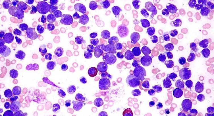
Chronic myeloproliferative neoplasms (CMPNs) are a group of diseases characterized by the excessive production of blood cells in the bone marrow, leading to an increased number of red blood cells, white blood cells, or platelets. These conditions typically progress slowly and can result in complications such as blood clots, bleeding issues, and an increased risk of developing acute leukemia.
Comparison of Clinical and Laboratory Findings of myeloproliferative syndrome
1. Chronic Myelogenous Leukemia (CML)
CML is a type of cancer that affects the blood and bone marrow. It is characterized by the overproduction of myeloid cells.
- Clinical Findings:
- Symptoms often include fatigue, night sweats, weight loss, and splenomegaly (enlarged spleen).
- Patients may experience abdominal discomfort due to splenic enlargement.
- Chronic phase symptoms can be mild, but progression to accelerated or blast phases can lead to more severe symptoms.
- Laboratory Findings:
- Blood tests typically show leukocytosis (elevated white blood cell count), often exceeding 100,000 cells/µL.
- The presence of immature myeloid cells (myeloblasts) in peripheral blood.
- A hallmark finding is the Philadelphia chromosome (BCR-ABL fusion gene) detected via cytogenetic analysis or PCR.
- Bone marrow biopsy reveals hypercellularity with increased myeloid lineage.
2. Polycythemia Vera (PV)
PV is a myeloproliferative neoplasm characterized by an increase in red blood cell mass.
- Clinical Findings:
- Symptoms may include headaches, dizziness, visual disturbances, and pruritus (itching), especially after bathing.
- Patients often present with ruddy complexion due to increased red blood cell mass.
- Splenomegaly is common, and there may be complications such as thrombosis.
- Laboratory Findings:
- Elevated hemoglobin (>16.5 g/dL in men; >16 g/dL in women) or hematocrit (>49% in men; >48% in women).
- Serum erythropoietin levels are typically low.
- Bone marrow biopsy shows hypercellularity with increased erythroid, granulocytic, and megakaryocytic lineages.
- JAK2 V617F mutation is present in approximately 95% of cases.
3. Essential Thrombocythemia (ET)
ET is another myeloproliferative neoplasm primarily affecting megakaryocytes leading to elevated platelet counts.
- Clinical Findings:
- Patients may be asymptomatic or present with thrombotic events such as stroke or myocardial infarction due to high platelet counts.
- Symptoms can include headaches, dizziness, and visual disturbances similar to PV.
- Splenomegaly may occur but is less common than in CML or PV.
- Laboratory Findings:
- Markedly elevated platelet count (>450 x10^9/L).
- Normal red blood cell mass; hemoglobin levels are usually normal.
- Bone marrow biopsy shows increased megakaryocytes with abnormal morphology.
- JAK2 V617F mutation is found in about half of the patients; other mutations like CALR or MPL may also be present.
4. Myelofibrosis with Myeloid Metaplasia (MMM)
MMM involves fibrosis of the bone marrow leading to extramedullary hematopoiesis.
- Clinical Findings:
- Symptoms include fatigue, weakness, night sweats, fever, weight loss, and significant splenomegaly due to extramedullary hematopoiesis.
- Patients may experience bone pain due to marrow infiltration and fibrosis.
- Laboratory Findings:
- Peripheral blood smear shows leukoerythroblastosis (immature white cells and nucleated red cells).
- Anemia is common with variable white blood cell counts; thrombocytopenia can also occur.
- Bone marrow biopsy reveals hypercellularity initially but progresses to fibrosis which can be assessed using reticulin stains.
- JAK2 V617F mutation occurs in about half of the cases; other mutations may also be identified.
In summary:
- CML primarily affects myeloid cells with characteristic findings including leukocytosis and Philadelphia chromosome positivity.
- PV results from increased red cell mass with low erythropoietin levels and JAK2 mutations being prevalent.
- ET features elevated platelets without significant changes in red blood cell mass but often has JAK2 mutations as well.
- MMM presents with anemia and leukoerythroblastosis alongside significant splenomegaly due to extramedullary hematopoiesis.
Philadelphia Chromosome: Overview
The Philadelphia chromosome is a specific genetic abnormality that is primarily associated with chronic myelogenous leukemia (CML). It was first discovered in 1960 by researchers David A. Hungerford and Peter C. Nowell at the Fox Chase Cancer Center and the University of Pennsylvania, respectively. The abnormality involves a translocation between chromosomes 9 and 22, specifically the fusion of the BCR gene on chromosome 22 with the ABL gene on chromosome 9, resulting in a new gene called BCR-ABL. This fusion gene produces a protein that has increased tyrosine kinase activity, which plays a crucial role in promoting cell proliferation and survival.
Disease Association
The Philadelphia chromosome is most commonly associated with CML, a type of cancer that affects the blood and bone marrow. In CML, the presence of this chromosomal abnormality leads to the production of the BCR-ABL fusion protein, which causes uncontrolled growth of myeloid cells. This results in an overproduction of white blood cells that can crowd out normal cells, leading to various symptoms such as fatigue, splenomegaly (enlarged spleen), and increased susceptibility to infections.
In addition to CML, the Philadelphia chromosome can also be found in some cases of acute lymphoblastic leukemia (ALL) and occasionally in acute myeloid leukemia (AML). Its presence in these diseases indicates a poor prognosis and often necessitates more aggressive treatment strategies.
Significance
The discovery of the Philadelphia chromosome marked a pivotal moment in cancer research as it provided one of the first clear links between genetics and cancer development. It demonstrated that specific chromosomal abnormalities could serve as biomarkers for certain malignancies. The identification of BCR-ABL not only advanced our understanding of CML but also led to targeted therapies aimed at inhibiting its activity.
One of the most significant advancements stemming from this discovery was the development of imatinib (Gleevec), a targeted therapy that specifically inhibits the BCR-ABL tyrosine kinase. This drug has transformed CML from a fatal disease into a manageable chronic condition for many patients. The success of imatinib paved the way for further research into targeted therapies for other cancers based on their unique genetic profiles.
Overall, the Philadelphia chromosome serves as an important model for understanding how genetic alterations can lead to cancer and how these insights can be translated into effective treatments.
Significance of the Placenta-Like Alkaline Phosphatase (PLAP) Score
1. Overview of PLAP and Its Expression in Tumors
Placenta-like alkaline phosphatase (PLAP), also known as alkaline phosphatase, placental type (ALPP), is an enzyme primarily expressed in the placenta during pregnancy. Its expression is not limited to normal tissues; it has been found in various tumors, particularly testicular germ cell tumors, ovarian cancer, and endometrial cancer. The significance of PLAP lies in its potential role as a biomarker for certain malignancies.
2. Diagnostic Utility of PLAP
The PLAP score is particularly valuable in the diagnostic process for specific types of cancers. High levels of PLAP expression are often associated with testicular germ cell tumors, where it can be detected in nearly all cases. This makes it a crucial marker for confirming diagnoses and differentiating between germ cell tumors and other neoplasms. In addition to testicular cancers, elevated PLAP levels have been observed in several other tumor types, including ovarian and endometrial cancers.
3. Prognostic Implications
Beyond its diagnostic utility, the PLAP score may also have prognostic significance. Studies have indicated that high PLAP expression correlates with advanced pathological tumor stages and metastasis in certain cancers such as colorectal cancer. Conversely, low levels of PLAP expression might indicate a less aggressive disease course in other malignancies like endometrial cancer. Therefore, assessing the PLAP score can provide insights into tumor behavior and patient outcomes.
4. Therapeutic Targeting
The unique expression pattern of PLAP presents opportunities for targeted immunotherapy. Since PLAP is predominantly expressed on malignant cells rather than normal tissues, it serves as a promising target for therapeutic interventions aimed at selectively attacking tumor cells while minimizing damage to healthy tissues. This specificity enhances the potential efficacy of treatments designed to target PLAP-expressing tumors.
5. Conclusion
In summary, the significance of the PLAP score extends from its role as a diagnostic marker to its implications for prognosis and therapy in various cancers. Understanding the expression patterns and clinical relevance of PLAP can aid clinicians in making informed decisions regarding diagnosis, treatment planning, and patient management.
Definition of Leukoerythroblastosis, Leukemoid Reaction, Dyserythropoiesis, Dysmyelopoiesis, Dysmegakaryopoiesis, Ringed Sideroblasts and Myelofibrosis
Leukoerythroblastosis
Leukoerythroblastosis is a hematological condition characterized by the presence of immature white blood cells (leukocytes) and red blood cell precursors (erythroblasts) in the peripheral blood. This phenomenon typically indicates a response to severe anemia or bone marrow infiltration by malignant processes, such as leukemia or metastatic cancer. The presence of these immature cells suggests that the bone marrow is under stress and attempting to produce more blood cells to compensate for an underlying pathology.
Leukemoid Reaction
A leukemoid reaction refers to an extreme increase in white blood cell count that mimics leukemia but occurs due to non-malignant causes. This reaction can be triggered by various factors, including infections, inflammation, or severe stress. In contrast to leukemia, where there is a clonal proliferation of abnormal leukocytes, a leukemoid reaction involves a reactive process with normal-appearing white blood cells. The differentiation between the two can often be made through clinical context and laboratory findings.
Dyserythropoiesis
Dyserythropoiesis describes abnormal erythropoiesis, which is the process of red blood cell production. In this condition, there are defects in the maturation and development of erythroid progenitor cells within the bone marrow. Dyserythropoiesis can manifest as ineffective erythropoiesis leading to anemia and may be associated with various disorders, including myelodysplastic syndromes (MDS), vitamin B12 deficiency, or other nutritional deficiencies.
Dysmyelopoiesis
Dysmyelopoiesis refers to abnormal myelopoiesis, which is the formation of myeloid lineage cells (such as granulocytes, monocytes, and platelets) from hematopoietic stem cells in the bone marrow. This condition often results in ineffective hematopoiesis and can lead to cytopenias (decreased cell counts) or dysplastic features in myeloid cells. Dysmyelopoiesis is commonly seen in conditions like myelodysplastic syndromes and certain types of acute myeloid leukemia (AML).
Dysmegakaryopoiesis
Dysmegakaryopoiesis involves abnormalities in megakaryocyte development and function within the bone marrow. Megakaryocytes are responsible for producing platelets through a process called thrombopoiesis. Dysmegakaryopoiesis can result in thrombocytopenia (low platelet counts) or dysfunctional platelets and may occur in various disorders such as essential thrombocythemia or myelodysplastic syndromes.
Ringed Sideroblasts
Ringed sideroblasts are erythroid precursor cells characterized by iron-loaded mitochondria that form a ring around the nucleus when stained with specific dyes. These cells are indicative of impaired heme synthesis and are commonly associated with conditions such as sideroblastic anemia. The presence of ringed sideroblasts reflects an inability to incorporate iron into hemoglobin properly despite adequate iron availability.
Myelofibrosis
Myelofibrosis is a type of chronic myeloproliferative neoplasm characterized by fibrosis (scarring) of the bone marrow tissue, leading to ineffective hematopoiesis and resultant cytopenias. Patients may experience symptoms such as splenomegaly (enlargement of the spleen), fatigue, night sweats, and weight loss. Myelofibrosis can be primary or secondary to other conditions like polycythemia vera or essential thrombocythemia. Diagnosis typically involves bone marrow biopsy showing increased collagen deposition along with peripheral blood findings.
Different Types of Myelodysplastic Syndromes
Myelodysplastic syndromes (MDS) are a group of disorders caused by poorly formed or dysfunctional blood cells. They occur when the bone marrow does not produce enough healthy blood cells, leading to various complications. The classification of MDS is essential for determining prognosis and treatment options. Below are the different types of MDS:
1. Refractory Anemia (RA)
Refractory anemia is characterized by a low red blood cell count (anemia) that does not improve with standard treatments. Patients may experience symptoms such as fatigue and weakness due to insufficient oxygen delivery to tissues.
2. Refractory Anemia with Ringed Sideroblasts (RARS)
This type features the presence of ringed sideroblasts, which are abnormal red blood cell precursors that accumulate iron in their mitochondria. RARS typically presents with anemia and can be associated with a better prognosis compared to other forms of MDS.
3. Refractory Cytopenia with Multilineage Dysplasia (RCMD)
RCMD involves deficiencies in multiple types of blood cells (red cells, white cells, and platelets) due to ineffective hematopoiesis. This type indicates dysplasia in more than one lineage, suggesting a more complex underlying pathology.
4. Refractory Cytopenia with Multilineage Dysplasia and Ringed Sideroblasts (RCMD-RS)
This subtype combines features of RCMD and RARS, indicating multilineage cytopenias along with the presence of ringed sideroblasts. It often has a varied prognosis based on individual patient factors.
5. Refractory Anemia with Excess Blasts (RAEB)
RAEB is characterized by an increased number of immature blood cells (blasts) in the bone marrow, which can indicate a progression towards acute myeloid leukemia (AML). RAEB is further classified into two subtypes: RAEB-1 and RAEB-2, depending on the percentage of blasts present.
6. Myelodysplastic Syndrome, Unclassified (MDS-U)
This category includes cases that do not fit neatly into any other classification but still exhibit dysplastic changes in the bone marrow or peripheral blood without meeting specific criteria for other types.
7. MDS Associated with Isolated Del(5q)
This type is characterized by a deletion on chromosome 5 and often leads to anemia that responds well to certain treatments like lenalidomide. Patients typically have a better prognosis compared to those with more complex chromosomal abnormalities.
In addition to these classifications, it’s important to note that MDS can be primary (de novo), occurring spontaneously without known risk factors, or secondary, arising from previous chemotherapy or radiation therapy exposure.
The classification helps healthcare providers determine appropriate treatment strategies and predict patient outcomes based on the specific characteristics of each type.


