Structure of the Cell Membrane
The cell membrane, also known as the plasma membrane, is a crucial component of all living cells, serving as a protective barrier that separates the internal contents of the cell from its external environment. The fundamental structure of the cell membrane is characterized by a bilayer of phospholipids, which plays a vital role in its function and properties.
Bi-Lipid Structure
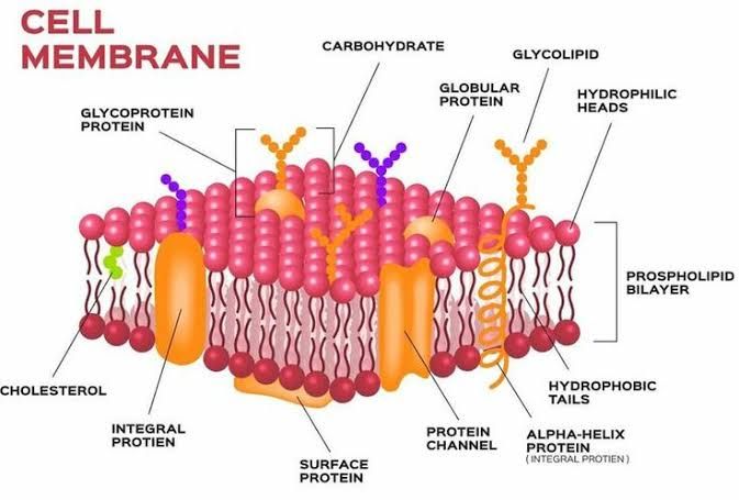
- Phospholipid Composition: The primary building blocks of the cell membrane are phospholipids. Each phospholipid molecule consists of two main parts: a hydrophilic (water-attracting) “head” and two hydrophobic (water-repelling) “tails.” The head contains a phosphate group that is polar and interacts favorably with water, while the tails consist of long fatty acid chains that are nonpolar and do not interact with water.
- Formation of the Bilayer: In an aqueous environment, phospholipids spontaneously arrange themselves into a bilayer. This occurs because the hydrophilic heads face outward towards the water on both sides (the intracellular fluid inside the cell and extracellular fluid outside), while the hydrophobic tails face inward, away from water. This arrangement creates a semi-permeable barrier that regulates what enters and exits the cell.
- Amphipathic Nature: Phospholipids are described as amphipathic molecules due to their dual nature—having both hydrophilic and hydrophobic regions. This property is essential for forming membranes because it allows for self-assembly into bilayers in aqueous environments.
- Fluid Mosaic Model: The structure of the cell membrane is often described by the fluid mosaic model, which suggests that while the phospholipid bilayer provides fluidity, various proteins are embedded within this layer, creating a mosaic-like appearance. These proteins can move laterally within the layer, contributing to membrane flexibility and functionality.
- Role of Cholesterol: Embedded within this phospholipid bilayer are cholesterol molecules that help to stabilize membrane fluidity across varying temperatures. Cholesterol molecules fit between phospholipids, preventing them from packing too closely together at lower temperatures and maintaining structural integrity at higher temperatures.
- Membrane Proteins: In addition to phospholipids and cholesterol, various proteins are integrated into or associated with the membrane. These include integral proteins that span across the bilayer (such as channel proteins for ion transport) and peripheral proteins that attach to either surface of the membrane (involved in signaling or structural support).
- Glycocalyx Formation: Some integral proteins have carbohydrate groups attached to them, forming glycoproteins which extend into the extracellular space. Collectively with glycolipids (carbohydrates attached to lipids), these structures form a fuzzy coating known as glycocalyx that plays roles in cell recognition and protection.
- Selective Permeability: The unique structure of the lipid bilayer contributes to its selective permeability; only certain substances can pass through unaided based on their size and polarity. Small nonpolar molecules like oxygen can diffuse freely through, whereas larger or polar molecules require specific transport mechanisms.
In summary, the bi-lipid structure of the cell membrane consists primarily of two layers of phospholipids arranged such that their hydrophilic heads face outward toward aqueous environments while their hydrophobic tails face inward, creating a selectively permeable barrier essential for cellular function.
Components of Membrane Structure explained
Lipid Bilayer
The lipid bilayer is the fundamental structure of cell membranes, primarily composed of phospholipids. Each phospholipid molecule consists of a hydrophilic (water-attracting) “head” and two hydrophobic (water-repelling) “tails.” In an aqueous environment, these molecules arrange themselves into a bilayer with the hydrophobic tails facing inward, shielded from water, while the hydrophilic heads face outward towards the water on both sides. This arrangement creates a semi-permeable membrane that allows certain substances to pass through while blocking others.
The lipid bilayer serves several critical functions:
- Barrier Function: It acts as a barrier to protect cellular components from the external environment.
- Fluidity: The fluid nature of the lipid bilayer allows for flexibility and movement within the membrane, enabling processes such as endocytosis and exocytosis.
- Selectivity: The composition of lipids can influence membrane permeability and fluidity, allowing cells to maintain homeostasis by regulating what enters and exits.
Cholesterol
Cholesterol is interspersed within the lipid bilayer and plays a crucial role in maintaining membrane integrity and fluidity. It is a type of sterol that fits between phospholipid molecules, preventing them from packing too closely together at lower temperatures, which would make the membrane too rigid. Conversely, at higher temperatures, cholesterol helps stabilize the membrane by restraining excessive movement of phospholipids.
Key functions of cholesterol include:
- Membrane Fluidity Regulation: Cholesterol maintains optimal fluidity across varying temperatures.
- Formation of Lipid Rafts: Cholesterol-rich microdomains known as lipid rafts serve as platforms for signaling molecules and proteins involved in various cellular processes.
- Barrier to Small Molecules: Cholesterol contributes to the overall impermeability of membranes to small polar molecules.
Peripheral Proteins
Peripheral proteins are loosely attached to the exterior or interior surfaces of the membrane and do not penetrate into the lipid bilayer. They can be associated with integral proteins or directly with phospholipids through ionic interactions or hydrogen bonds.
Functions of peripheral proteins include:
- Structural Support: They help maintain cell shape by anchoring cytoskeletal elements to the plasma membrane.
- Signaling Pathways: Many peripheral proteins are involved in signal transduction pathways; they can act as enzymes or receptors that transmit signals from outside to inside the cell.
- Cell Recognition: Some peripheral proteins play roles in cell recognition and communication by interacting with glycoproteins on neighboring cells.
Integral Proteins
Integral proteins span across the entire lipid bilayer and are embedded within it, often extending from one side of the membrane to another. These proteins can be classified into two categories: transmembrane proteins (which traverse both layers) and monotopic proteins (which only penetrate one layer).
Functions of integral proteins include:
- Transport Channels/Carriers: Integral proteins facilitate transport across membranes by forming channels or carriers for ions and molecules that cannot diffuse freely through the lipid bilayer.
- Receptors: Many integral proteins function as receptors that bind specific ligands (such as hormones), triggering cellular responses.
- Enzymatic Activity: Some integral proteins have enzymatic functions that catalyze reactions directly at the membrane surface.
In summary, each component—lipid bilayer, cholesterol, peripheral proteins, and integral proteins—plays distinct yet interconnected roles in maintaining cellular structure and function.
Signal Transduction and Ligand-Receptor Interaction Across the Membrane
Introduction to Signal Transduction
Signal transduction is a fundamental biological process that allows cells to respond to external stimuli. It begins at the cell membrane, where receptors interact with specific ligands. This interaction triggers a cascade of intracellular events that ultimately lead to a cellular response. The complexity of signal transduction pathways reflects the diverse roles that cells play in multicellular organisms.
Ligand-Receptor Interaction
The initial step in signal transduction involves ligand-receptor interactions. Receptors are specialized proteins located on the cell surface or within the cell, which bind specific molecules known as ligands. These ligands can be hormones, neurotransmitters, or other signaling molecules. The binding of a ligand to its receptor induces conformational changes in the receptor, activating it and initiating downstream signaling pathways.
- Types of Receptors: There are several types of receptors based on their structure and mechanism of action:
- G-Protein Coupled Receptors (GPCRs): These receptors activate intracellular G-proteins upon ligand binding, leading to various signaling pathways.
- Receptor Tyrosine Kinases (RTKs): Upon ligand binding, these receptors undergo dimerization and autophosphorylation, activating downstream signaling cascades.
- Ion Channel Receptors: These receptors open or close ion channels in response to ligand binding, altering the membrane potential and initiating cellular responses.
- Mechanism of Action: The interaction between ligands and receptors can be characterized by several key processes:
- Avidity and Consumption: Avidity refers to the overall strength of binding between a receptor and its ligand, while consumption describes how effectively a receptor internalizes bound ligands through endocytosis.
- Endocytosis: Following activation, many receptors undergo endocytosis, where they are internalized into the cell along with their bound ligands. This process not only regulates receptor availability but also enhances signaling accuracy by controlling ligand concentration at the receptor site.
Membrane Dynamics in Signal Transduction
The membrane environment plays a crucial role in signal transduction processes:
- Local Composition Heterogeneities: Membranes are not uniform; they contain various lipids and proteins that can create microdomains influencing receptor activity.
- Mechanical Effects: The physical properties of membranes can affect how receptors cluster together and interact with each other, which is essential for effective signal transduction.
- Biophysical Interactions: Recent studies have highlighted how membrane components actively participate in signaling processes by affecting protein-protein interactions at the membrane interface.
Conclusion
Understanding signal transduction across membranes requires an appreciation for both ligand-receptor interactions and the complex dynamics of membrane environments. By studying these processes, researchers can gain insights into how cells communicate and respond to their surroundings.
Understanding the Processes of Diffusion, Endocytosis, and Exocytosis Across the Membrane
Diffusion
Diffusion is a passive transport process that involves the movement of molecules from an area of higher concentration to an area of lower concentration. This process occurs due to the random motion of particles and does not require energy input from the cell. In biological systems, diffusion is crucial for maintaining homeostasis and facilitating the exchange of substances between cells and their environment.
- Simple Diffusion: Small nonpolar molecules (e.g., oxygen, carbon dioxide) can pass directly through the lipid bilayer of cell membranes without assistance.
- Facilitated Diffusion: Larger or polar molecules (e.g., glucose, ions) require specific transport proteins to help them cross the membrane. These proteins can be channels or carriers that facilitate movement down their concentration gradient.
Endocytosis
Endocytosis is an active transport mechanism that allows cells to import large molecules or particles by engulfing them in vesicles formed from the plasma membrane. This process requires energy in the form of ATP.
- Types of Endocytosis:
- Phagocytosis: Often referred to as “cell eating,” this process involves immune cells engulfing large particles such as bacteria or dead cells. The plasma membrane extends around the particle, forming a vesicle called a phagosome, which then fuses with lysosomes for degradation.
- Pinocytosis: Known as “cell drinking,” pinocytosis involves the uptake of extracellular fluid and dissolved solutes. The membrane invaginates to form small vesicles that bring in liquid along with any solutes present.
- Receptor-Mediated Endocytosis: This specialized form involves receptors on the cell surface binding specific ligands (e.g., hormones, nutrients). Once bound, these receptors cluster together and initiate invagination to form a vesicle containing the ligand.
Exocytosis
Exocytosis is another active transport mechanism used by cells to export materials out into the extracellular space. This process also requires energy and involves vesicles that fuse with the plasma membrane.
- Steps of Exocytosis:
- Vesicles containing substances (e.g., hormones, neurotransmitters) are transported to the plasma membrane.
- The vesicle membrane fuses with the cell membrane, resulting in the release of its contents outside the cell.
- There are two main types:
- Constitutive Exocytosis: This occurs continuously and is responsible for delivering lipids and proteins to maintain cellular functions.
- Regulated Exocytosis: This type occurs in response to specific signals (e.g., neurotransmitter release at synapses), where vesicles only fuse with the membrane when triggered by certain stimuli.
In summary, diffusion allows for passive movement across membranes based on concentration gradients, while endocytosis and exocytosis are active processes that enable cells to import and export larger molecules through vesicular transport mechanisms.
Channels and Their Role in Trafficking Across the Membrane
Introduction to Membrane Channels
Membrane channels are integral membrane proteins that facilitate the transport of ions and molecules across cellular membranes. They play a crucial role in maintaining cellular homeostasis, signaling, and various physiological processes. The presence of these channels is particularly significant in epithelial cells, where they contribute to vectorial transport—meaning the directional movement of substances across the cell layer.
Types of Channels Involved in Trafficking
- Ion Channels: These channels allow specific ions (such as Na+, K+, Ca2+, and Cl-) to pass through the membrane in response to electrochemical gradients. Ion channels can be gated by various stimuli, including voltage changes, ligand binding, or mechanical stress. For example, Transient Receptor Potential (TRP) channels are a superfamily of cation channels that not only function at the plasma membrane but also localize to intracellular membranes, influencing both ion homeostasis and membrane trafficking.
- Solute Transporters: These proteins facilitate the movement of solutes across membranes either via passive diffusion or active transport mechanisms. They can be categorized into uniporters, symporters, and antiporters based on their transport mechanism. The polarized expression of these transporters at either the apical or basolateral membrane domains is essential for proper epithelial function.
Mechanisms of Channel Trafficking
The trafficking of channels involves several key processes:
- Biosynthesis and Delivery: Channels are synthesized in the endoplasmic reticulum (ER) and transported to the Golgi apparatus for post-translational modifications before being delivered to their final destinations—either apical or basolateral membranes.
- Polarized Distribution: Epithelial cells exhibit a polarized structure where different sets of channels are localized to distinct membrane domains. This polarization is critical for their function in secretion and absorption processes.
- Endocytosis and Recycling: Once at the plasma membrane, channels can be internalized through endocytosis. This process allows for dynamic regulation of channel numbers on the surface; recycled channels can return to their original location or be targeted for degradation.
- Regulation by Physiological Stimuli: The activity of these channels can be modulated by hormones or neurotransmitters that influence their trafficking pathways. For instance, an increase in intracellular calcium levels may promote the translocation of certain TRP channels from intracellular stores to the plasma membrane.
Role in Cellular Functions
Channels significantly impact various cellular functions:
- Signal Transduction: Many ion channels participate in signal transduction pathways that regulate cellular responses to external stimuli.
- Homeostasis Maintenance: By controlling ion flow across membranes, these channels help maintain ionic balance within cells and tissues.
- Fluid Transport Regulation: In epithelial tissues, proper channel function is essential for fluid secretion and absorption processes critical for organ function (e.g., kidney filtration).
Conclusion
The presence and functionality of membrane channels are vital for effective trafficking across cellular membranes. Their ability to dynamically adjust localization and activity in response to physiological cues underscores their importance in maintaining homeostasis and facilitating communication within cells.
Interaction of Integral Membrane Proteins with Cytoskeletal Elements to Maintain Cell Shape
The maintenance of cell shape is a complex process that involves the interplay between integral membrane proteins and the cytoskeleton. Integral membrane proteins are embedded within the lipid bilayer of the plasma membrane and play crucial roles in various cellular functions, including signaling, transport, and maintaining structural integrity. The cytoskeleton, composed of filamentous polymers such as microtubules, actin filaments, and intermediate filaments, provides mechanical support and facilitates cellular movement.
1. Role of Integral Membrane Proteins
Integral membrane proteins can interact with cytoskeletal elements through several mechanisms:
- Anchoring: Many integral proteins have specific domains that allow them to bind directly to cytoskeletal components. For example, integrins are a class of integral membrane proteins that connect the extracellular matrix to actin filaments inside the cell. This anchoring helps stabilize cell shape by providing a physical link between the external environment and the internal cytoskeletal framework.
- Signal Transduction: Integral membrane proteins often function as receptors that transmit signals from outside the cell to the cytoplasm. When these receptors bind ligands (such as hormones or growth factors), they can activate intracellular signaling pathways that lead to changes in cytoskeletal dynamics, promoting alterations in cell shape or motility.
- Formation of Membrane Domains: Some integral proteins cluster together in specific regions of the membrane known as lipid rafts or membrane microdomains. These clusters can influence local cytoskeletal organization by recruiting additional proteins that interact with both the membrane and the underlying cytoskeleton.
2. Interaction with Cytoskeletal Elements
The interaction between integral membrane proteins and cytoskeletal elements is vital for maintaining cell shape:
- Cytoskeletal Support: The cytoskeleton provides a scaffold that supports the plasma membrane. By anchoring integral proteins to this scaffold, cells can maintain their shape against mechanical stress. For instance, during cellular stretching or deformation, anchored integral proteins help distribute forces across the membrane.
- Dynamic Remodeling: The cytoskeleton is not static; it undergoes constant remodeling in response to various stimuli. This dynamic nature allows cells to adapt their shape according to their environment or functional needs (e.g., during migration). Integral membrane proteins play a role in this remodeling process by facilitating interactions with regulatory proteins that modulate cytoskeletal assembly and disassembly.
- Mechanical Signal Transmission: Both internal and external mechanical forces can be transmitted through integral membrane proteins to the cytoskeleton. This transmission is essential for processes such as mechanotransduction, where cells convert mechanical stimuli into biochemical responses. For example, when a cell experiences shear stress from fluid flow, integrins can relay this information through their connections with actin filaments, leading to changes in cell shape or behavior.
3. Conclusion
In summary, integral membrane proteins interact intricately with cytoskeletal elements to maintain cell shape through anchoring mechanisms, signal transduction pathways, and dynamic remodeling processes. These interactions ensure that cells remain structurally stable while also being capable of responding flexibly to environmental changes.


