Upper and Lower Motor Neurons
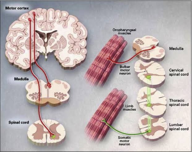
Definition of Upper Motor Neurons: Upper motor neurons (UMNs) are a type of motor neuron that originate in the cerebral cortex or brainstem and send signals down to lower motor neurons. They play a crucial role in the voluntary control of movement by transmitting impulses that initiate and modulate muscle contractions. UMNs are involved in both conscious movements, such as walking or writing, and subconscious processes, like maintaining posture.
Examples of Upper Motor Neurons:
- Pyramidal Tract Neurons: These neurons originate in the primary motor cortex (located in the frontal lobe) and descend through the brainstem into the spinal cord. They are primarily responsible for voluntary movements.
- Extrapyramidal Tract Neurons: These include pathways such as the rubrospinal tract, which is involved in involuntary movements related to balance and posture.
Definition of Lower Motor Neurons: Lower motor neurons (LMNs) are located in the spinal cord and brainstem, where they receive signals from upper motor neurons. LMNs directly innervate skeletal muscles, causing them to contract and produce movement. They are essential for executing voluntary movements initiated by UMNs.
Examples of Lower Motor Neurons:
- Alpha Motor Neurons: These are the primary type of LMN that innervate extrafusal muscle fibers, leading to muscle contraction. They reside in the anterior horn of the spinal cord.
- Gamma Motor Neurons: These neurons innervate intrafusal fibers within muscle spindles, helping regulate muscle tone by adjusting sensitivity to stretch.
In summary, upper motor neurons originate in higher brain centers and control lower motor neurons, which directly activate muscles to produce movement.
Corticospinal (Pyramidal) Tract
The corticospinal tract (CST), also known as the pyramidal tract, is a crucial neural pathway that conveys movement-related information from the cerebral cortex to the spinal cord. This tract is primarily responsible for voluntary motor control, particularly fine movements of the limbs and trunk. The CST consists of approximately 1 million nerve fibers, which transmit signals at an average conduction velocity of about 60 m/s using glutamate as their neurotransmitter.
Anatomy and Course of the CST
The CST originates from several areas in the cerebral cortex, with about half of its axons extending from neurons in the primary motor cortex (M1). Other contributing regions include nonprimary motor areas and parts of the parietal lobe, such as the somatosensory cortex. The axons descend through large fiber bundles called cerebral peduncles into the brainstem, where they form two prominent ridges known as pyramids in the medulla oblongata.
At the base of these pyramids, approximately 90% of CST fibers decussate (cross over) to form the lateral corticospinal tract. This crossing means that motor commands originating in one hemisphere of the brain will control muscles on the opposite side of the body. The remaining 10% of fibers continue down into the ipsilateral spinal cord as part of the anterior (or ventral) corticospinal tract.
Functionality and Control
The lateral corticospinal tract primarily governs limb muscle movements, while the anterior corticospinal tract is involved with controlling muscles in the trunk, neck, and shoulders. The CST plays a significant role in mediating voluntary distal movements—those involving finer control—such as writing or typing. It also contributes to reflex actions and modulates spinal reflexes by influencing lower motor neurons located in the spinal cord’s ventral horn.
Direct Motor Pathways from Cortex to Trunk and Limbs
The direct pathways from the cortex to trunk and limb muscles are facilitated by both branches of the corticospinal tract:
- Lateral Corticospinal Tract: This pathway is essential for controlling voluntary movements in distal muscles, particularly those found in limbs. It allows for precise movements such as grasping or manipulating objects.
- Anterior Corticospinal Tract: While this pathway also contributes to voluntary movement, it primarily influences proximal muscles associated with posture and balance. Most fibers from this tract decussate within the spinal cord just before synapsing with lower motor neurons.
In summary, both branches work together to ensure coordinated movement across various muscle groups throughout the body, allowing for complex actions that require both fine motor skills and gross motor control.
Indirect Motor Pathways from the Cortex to the Trunk and Limbs through Extrapyramidal Tracts
Overview of Indirect Motor Pathways
The motor pathways in the central nervous system can be categorized into two main types: pyramidal and extrapyramidal. The pyramidal tracts, primarily the corticospinal tract, are responsible for voluntary motor control, particularly fine movements. In contrast, the extrapyramidal tracts, including the rubrospinal and reticulospinal tracts, play a crucial role in involuntary and automatic movements, as well as in regulating posture and muscle tone.
Rubrospinal Tract
The rubrospinal tract originates from the red nucleus located in the midbrain. This tract is involved mainly in facilitating flexor muscle activity while inhibiting extensor muscles. The pathway can be described as follows:
- Origin: Neurons in the red nucleus receive input from the motor cortex and cerebellum.
- Decussation: The axons of these neurons cross over (decussate) to the opposite side at the level of the midbrain.
- Descend through Brainstem: The decussated fibers descend through the brainstem alongside other descending pathways.
- Termination: The rubrospinal tract terminates primarily in cervical spinal cord segments where it influences upper limb musculature by synapsing on interneurons that project to motoneurons controlling flexor muscles.
This pathway is particularly important for coordinating movement of the upper limbs and is more pronounced in non-human primates than in humans.
Reticulospinal Tract
The reticulospinal tract arises from various nuclei within the reticular formation of the brainstem, which integrates sensory information and modulates motor output. It has two main components:
- Medial Reticulospinal Tract:
- Originates from pontine reticular formation.
- Facilitates extensor muscle activity and promotes postural stability.
- Descends ipsilaterally (on the same side) through the medulla and spinal cord.
- Terminates primarily on interneurons that influence motoneurons controlling axial muscles (trunk) and proximal limb muscles.
- Lateral Reticulospinal Tract:
- Originates from medullary reticular formation.
- Inhibits extensor activity while facilitating flexor activity.
- Also descends ipsilaterally but has a broader influence across multiple spinal cord levels.
Both components of this tract contribute significantly to maintaining posture during movement and adjusting muscle tone based on sensory feedback.
Integration with Other Systems
The indirect pathways do not operate in isolation; they integrate with other systems such as:
- Cerebellum: Provides feedback for coordination and balance.
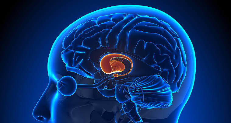
- Basal Ganglia: Modulates movement initiation and inhibition, influencing both direct (pyramidal) and indirect (extrapyramidal) pathways.
- Sensory Inputs: Proprioceptive information helps adjust motor output via these tracts to maintain balance and posture during dynamic activities.
Overall, these indirect motor pathways are essential for executing complex movements that require coordination between different muscle groups, especially during tasks that involve postural adjustments or reflexive actions.
In summary, both rubrospinal and reticulospinal tracts serve critical roles in modulating voluntary movements initiated by cortical areas while also contributing to involuntary reflexes necessary for maintaining posture and balance during locomotion or other activities.
Motor Pathways to the Face Muscles
The motor pathways that control the muscles of the face are primarily mediated by cranial nerves, specifically the facial nerve (cranial nerve VII). Understanding these pathways involves examining both the anatomical structures involved and the functional processes that govern facial movement.
1. Cranial Nerve Anatomy
The facial nerve is responsible for innervating most of the muscles of facial expression. It emerges from the brainstem at the level of the pons and travels through several anatomical regions before branching out to its target muscles. The pathway can be divided into several key components:
- Origin in the Brainstem: The facial nerve originates from motor nuclei located in the pons. These nuclei receive input from various brain regions, including cortical areas involved in planning and executing movements.
- Pathway Through the Skull: After originating in the pons, the facial nerve traverses through a bony canal (the facial canal) within the temporal bone. During this journey, it gives off several branches that serve different functions.
2. Branching and Innervation
Once it exits the skull via the stylomastoid foramen, the facial nerve divides into five major branches that innervate specific regions of the face:
- Temporal Branches: These branches innervate muscles around the forehead and upper eyelids, allowing for movements such as raising eyebrows and closing eyes.
- Zygomatic Branches: These control muscles around the cheeks, facilitating actions like smiling or puffing out cheeks.
- Buccal Branches: Responsible for innervating muscles around the mouth, these branches enable movements such as puckering lips or frowning.
- Marginal Mandibular Branch: This branch controls muscles of the lower lip and chin, allowing for expressions like grimacing or lowering lip.
- Cervical Branch: This branch innervates platysma muscle in the neck region, contributing to broader expressions involving neck movement.
3. Central Nervous System Control
The initiation of movement in facial muscles is influenced by higher brain centers:
- Motor Cortex Involvement: The primary motor cortex (located in the frontal lobe) plays a crucial role in planning voluntary movements. Neurons from this area project down to brainstem nuclei where they synapse with lower motor neurons of cranial nerves.
- Bilateral Control: Facial muscle control is somewhat unique because many upper face muscles receive bilateral input from both hemispheres of the brain. This means that even if one side of the motor cortex is damaged, some function may still be preserved due to redundancy in neural pathways.
4. Reflexive Actions
In addition to voluntary control, certain reflexive actions involving facial muscles are mediated through different pathways:
- Facial Reflexes: For example, when an object approaches your eye rapidly, a reflexive blink occurs via connections between sensory inputs and motor outputs without conscious thought.
5. Integration with Other Systems
Facial expressions are not solely controlled by motor pathways; they also involve complex interactions with emotional processing systems:
- Amygdala Influence: The amygdala plays a significant role in processing emotions and can influence how facial expressions are executed based on emotional states.
In summary, motor pathways to face muscles involve intricate networks starting from brainstem nuclei through cranial nerves that branch out to various facial regions while being modulated by higher cognitive functions related to emotion and social interaction.
Overview of Upper and Lower Motor Neuron Lesions
Upper Motor Neuron Lesions
Upper motor neuron (UMN) lesions are characterized by damage to the neural pathways that originate in the brain and travel down to the spinal cord. The typical signs and symptoms associated with UMN lesions include:
- Weakness: There is a pyramidal pattern of weakness, where extensors are weaker than flexors in the arms, while the reverse is true in the legs.
- Muscle Wasting: Disuse atrophy is minimal or absent; however, contractures may develop over time.
- Tone: Increased muscle tone, often presenting as spasticity or rigidity, which may be accompanied by ankle clonus.
- Reflexes: Hyperreflexia is common, indicating an exaggerated reflex response.
- Babinski Sign: A positive Babinski sign is observed, where there is an extension of the hallux (big toe) and fanning of the other toes when the sole of the foot is stimulated.
- Other Signs: Additional signs may include a positive Hoffmann’s sign and pronator drift during specific tests.
Lower Motor Neuron Lesions
Lower motor neuron (LMN) lesions affect nerve fibers that travel from the anterior horn of the spinal cord to muscles. The signs and symptoms associated with LMN lesions include:
- Weakness: Weakness typically follows a focal or root-innervated pattern, affecting specific muscle groups rather than showing a generalized weakness.
- Muscle Wasting: Marked muscle atrophy occurs due to denervation of muscles innervated by affected lower motor neurons.
- Tone: Reduced muscle tone leads to hypotonia; muscles may feel floppy or weak upon examination.
- Reflexes: Reflexes are reduced or absent, indicating a loss of reflex activity in affected areas.
- Babinski Sign: A negative Babinski sign is present; instead of extension, there is a downward movement of digits when stimulating the sole of the foot.
- Fasciculations: Fasciculations (involuntary muscle twitches) can be observed in affected muscle groups.
In summary, upper motor neuron lesions present with hypertonia, spastic paralysis, hyperreflexia, and positive Babinski signs, whereas lower motor neuron lesions are characterized by hypotonia, flaccid paralysis, reduced reflexes, and marked atrophy.


