Description of the Cubital Fossa
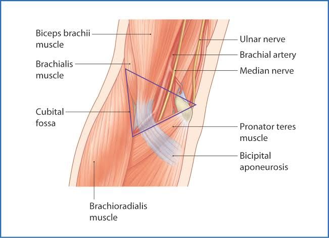
The cubital fossa, also known as the antecubital fossa, is a triangular-shaped depression located on the anterior aspect of the elbow joint. It serves as a critical anatomical area that facilitates the transition between the upper arm and forearm. The apex of this triangle points distally towards the forearm, while its base is formed by an imaginary horizontal line connecting the medial and lateral epicondyles of the humerus.
Borders of the Cubital Fossa
The cubital fossa is defined by three borders:
- Lateral Border: Formed by the medial border of the brachioradialis muscle.
- Medial Border: Defined by the lateral border of the pronator teres muscle.
- Superior Border: An imaginary horizontal line drawn between the epicondyles of the humerus.
Additionally, it has a roof and a floor:
- Roof: Composed of skin, subcutaneous fat, fascia, and bicipital aponeurosis.
- Floor: Formed by two muscles—the brachialis muscle proximally and supinator muscle distally.
Contents of the Cubital Fossa
The cubital fossa contains several important structures that pass between the arm and forearm. These contents are arranged from lateral to medial:
- Radial Nerve:
- The radial nerve travels along the lateral border of the cubital fossa before dividing into superficial and deep branches. It plays a crucial role in providing motor function to muscles in the posterior forearm and sensory function to parts of the hand.
- Biceps Tendon:
- The tendon of biceps brachii runs centrally through the cubital fossa, attaching to the radial tuberosity just distal to where it meets with the radius. This tendon contributes to flexion at both shoulder and elbow joints.
- Brachial Artery:
- The brachial artery bifurcates into radial and ulnar arteries at the apex of the cubital fossa. It is responsible for supplying blood to structures in both arms and forearms. The pulse can be palpated in this area just medial to where it lies beneath biceps tendon.
- Median Nerve:
- The median nerve passes medially through this space, exiting by passing between two heads of pronator teres muscle. This nerve provides motor innervation to most flexor muscles in the forearm as well as sensory innervation to parts of hand.
- Superficial Veins:
- The roof also contains several superficial veins, notably including the median cubital vein which connects basilic and cephalic veins. This vein is commonly accessed for venipuncture due to its superficial location.
In summary, these structures within or passing through the cubital fossa are vital for both movement and sensation in upper limb functionality.
Clinical Importance of the Cubital Fossa
The cubital fossa is clinically significant for several reasons, primarily due to its anatomical features and the structures it contains. Here are the key points regarding its clinical importance:
- Venipuncture Site: The cubital fossa is a common site for venipuncture, which is the process of obtaining intravenous access for blood sampling or transfusion. The median cubital vein, located in this area, is easily accessible and often used because it lies directly on the deep fascia and crosses the bicipital aponeurosis, providing a safe distance from deeper structures such as the brachial artery and median nerve.
- Brachial Pulse Measurement: The brachial pulse can be palpated in the cubital fossa, specifically medial to the biceps tendon. This location is crucial for assessing circulation in the arm and is commonly used when measuring blood pressure manually. A stethoscope is placed over this area to auscultate Korotkoff sounds during blood pressure measurement.
- Injury Assessment: The cubital fossa’s contents can be at risk during injuries such as supracondylar humeral fractures, which are particularly common in children. Such fractures may lead to damage of important structures within the fossa, including the brachial artery and various nerves (median, ulnar, and radial). If these structures are compromised, it can result in serious complications like Volkmann’s ischemic contracture due to impaired blood flow.
- Surgical Considerations: Knowledge of the anatomy of the cubital fossa is essential for surgeons performing procedures in this region. Understanding its boundaries and contents helps prevent inadvertent injury to nerves and vessels during surgical interventions.
- Nerve Compression Syndromes: Conditions such as pronator teres syndrome can occur when there is compression of the median nerve as it passes through or near the cubital fossa. Recognizing symptoms related to this area can aid in diagnosing nerve entrapment syndromes.
In summary, the cubital fossa plays a vital role in clinical practice due to its involvement in venipuncture, pulse measurement, potential injury implications, surgical relevance, and association with nerve compression syndromes.
Description of the Popliteal Fossa
The popliteal fossa is a diamond-shaped anatomical space located at the posterior aspect of the knee joint. This region serves as a crucial pathway for neurovascular structures that traverse between the thigh and leg. The fossa is bordered by specific muscles from both the thigh and leg, creating a defined area where important nerves and blood vessels are situated.
Boundaries of the Popliteal Fossa
The boundaries of the popliteal fossa are formed by several key muscles:
- Superomedial Boundary: Formed by the semimembranosus muscle.
- Superolateral Boundary: Formed by the biceps femoris muscle.
- Inferomedial Boundary: Created by the medial head of the gastrocnemius muscle.
- Inferolateral Boundary: Formed by the lateral head of the gastrocnemius muscle and sometimes includes the plantaris muscle.
The floor of this fossa consists of several structures including:
- The popliteal surface of the femur.
- The capsule of the knee joint, which is reinforced by the oblique popliteal ligament.
- The popliteus muscle, which is covered by its fascia.
The roof is composed of:
- The fascia lata, which is reinforced with transverse fibers, providing a protective covering over the contents within.
Content of the Popliteal Fossa
The popliteal fossa contains several important neurovascular structures, which can be listed and explained as follows:
- Popliteal Artery:
- This artery is a continuation of the femoral artery and is located deep within the fossa. It supplies blood to various structures in the leg and foot. Its position makes it vulnerable to injury or compression from surrounding structures.
- Popliteal Vein:
- Situated just superficial to the popliteal artery, this vein collects deoxygenated blood from lower extremities and drains into it. It plays a critical role in venous return from the leg.
- Tibial Nerve:
- This nerve branches off from the sciatic nerve at the superior angle of the popliteal fossa. It runs medially within this space and innervates muscles in both compartments (posterior compartment of thigh and leg) as well as providing sensory innervation to parts of the foot.
- Common Fibular Nerve (Common Peroneal Nerve):
- Also branching from the sciatic nerve, this nerve travels laterally along with its accompanying structures in this region. It innervates muscles in both anterior and lateral compartments of the leg, contributing to movement and sensation in these areas.
- Small Saphenous Vein:
- This vein pierces through fascia lata to drain into the popliteal vein after running up along with its accompanying sural nerve on posterior aspect of calf. It plays an essential role in venous drainage from superficial tissues in that area.
- Posterior Femoral Cutaneous Nerve:
- This nerve provides sensory innervation to skin on posterior thigh and part of leg; it also traverses through this fossa beneath fascia lata.
These contents are vital for both vascular supply and neural function in lower limbs, making any pathology affecting them clinically significant.
The clinical importance of the popliteal fossa can be understood through several key aspects:
1. Baker’s Cyst:
A Baker’s cyst, also known as a popliteal cyst, is a fluid-filled sac that forms behind the knee. It is often associated with conditions that cause knee joint swelling, such as arthritis or meniscus tears. The cyst can lead to discomfort and may restrict movement if it becomes large enough.
2. Popliteal Pulse:
The popliteal fossa contains the popliteal artery, which is crucial for assessing blood flow to the lower leg and foot. Palpating the popliteal pulse is an important clinical skill used to evaluate circulation in patients with vascular diseases or after trauma.
3. Popliteal Aneurysm:
A popliteal aneurysm occurs when there is an abnormal dilation of the popliteal artery. This condition can lead to complications such as thrombosis (blood clot formation) or embolism (where a clot travels to other parts of the body), potentially resulting in limb ischemia or loss.
4. Popliteal Nerve Block:
The popliteal fossa is a common site for administering nerve blocks, particularly for surgical procedures involving the foot and ankle. A popliteal nerve block targets the tibial and common fibular nerves, providing effective anesthesia during surgeries on the lower extremity.
5. Lymphatic Drainage:
The lymph nodes located within the popliteal fossa play a significant role in draining lymph from the lower leg and foot. Understanding this drainage pathway is essential for diagnosing infections or malignancies that may affect lymphatic function.
In summary, the clinical significance of the popliteal fossa encompasses its involvement in various medical conditions and procedures, making it an important anatomical region in both diagnosis and treatment.


