Definition of Bronchiectasis
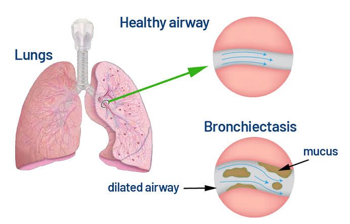
Bronchiectasis is a chronic lung condition characterized by the abnormal and permanent dilation of the bronchi, which are the large air passages from the trachea to the lungs. This dilation leads to a cycle of infection and inflammation, resulting in damage to the bronchial walls. The condition can cause significant respiratory symptoms, including chronic cough, sputum production, and recurrent respiratory infections.
General Classification of Bronchiectasis
Bronchiectasis can be classified into several categories based on different criteria:
- Etiological Classification:
- Cystic Fibrosis (CF): This is one of the most common causes of bronchiectasis in children and young adults. It is a genetic disorder that affects the lungs and digestive system.
- Non-Cystic Fibrosis Bronchiectasis (NCFB): This includes all other forms not related to cystic fibrosis. Causes can include:
- Infectious Causes: Such as post-infectious bronchiectasis following pneumonia or tuberculosis.
- Immune Deficiencies: Conditions that impair immune function can lead to recurrent infections and subsequent bronchiectasis.
- Environmental Factors: Exposure to toxic substances like chlorine gas or ammonia can also result in bronchial damage leading to bronchiectasis.
- Genetic Disorders: Other than CF, such as primary ciliary dyskinesia.
- Morphological Classification:
- Cylindrical Bronchiectasis: The bronchi are uniformly dilated and appear cylindrical.
- Varicose Bronchiectasis: The bronchi have an irregular shape resembling varicose veins.
- Cystic Bronchiectasis: The bronchi are markedly dilated with cyst-like formations.
- Radiological Classification:
- Based on imaging studies (like CT scans), bronchiectasis can be categorized according to its appearance:
- Focal Bronchiectasis: Limited to one area of the lung.
- Diffuse Bronchiectasis: Involves multiple areas throughout both lungs.
- Based on imaging studies (like CT scans), bronchiectasis can be categorized according to its appearance:
- Functional Classification:
- This classification considers how bronchiectasis affects lung function, often assessed through pulmonary function tests (PFTs). Patients may experience varying degrees of airflow obstruction depending on the severity and extent of their condition.
- Age-Related Classification:
- Children may present with different causes compared to adults, with CF being more prevalent in younger populations while older adults might have more cases linked to environmental exposures or infections.
Understanding these classifications helps healthcare providers determine appropriate management strategies for patients with bronchiectasis, as treatment may vary significantly based on underlying causes and disease characteristics.
The complexity of bronchiectasis necessitates a comprehensive approach for diagnosis and treatment, taking into account individual patient history, clinical presentation, and specific etiologies involved.
General Features of Bronchiectasis
- Pathophysiology: The primary feature of bronchiectasis is the irreversible widening and thickening of the bronchial walls. This occurs due to chronic inflammation and damage, often resulting from repeated infections or other lung conditions.
- Symptoms: Common symptoms include a persistent cough that produces large amounts of sputum, shortness of breath, wheezing, chest pain, and fatigue. Patients may also experience exacerbations where symptoms worsen significantly.
- Causes: The causes of bronchiectasis can vary widely but often include previous lung infections (such as pneumonia or tuberculosis), cystic fibrosis, autoimmune diseases, and exposure to environmental toxins. In many cases (approximately 40%), the cause remains unknown (idiopathic bronchiectasis).
- Diagnosis: Diagnosis typically involves a combination of patient history, physical examination, imaging studies such as high-resolution computed tomography (HRCT) scans, and pulmonary function tests to assess lung function.
- Management: While there is no cure for bronchiectasis, treatment focuses on managing symptoms and preventing complications. This may include antibiotics for infections, bronchodilators to open airways, mucolytics to help clear mucus, and pulmonary rehabilitation programs.
- Complications: Patients with bronchiectasis are at increased risk for respiratory infections, hemoptysis (coughing up blood), respiratory failure in severe cases, and reduced quality of life due to chronic symptoms.
- Epidemiology: Bronchiectasis can affect individuals of all ages but is more common in adults over 60 years old and tends to be more prevalent in women than men.
Common Types of Bronchiectasis and Focus on Cystic Fibrosis
Common Types of Bronchiectasis
- Cystic Fibrosis (CF) Bronchiectasis:
- Cystic fibrosis is a genetic disorder caused by mutations in the CFTR gene, leading to thick, sticky mucus production. This mucus obstructs airways and creates an environment conducive to bacterial infections, particularly with organisms like Pseudomonas aeruginosa. In CF patients, bronchiectasis often develops early in life and is one of the most common pulmonary complications associated with the disease.
- Post-Infectious Bronchiectasis:
- This type occurs following severe respiratory infections such as pneumonia or tuberculosis. The damage caused by these infections can lead to scarring and dilation of the bronchi.
- Congenital Bronchiectasis:
- Some individuals are born with structural abnormalities that predispose them to bronchiectasis. Conditions such as primary ciliary dyskinesia or immunodeficiencies can lead to congenital forms of this disease.
- Obstructive Bronchiectasis:
- This form arises due to obstruction in the airway from tumors, foreign bodies, or other lesions that prevent normal airflow and drainage of secretions.
- Idiopathic Bronchiectasis:
- In some cases, no clear cause can be identified despite thorough investigation; this is termed idiopathic bronchiectasis.
Focus on Cystic Fibrosis
Cystic fibrosis-related bronchiectasis has unique characteristics that set it apart from other types:
- Pathophysiology: The defective CFTR protein results in impaired chloride ion transport across epithelial cells, leading to dehydration of airway surfaces and thickened mucus production. This thick mucus obstructs airways and traps bacteria, resulting in chronic inflammation and recurrent lung infections.
- Clinical Manifestations: Patients typically present with chronic productive cough, frequent respiratory infections (often with Pseudomonas aeruginosa), wheezing, and progressive decline in lung function over time. Symptoms may begin in early childhood but can vary significantly among individuals.
- Diagnosis: Diagnosis involves imaging studies such as high-resolution computed tomography (HRCT) scans that reveal characteristic features like dilated bronchi with wall thickening. Pulmonary function tests may show obstructive patterns indicative of airflow limitation.
- Management Strategies: Treatment focuses on managing symptoms and preventing complications:
- Airway Clearance Techniques (ACTs): These include chest physiotherapy methods like percussion or devices designed to help clear mucus.
- Inhaled Antibiotics: To target specific pathogens commonly found in CF patients.
- Bronchodilators: To improve airflow.
- Nutritional Support: Given the malabsorption issues associated with CF.
- Lung Transplantation: Considered for advanced cases where lung function deteriorates significantly despite aggressive management.
- Prognosis: With advancements in treatment strategies for cystic fibrosis over recent decades, including new therapies targeting the underlying genetic defect (e.g., CFTR modulators), outcomes have improved significantly for many patients; however, bronchiectasis remains a significant cause of morbidity and mortality within this population.
In summary, while bronchiectasis can arise from various causes, cystic fibrosis represents a distinct category due to its genetic basis and specific pathophysiological mechanisms that lead to significant pulmonary complications requiring targeted management approaches.
Clinical Evaluation for Patients with Bronchiectasis Exacerbation
Patients with bronchiectasis often experience exacerbations, which are acute worsening of symptoms that can significantly impact their quality of life and overall health status. Clinical evaluation during these exacerbations is critical for appropriate management.
Step 1: Patient History
The first step in evaluating a patient with suspected bronchiectasis exacerbation involves a comprehensive medical history. Key aspects include:
- Symptom Assessment: Documenting the onset, duration, and severity of symptoms such as increased cough, sputum production (including changes in color or volume), hemoptysis (coughing up blood), chest pain, dyspnea (shortness of breath), and fever.
- Previous Exacerbations: Understanding the frequency and severity of past exacerbations can provide insight into disease progression and treatment efficacy.
- Medication Review: Evaluating current medications, including inhaled therapies, antibiotics, corticosteroids, and any recent changes in treatment regimens.
- Comorbid Conditions: Identifying other health issues such as asthma, COPD (Chronic Obstructive Pulmonary Disease), or heart disease that may complicate management.
Step 2: Physical Examination
A thorough physical examination is essential to assess the patient’s current health status. Important components include:
- Vital Signs: Monitoring temperature for fever, respiratory rate for tachypnea (rapid breathing), heart rate for tachycardia (rapid heartbeat), and oxygen saturation levels.
- Respiratory Examination: Auscultation of lung sounds to identify wheezing or crackles that may indicate airway obstruction or infection. Observing for signs of respiratory distress or use of accessory muscles during breathing.
- General Appearance: Assessing overall appearance for signs of systemic illness such as cyanosis (bluish discoloration) or clubbing of fingers.
Step 3: Diagnostic Testing
To confirm an exacerbation and guide treatment decisions, various diagnostic tests may be performed:
- Sputum Culture: Collecting sputum samples to identify pathogens responsible for infection. This helps tailor antibiotic therapy based on culture results.
- Chest Imaging: A chest X-ray or CT scan can provide information about structural changes in the lungs and rule out other complications such as pneumonia or lung abscesses.
- Pulmonary Function Tests (PFTs): These tests measure lung function to determine the extent of airflow obstruction and help assess baseline pulmonary status.
Step 4: Laboratory Tests
Laboratory evaluations are crucial in managing exacerbations:
- Complete Blood Count (CBC): To check for leukocytosis (increased white blood cells) indicating infection or inflammation.
- C-Reactive Protein (CRP): Elevated levels can suggest active inflammation associated with an exacerbation.
- Arterial Blood Gases (ABGs): In cases where patients exhibit significant respiratory distress, ABGs can help assess gas exchange efficiency.
Step 5: Assessing Severity and Treatment Plan
Once the clinical evaluation is complete, determining the severity of the exacerbation is vital. This assessment guides treatment decisions:
- Mild Exacerbations: May be managed with oral antibiotics and increased bronchodilator therapy.
- Moderate to Severe Exacerbations: Often require hospitalization for intravenous antibiotics, systemic corticosteroids, oxygen therapy if needed, and close monitoring.
Conclusion
Effective clinical evaluation during bronchiectasis exacerbations involves a systematic approach encompassing patient history, physical examination, diagnostic testing, laboratory evaluations, and severity assessment. This comprehensive evaluation enables healthcare providers to implement timely interventions that improve patient outcomes.
Required Investigations for Patients with Bronchiectasis
Below are the key investigations required for patients with bronchiectasis:
1. Clinical History and Physical Examination
Before any diagnostic tests, a thorough clinical history is essential. This includes:
- Symptoms: Chronic cough, sputum production (often purulent), hemoptysis, dyspnea, and wheezing.
- Medical History: Previous respiratory infections, history of cystic fibrosis or other genetic conditions, autoimmune diseases, or exposure to environmental toxins.
- Family History: Genetic predispositions that may contribute to bronchiectasis.
A physical examination will focus on respiratory signs such as crackles or wheezing and may reveal clubbing of fingers in some cases.
2. Chest Imaging
- High-Resolution Computed Tomography (HRCT) Scan: This is the gold standard for diagnosing bronchiectasis. HRCT can visualize bronchial dilation, wall thickening, and mucus plugging. It helps classify bronchiectasis into different types (cylindrical, varicose, cystic) based on the pattern observed.
- Chest X-ray: While less sensitive than HRCT, chest X-rays can provide initial information about lung structure and rule out other conditions like pneumonia or lung tumors.
3. Sputum Analysis
- Sputum Culture: Identifying pathogens in sputum is crucial for managing infections associated with bronchiectasis. Common organisms include Pseudomonas aeruginosa, Haemophilus influenzae, and Staphylococcus aureus. Cultures should be performed during exacerbations to guide antibiotic therapy.
- Sputum Cytology: This can help identify inflammatory cells or malignancy if there are concerning features.
4. Pulmonary Function Tests (PFTs)
PFTs assess lung function and help determine the severity of airflow obstruction or restriction. Key measurements include:
- Forced Vital Capacity (FVC): Measures the total amount of air exhaled forcefully after taking a deep breath.
- Forced Expiratory Volume in 1 second (FEV1): Assesses how much air can be forcibly exhaled in one second; reduced values indicate obstructive patterns typical in bronchiectasis.
These tests help monitor disease progression over time.
5. Blood Tests
Routine blood tests can provide additional information:
- Complete Blood Count (CBC): To check for signs of infection or inflammation.
- Immunological Tests: Assessing immunoglobulin levels (IgG, IgA) can identify underlying immune deficiencies that may contribute to recurrent infections.
- Cystic Fibrosis Testing: In younger patients or those with a family history suggestive of cystic fibrosis, sweat chloride testing or genetic testing may be warranted.
6. Additional Investigations
Depending on clinical suspicion for specific causes of bronchiectasis:
- Bronchoscopy: This procedure allows direct visualization of the airways and collection of biopsies or secretions if needed. It is particularly useful if there is suspicion of obstruction due to foreign bodies or tumors.
- Allergy Testing: If allergic bronchopulmonary aspergillosis (ABPA) is suspected due to asthma-like symptoms alongside bronchiectasis, specific IgE testing for Aspergillus species may be indicated.
- Imaging Studies Beyond HRCT: In certain cases where complications such as abscesses are suspected, further imaging like MRI may be necessary.
In summary, diagnosing and managing bronchiectasis requires a multifaceted approach involving detailed clinical assessment followed by targeted investigations including imaging studies like HRCT scans, sputum analysis for pathogen identification, pulmonary function tests to evaluate lung capacity and function, blood tests for immune status evaluation, and potentially bronchoscopy when indicated.
The combination of these investigations helps establish an accurate diagnosis while guiding effective treatment strategies tailored to individual patient needs.
Management Outlines for Bronchiectasis
The management of bronchiectasis involves a multidisciplinary approach that includes pharmacological treatments, airway clearance techniques, and addressing underlying causes.
1. Diagnosis and Assessment
Before initiating treatment, a thorough assessment is essential. This includes:
- Clinical History: Understanding symptoms such as chronic cough, sputum production, hemoptysis, and dyspnea.
- Imaging Studies: High-resolution computed tomography (HRCT) scans are the gold standard for diagnosing bronchiectasis.
- Pulmonary Function Tests (PFTs): These tests assess the degree of airflow obstruction and overall lung function.
- Microbiological Evaluation: Sputum cultures help identify pathogens that may be contributing to exacerbations.
2. Treatment Goals
The primary goals in managing bronchiectasis include:
- Reducing symptoms
- Preventing exacerbations
- Improving quality of life
- Addressing any underlying conditions
3. Pharmacological Management
Pharmacological interventions can be categorized into several classes:
- Antibiotics: Long-term macrolide antibiotics (e.g., azithromycin) are often used to reduce exacerbation frequency in patients with frequent infections. Short courses of antibiotics are prescribed during acute exacerbations based on sputum culture results.
- Bronchodilators: Inhaled beta-agonists or anticholinergics may be used to relieve symptoms related to airflow obstruction.
- Corticosteroids: In cases where there is significant inflammation or asthma overlap, inhaled corticosteroids may be beneficial.
- Mucolytics: Agents like N-acetylcysteine can help thin mucus secretions, making it easier for patients to clear their airways.
4. Airway Clearance Techniques
Effective airway clearance is vital in managing bronchiectasis:
- Chest Physiotherapy (CPT): Techniques such as postural drainage and percussion can help mobilize secretions.
- Positive Expiratory Pressure (PEP) Devices: These devices create back pressure during exhalation, helping keep airways open and facilitating mucus clearance.
- High-Frequency Chest Wall Oscillation (HFCWO): This technique uses an inflatable vest that vibrates at high frequencies to loosen mucus.
5. Vaccination and Preventive Measures
Vaccinations play a critical role in preventing respiratory infections:
- Influenza Vaccine: Annual vaccination is recommended for all patients with bronchiectasis.
- Pneumococcal Vaccine: Patients should receive pneumococcal vaccinations according to guidelines to prevent pneumonia caused by Streptococcus pneumoniae.
Additionally, smoking cessation programs are essential for smokers as tobacco use can exacerbate lung damage.
6. Management of Exacerbations
Patients should have a clear action plan for managing exacerbations:
- Recognizing early signs of an exacerbation (increased cough or sputum production).
- Prompt initiation of antibiotics based on previous culture results or empirical therapy if cultures are not available.
Regular follow-up appointments should be scheduled to monitor lung function and adjust treatment plans as necessary.
7. Addressing Underlying Causes
Identifying and treating any underlying conditions contributing to bronchiectasis is crucial:
- Conditions such as cystic fibrosis, immune deficiencies, or allergic bronchopulmonary aspergillosis must be managed appropriately.
- Referral to specialists may be necessary for comprehensive care.
Conclusion
Management outlines for bronchiectasis emphasize a combination of pharmacological treatment, airway clearance techniques, preventive measures through vaccination, and addressing underlying causes. A personalized approach tailored to each patient’s needs can significantly enhance outcomes and quality of life for individuals living with this chronic condition.


