Dealing with Emergency Situations in ENT
1. Understanding Common ENT Emergencies
Emergency situations in the ear, nose, and throat (ENT) can arise from various conditions, including epistaxis (nosebleeds), sudden hearing loss, foreign bodies in the ear or nose, and infections such as epiglottitis or peritonsillar abscesses. Recognizing these emergencies is crucial for timely intervention.
2. Management of Epistaxis
- Step 1: Visualize the Bleed
Begin by ensuring proper orientation of the nasal speculum. If bleeding is present, instruct the patient to gently blow their nose to clear any clots. - Step 2: Anesthetize
Use a cotton pledget soaked in a 1:1 mixture of oxymetazoline (a vasoconstrictor) and lidocaine (an anesthetic). This combination may be more effective than cocaine and has fewer side effects. Leave it in place for 5–10 minutes while applying firm pressure to the nose. - Step 3: Cauterize
After anesthesia, cauterize the dry edges around the bleeding site using silver nitrate for no longer than 10–15 seconds. Ensure eye protection is used as this can cause sneezing. - Step 4: Tamponade if Necessary
If bleeding persists, consider tamponade with a balloon-type device wrapped in gelfoam or surgicel to promote clotting. Nasal balloons are generally preferred over traditional tampons due to higher patient satisfaction. - Management Pearls:
Applying ice to the palate can reduce nasal blood flow significantly. Tranexamic acid (TXA) may also be beneficial for anterior bleeds but should be used judiciously based on recent studies indicating limited benefit when added after initial treatment. - Antibiotic Considerations:
Antibiotics are not routinely required for immunocompetent patients unless nasal packing is expected to remain for more than 72 hours or if the patient is immunocompromised.
3. Handling Sudden Hearing Loss
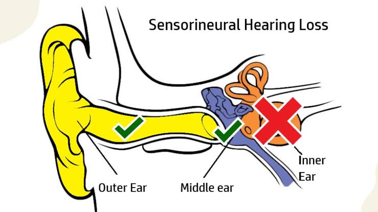
Sudden sensorineural hearing loss requires immediate attention. It may result from various causes including viral infections or vascular issues affecting the inner ear. Patients presenting with this condition should undergo prompt evaluation and management to prevent permanent hearing loss.
4. Foreign Body Removal
For nasal foreign bodies:
- Attempt positive pressure techniques such as the “parent’s kiss” method.
- If unsuccessful, use a mixture of oxymetazoline and lidocaine to reduce swelling before attempting removal.
For ear foreign bodies:
- Pulling posteriorly on the pinna while stabilizing the head can help facilitate removal without pushing the object further into the canal.
5. Recognizing and Managing Infections
Conditions like epiglottitis and peritonsillar abscesses require urgent care:
- Epiglottitis: Look for symptoms such as difficulty breathing, drooling, and stridor; imaging may be necessary.
- Peritonsillar Abscess: Symptoms include severe sore throat, fever, and trismus; drainage may be required along with antibiotics.
In all cases of suspected infection or significant airway compromise, immediate consultation with an ENT specialist is warranted.
6. Conclusion
In summary, managing ENT emergencies involves a systematic approach that includes assessment of symptoms, appropriate interventions such as cauterization for epistaxis or removal techniques for foreign bodies, and timely referral for serious conditions like infections or sudden hearing loss. Prompt recognition and treatment are essential to prevent complications.
Blood Supply of the Nose
The blood supply of the nose is derived from both the external and internal carotid arteries, which provide a rich vascular network essential for its functions. Understanding this blood supply involves examining the specific arteries involved and their respective contributions to different regions of the nasal cavity.
External Carotid Artery Contributions
The external carotid artery plays a significant role in supplying blood to the nose through several branches:
- Sphenopalatine Artery: This artery is a major contributor to the nasal cavity’s blood supply. It enters the nasal cavity through the sphenopalatine foramen and supplies the posterior part of the nasal septum and lateral wall.
- Greater Palatine Artery: This artery primarily supplies the hard palate but also contributes to the vascularization of the nasal cavity, particularly in its lower regions.
- Superior Labial Artery: A branch of the facial artery, it supplies blood to the upper lip and also provides some vascular support to the anterior part of the nasal cavity.
- Lateral Nasal Arteries: These arteries arise from branches of both the facial and maxillary arteries, supplying blood to parts of the external nose and contributing to areas around the nares.
These branches anastomose (connect) with each other within a specific area known as Kiesselbach’s plexus or Little’s area, located on the anterior part of the nasal septum. This plexus is clinically significant because it is a common site for epistaxis (nosebleeds).
Internal Carotid Artery Contributions
The internal carotid artery also contributes to nasal blood supply through its branches:
- Anterior Ethmoidal Artery: This artery supplies blood to parts of the upper nasal cavity and contributes to surrounding structures, including portions of the external nose.
- Posterior Ethmoidal Artery: Similar to its anterior counterpart, this artery supplies deeper structures within the nasal cavity as well as adjacent sinuses.
Both these ethmoidal arteries are crucial for supplying blood to areas that are not reached by branches from the external carotid artery.
Venous Drainage
The venous drainage system complements this arterial supply. The veins draining from these regions include:
- Ophthalmic Veins: These drain areas supplied by ethmoidal arteries.
- Facial Vein: Drains superficial structures.
- Pterygoid Plexus and Pharyngeal Plexus: These networks allow for drainage from deeper structures in relation to potential spread of infections due to their connections with cranial venous sinuses, notably allowing for possible complications such as cavernous sinus thrombosis.
In summary, understanding both arterial supply and venous drainage is essential for comprehending how blood circulates through this complex anatomical region, which is vital for respiratory function, olfaction, and overall health.
Causes of Epistaxis and Stridor
Causes of Epistaxis
Epistaxis, commonly known as a nosebleed, occurs when small blood vessels or capillaries in the nasal septum rupture or tear. The causes can be classified into common and abnormal conditions:
- Common Causes:
- Dry Air: High temperatures and low humidity can dry out the nasal mucosa, leading to cracking and bleeding.
- Nose Picking: This action can directly injure the delicate blood vessels in the nose.
- Excessive Sneezing or Nose Blowing: Forceful actions can cause trauma to the nasal lining.
- Injury: Trauma from accidents affecting the nose or face can lead to bleeding.
- Medications: Certain medications, such as intranasal steroids, anticoagulants (like warfarin), and antihistamines, may dry out the nasal passages or affect clotting.
- Medical Devices: Use of CPAP machines for sleep apnea may irritate the nasal lining.
- Infections: Conditions like acute sinusitis or upper respiratory infections can inflame and damage blood vessels.
- Abnormal Conditions:
- Septal Deviation or Perforation: Structural abnormalities in the nasal septum can predispose individuals to bleeding.
- Clotting Disorders: Conditions such as hemophilia or immune thrombocytopenia (ITP) affect the body’s ability to stop bleeding.
- Nasal Polyps or Tumors: Growths in the nasal cavity may cause obstruction and bleeding.
- Cancer: Nasopharyngeal carcinoma and other malignancies can lead to epistaxis.
Causes of Stridor
Stridor is a high-pitched wheezing sound caused by disrupted airflow in the upper airway. It indicates an obstruction that may be due to various factors:
- Obstruction:
- Foreign Bodies: Inhalation of objects can block airways, leading to stridor.
- Tumors: Benign or malignant growths in the airway can obstruct airflow.
- Inflammation:
- Anatomical Abnormalities:
- Congenital Conditions: Conditions like laryngomalacia (softening of tissues above the vocal cords) are common causes of stridor in infants.
- Vocal Cord Paralysis: Loss of function in vocal cords affects airflow and produces stridor.
- Infections:
- Epiglottitis: Bacterial infection causing inflammation of the epiglottis leads to severe airway obstruction and stridor.
- Trauma:
- Injuries to the neck or throat area may cause swelling or direct obstruction resulting in stridor.
In summary, both epistaxis and stridor have multiple causes ranging from environmental factors and physical trauma to underlying medical conditions that require careful evaluation for appropriate management.
Indications for Tracheostomy
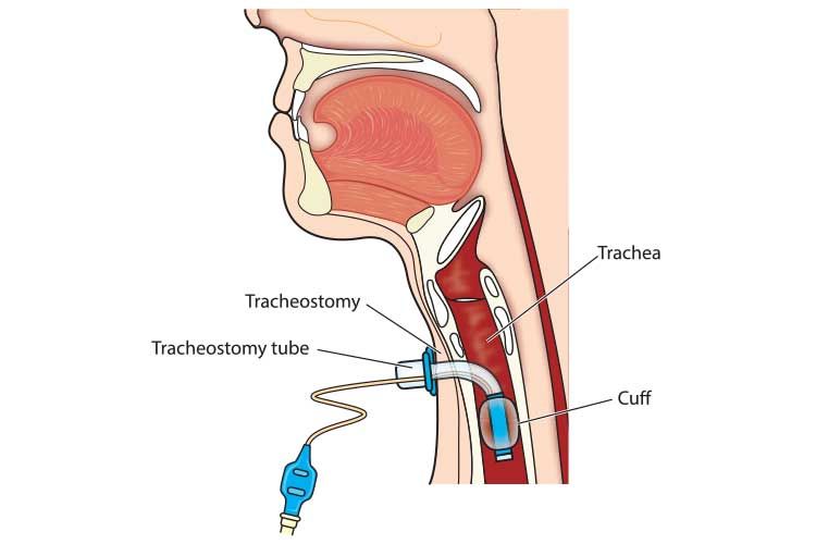
Tracheostomy is a surgical procedure that involves creating an opening in the trachea, typically to facilitate breathing when normal airflow is obstructed or compromised. The indications for performing a tracheostomy can be categorized into several key areas:
- Airway Obstruction:
- Tracheostomy is indicated when there is an airway obstruction above the level of the trachea, which may be present or anticipated. This could include conditions such as tumors, severe inflammation, or foreign body aspiration.
- In cases where there is obstruction in the upper or mid-trachea that requires stenting, a tracheostomy tube can be placed to maintain airway patency.
- Prolonged Intubation:
- When patients require prolonged mechanical ventilation (generally considered to be more than 10 days), tracheostomy offers several advantages over traditional endotracheal intubation. These advantages include improved patient comfort, reduced risk of injury to laryngeal structures (such as posterior glottic stenosis), and better management of airway secretions.
- Studies have shown that early tracheostomy performed within 7 days of intubation can lead to decreased length of stay in intensive care units (ICU).
- Inability to Intubate:
- In situations where intubation cannot be successfully performed and general anesthesia is required, a tracheostomy may be necessary to secure the airway.
- Adjunct to Major Surgery:
- Tracheostomy may also serve as an adjunct procedure during major head and neck surgeries or trauma management, providing better access and control over the airway.
- Airway Protection:
- Patients with neurologic diseases or traumatic brain injuries may require tracheostomy for airway protection due to impaired protective reflexes.
- Improved Communication and Oral Intake:
- A tracheostomy can enhance a patient’s ability to speak and resume oral intake by bypassing obstructions caused by endotracheal tubes.
- Decreased Incidence of Complications:
- Performing a tracheostomy can reduce complications associated with prolonged endotracheal intubation, such as sinusitis and laryngotracheal injury.
In summary, the decision to perform a tracheostomy is based on various clinical indications primarily related to maintaining airway patency, managing prolonged ventilation needs, protecting the airway in vulnerable patients, and improving overall patient comfort and outcomes.
How to Perform an Emergent Tracheotomy
Performing an emergent tracheotomy is a critical procedure that should only be undertaken in life-threatening situations when the airway is obstructed, and other methods of relief have failed. This procedure involves creating an opening in the trachea (windpipe) to allow air to enter the lungs directly. Below are the steps to perform an emergent tracheotomy:
1. Assess the Situation
- Confirm that the individual is indeed choking and unable to breathe, speak, or cough effectively.
- Look for signs of severe distress, such as cyanosis (bluish skin), decreased consciousness, or inability to make sounds.
2. Call for Emergency Help
- Before proceeding with any invasive procedure, ensure that emergency medical services (EMS) have been contacted. This can be done while preparing for the procedure.
3. Prepare for the Procedure
- If possible, gather necessary materials: a sterile scalpel or knife, a tracheostomy tube or a hollow needle if available, and gloves if time permits.
- Position the person lying down on their back if they are conscious; otherwise, position them safely.
4. Locate the Trachea
- Identify the landmarks on the neck:
- Find the Adam’s apple (thyroid cartilage) which is prominent in adults.
- Slide your fingers down until you reach the cricoid cartilage; there will be a soft area between these two structures known as the cricothyroid membrane.
5. Make an Incision
- Using a scalpel or knife, make a horizontal incision about 1-2 cm long through the skin overlying the cricothyroid membrane.
- Cut carefully through this membrane to avoid damaging surrounding structures.
6. Insert a Tube
- Once you have accessed the airway by puncturing through the cricothyroid membrane, insert a tracheostomy tube or hollow needle into this opening.
- Ensure that it is positioned correctly within the trachea to allow airflow.
7. Secure and Monitor
- If using a tube, secure it in place with tape or ties.
- Monitor for signs of effective ventilation and ensure that emergency services are en route and will take over care as soon as they arrive.
8. Provide Ongoing Care
- Continue monitoring vital signs and provide any necessary support until professional medical help arrives.
- Be prepared to perform CPR if needed.
It is crucial to note that performing a tracheotomy carries significant risks including injury to surrounding structures (like blood vessels and nerves), bleeding, infection, and potential complications from improper placement of tubes. Therefore, this procedure should only be performed by trained individuals when absolutely necessary.


