Structure of Cell Membrane Receptors and Intracellular Receptors for Different Hormones
Introduction to Hormone Receptors
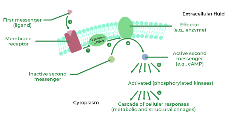
Hormones exert their effects on target cells by binding to specific receptors, which can be classified into two main categories: cell membrane receptors and intracellular receptors. The structure of these receptors is crucial for their function in signal transduction.
Cell Membrane Receptors
Cell membrane receptors are typically proteins that span the lipid bilayer of the cell membrane. They are primarily involved in the signaling pathways for hydrophilic hormones, such as peptide hormones (e.g., insulin, glucagon) and catecholamines (e.g., epinephrine). The structure of these receptors can be described in several key components:
- Extracellular Domain: This portion of the receptor extends outside the cell and is responsible for hormone binding. It often contains specific binding sites that recognize and bind to particular hormones with high affinity.
- Transmembrane Domain: This part consists of hydrophobic amino acid sequences that traverse the lipid bilayer. Typically, these domains are composed of alpha-helices or beta-sheets that stabilize the receptor within the membrane.
- Intracellular Domain: Located inside the cell, this domain interacts with various intracellular signaling molecules upon hormone binding. It may have intrinsic enzymatic activity (as seen in receptor tyrosine kinases) or interact with G-proteins (as seen in G-protein coupled receptors).
- Types of Membrane Receptors:
- G-Protein Coupled Receptors (GPCRs): These are a large family of receptors that activate intracellular signaling cascades through G-proteins upon ligand binding.
- Receptor Tyrosine Kinases (RTKs): These receptors undergo autophosphorylation on tyrosine residues when activated by their ligands, leading to downstream signaling.
- Ion Channel Receptors: These open or close in response to hormone binding, allowing ions to flow across the membrane and alter cellular activity.
Intracellular Receptors
Intracellular receptors are located within the cytoplasm or nucleus and primarily bind lipophilic hormones such as steroid hormones (e.g., cortisol, estrogen) and thyroid hormones (e.g., thyroxine). Their structure includes:
- Ligand-Binding Domain: This region binds to specific hormones and is often highly conserved among different types of intracellular receptors.
- DNA-Binding Domain: After hormone binding, many intracellular receptors translocate to the nucleus where they bind to specific DNA sequences known as hormone response elements (HREs). This interaction regulates gene transcription.
- Transactivation Domain: This part interacts with other transcription factors and co-regulators to modulate gene expression positively or negatively.
- Types of Intracellular Receptors:
- Steroid Hormone Receptors: These include glucocorticoid, mineralocorticoid, androgen, estrogen, and progesterone receptors.
- Thyroid Hormone Receptors: These regulate metabolism and development by modulating gene expression in response to thyroid hormones.
- Retinoic Acid Receptors: Involved in regulating gene expression during development and differentiation processes.
The activation mechanism for intracellular receptors generally involves hormone diffusion across the plasma membrane due to their lipophilicity, followed by binding to their respective receptor which then influences gene transcription directly.
Conclusion
Understanding the structural differences between cell membrane receptors and intracellular receptors is essential for comprehending how various hormones exert their physiological effects on target cells. While cell membrane receptors facilitate rapid responses through second messenger systems, intracellular receptors mediate longer-term changes through alterations in gene expression.
Different Types of Second Messengers
Second messengers are crucial molecules that relay signals from cell-surface receptors to target proteins within the cell, facilitating various physiological responses. They can be classified into several categories based on their chemical nature and function. The primary types of second messengers include:
- Cyclic Nucleotides
- Examples: Cyclic adenosine monophosphate (cAMP) and cyclic guanosine monophosphate (cGMP).
- Function: These molecules are synthesized from ATP and GTP, respectively, by specific enzymes (adenylyl cyclase for cAMP and guanylate cyclase for cGMP). They play significant roles in signaling pathways, influencing processes such as metabolism, gene expression, and muscle contraction.
- Lipid Derivatives
- Examples: Diacylglycerol (DAG) and phosphatidylinositol 3,4,5-trisphosphate (PIP3).
- Function: These lipids are generated through the action of phospholipases on membrane phospholipids. DAG remains in the membrane and activates protein kinase C (PKC), while PIP3 is involved in signaling pathways related to cell growth and survival.
- Ions
- Gases and Free Radicals
- Examples: Nitric oxide (NO) and carbon monoxide (CO).
- Function: These gaseous molecules can diffuse across membranes rapidly and influence various signaling pathways. Nitric oxide is particularly important in vasodilation and neurotransmission.
Each type of second messenger plays a distinct role in cellular communication, allowing cells to respond appropriately to external stimuli through complex signaling networks.
Intracellular Actions of Second Messengers
Below is a detailed explanation of the intracellular actions associated with each class of second messenger.
1. Cyclic Nucleotides
Cyclic nucleotides, such as cyclic adenosine monophosphate (cAMP) and cyclic guanosine monophosphate (cGMP), play significant roles in mediating cellular responses:
- cAMP:
- Activation of Protein Kinase A (PKA): cAMP binds to the regulatory subunits of PKA, causing a conformational change that releases the active catalytic subunits. These active subunits phosphorylate serine and threonine residues on target proteins, leading to various effects such as increased glycogen breakdown in liver cells or enhanced heart muscle contraction.
- Regulation of Ion Channels: cAMP can also modulate ion channels, such as those for calcium and potassium, affecting excitability and signaling in neurons and muscle cells.
- Gene Expression: cAMP influences gene transcription by activating transcription factors like CREB (cAMP response element-binding protein), which binds to specific DNA sequences to promote gene expression.
- cGMP:
- Activation of Protein Kinase G (PKG): Similar to cAMP, cGMP activates PKG, which phosphorylates target proteins involved in smooth muscle relaxation and vasodilation.
- Regulation of Ion Channels: cGMP can also affect ion channels, particularly those involved in neuronal signaling.
- Modulation of Phosphodiesterases: cGMP levels are regulated by phosphodiesterases that degrade it; thus, its action is tightly controlled within the cell.
2. Lipid Messengers
Lipid-derived second messengers include diacylglycerol (DAG) and phosphatidylinositol trisphosphate (PIP3):
- DAG:
- Activation of Protein Kinase C (PKC): DAG serves as a cofactor for PKC activation. Once activated by DAG and calcium ions, PKC phosphorylates various substrates involved in processes like cell growth, differentiation, and apoptosis.
- PIP3:
- Recruitment of Signaling Proteins: PIP3 acts as a docking site for proteins with pleckstrin homology (PH) domains. This recruitment activates downstream signaling pathways such as the Akt pathway, which is critical for cell survival and metabolism.
- Calcium Release from Endoplasmic Reticulum: PIP3 can also stimulate the release of calcium from the endoplasmic reticulum through the activation of IP3 receptors.
3. Ions
Ionic second messengers primarily include calcium ions (Ca²⁺) and other ions like sodium (Na⁺) or potassium (K⁺):
- Calcium Ions (Ca²⁺):
- Calcium Release from Stores: Calcium is released from intracellular stores such as the endoplasmic reticulum upon stimulation by second messengers like IP3. This increase in intracellular Ca²⁺ concentration activates various calcium-dependent processes including muscle contraction, neurotransmitter release in neurons, and enzyme activation.
- Binding to Calmodulin: Ca²⁺ binds to calmodulin, a calcium-binding messenger protein that then activates various enzymes such as myosin light chain kinase (MLCK), influencing muscle contraction.
- Other Ions:
- Changes in Na⁺ or K⁺ concentrations can affect membrane potential and excitability in neurons and muscle cells.
4. Gases/Free Radicals
Gasotransmitters like nitric oxide (NO) and carbon monoxide (CO), along with free radicals such as reactive oxygen species (ROS), serve important signaling roles:
- Nitric Oxide (NO):
- Vasodilation: NO diffuses across membranes and activates guanylate cyclase to produce cGMP from GTP. This leads to relaxation of smooth muscles in blood vessels.
- Neurotransmission: In neurons, NO acts as a retrograde messenger that modulates synaptic transmission.
- Reactive Oxygen Species (ROS):
- ROS can act as signaling molecules that influence pathways related to inflammation, apoptosis, and cellular stress responses.
In summary, second messengers facilitate complex intracellular signaling cascades that regulate diverse physiological processes ranging from metabolism to gene expression. Their precise control is essential for maintaining cellular homeostasis.
Understanding the Mechanism of Second Messenger Actions Including PIP2 Turnover, Ca²⁺/Protein Kinase C Systems, Diacylglycerol (DAG), and Nitric Oxide (NO)
1. Introduction to Second Messengers
Second messengers are intracellular signaling molecules released by the cell in response to exposure to extracellular signaling molecules (first messengers). They play a crucial role in amplifying the signal from first messengers and facilitating various cellular responses. Common second messengers include cyclic AMP (cAMP), calcium ions (Ca²⁺), diacylglycerol (DAG), and inositol trisphosphate (IP3).
2. Phosphatidylinositol 4,5-bisphosphate (PIP2) Turnover
PIP2 is a phospholipid found in the inner leaflet of the plasma membrane. It serves as a substrate for phospholipase C (PLC), an enzyme activated by various receptors, including G protein-coupled receptors (GPCRs). Upon activation, PLC hydrolyzes PIP2 into two important second messengers: diacylglycerol (DAG) and inositol trisphosphate (IP3).
- Inositol Trisphosphate (IP3): IP3 is soluble and diffuses through the cytoplasm to bind to IP3 receptors on the endoplasmic reticulum (ER). This binding leads to the release of Ca²⁺ ions from the ER into the cytosol.
- Diacylglycerol (DAG): DAG remains embedded in the plasma membrane and activates protein kinase C (PKC). PKC is a family of serine/threonine kinases that phosphorylate various target proteins, leading to diverse cellular responses.
3. Calcium Ions and Protein Kinase C Systems
The increase in intracellular Ca²⁺ concentration is a critical aspect of many signaling pathways. The released Ca²⁺ ions can bind to calmodulin, a calcium-binding messenger protein, which then activates various downstream targets.
- Activation of Protein Kinase C: DAG works synergistically with Ca²⁺ to activate PKC. The binding of DAG to PKC induces a conformational change that allows PKC to translocate from the cytosol to the plasma membrane where it can interact with its substrates.
- Functional Outcomes: The activation of PKC leads to phosphorylation events that regulate numerous cellular functions such as gene expression, cell growth, differentiation, and apoptosis.
4. Role of Nitric Oxide (NO)
Nitric oxide is another important second messenger involved in various physiological processes including vasodilation and neurotransmission.
- Synthesis of NO: NO is synthesized from L-arginine by nitric oxide synthase (NOS) enzymes. Once produced, NO diffuses across membranes due to its gaseous nature.
- Mechanism of Action: NO primarily exerts its effects by activating guanylate cyclase, which converts GTP into cyclic GMP (cGMP). cGMP acts as a second messenger that mediates many actions of NO including smooth muscle relaxation and modulation of neuronal signaling.
- Interaction with Other Pathways: cGMP can also influence other signaling pathways by activating protein kinases such as protein kinase G (PKG), which further propagates cellular responses similar to those mediated by DAG/PKC systems.
5. Conclusion
The turnover of PIP2 leading to the production of DAG and IP3 exemplifies how cells utilize second messengers for signal transduction. The interplay between Ca²⁺ dynamics and PKC activation plays a pivotal role in modulating various cellular functions. Additionally, nitric oxide serves as an essential gaseous signaling molecule that complements these pathways through cGMP production.
The understanding of these mechanisms is crucial for elucidating how cells respond to external stimuli and maintain homeostasis within biological systems.


