Ectopic Foci of Excitation and Re-entry Phenomena
Ectopic Foci of Excitation
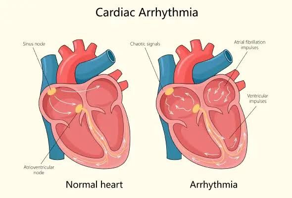
Ectopic foci are abnormal pacemaker sites in the heart that can initiate electrical impulses outside the normal conduction pathway, which is primarily governed by the sinoatrial (SA) node. These foci can arise from various locations within the heart, leading to arrhythmias. The primary types of ectopic foci include:
- Atrial Ectopic Foci: These originate in the atria and can lead to conditions such as atrial fibrillation or atrial flutter. Common sites include the pulmonary veins, where ectopic beats may be triggered by myocardial tissue that has properties similar to pacemaker cells.
- Junctional Ectopic Foci: These arise from the atrioventricular (AV) junction area. When the SA node fails to fire or if there is a block in conduction, junctional foci can take over as pacemakers, typically resulting in junctional rhythms.
- Ventricular Ectopic Foci: These originate in the ventricles and can cause premature ventricular contractions (PVCs). They may arise from various locations within the ventricular myocardium and are often associated with underlying heart disease or ischemia.
- Multifocal Atrial Tachycardia (MAT): This condition involves multiple ectopic foci within the atria firing at different rates, leading to an irregular rhythm that is often seen in patients with chronic lung disease.
The mechanisms behind ectopic focus formation generally involve changes in ion channel function, increased automaticity due to enhanced sympathetic tone, or re-entry circuits formed by altered conduction pathways.
Mechanism of Re-entry Phenomena
Re-entry is a critical mechanism underlying many types of cardiac arrhythmias. It occurs when an electrical impulse travels around a circuit of myocardial tissue that has been altered either by injury or by changes in conduction velocity. The basic requirements for re-entry include:
- Two Pathways: There must be two distinct conduction pathways for the impulse to travel—one that conducts impulses more slowly than normal and another that conducts impulses normally or faster.
- Unidirectional Block: A unidirectional block must occur in one of these pathways so that when an impulse travels down one pathway, it cannot return through it but can continue around the circuit via another route.
- Circus Movement: Once initiated, this re-entrant circuit allows for continuous propagation of impulses around a loop until interrupted by factors such as fatigue of myocardial cells or changes in electrolyte concentrations.
Re-entry phenomena are commonly observed in conditions such as atrial flutter, ventricular tachycardia, and certain forms of supraventricular tachycardia (SVT). For example:
- In atrial flutter, a re-entrant circuit often forms around anatomical barriers like valves or scar tissue.
- In ventricular tachycardia, re-entry can occur due to scarred myocardium from previous infarctions creating areas where conduction is slowed.
In summary, ectopic foci represent abnormal pacemaker activity arising from various locations within the heart muscle due to pathological changes, while re-entry phenomena describe a specific mechanism whereby electrical impulses perpetuate themselves around a circuit created by altered conduction properties.
Types of Arrhythmia and Their ECG Appearances
Arrhythmias are irregular heartbeats that can be classified into various types based on their origin and characteristics. Below, we will discuss five common types of arrhythmias: atrial fibrillation, atrial flutter, supraventricular tachycardia, ventricular tachycardia, and ventricular fibrillation. For each type, we will describe the ECG (electrocardiogram) appearance.
1. Atrial Fibrillation (AF)
Atrial fibrillation is characterized by rapid and chaotic electrical signals in the atria, leading to an irregular and often rapid heart rate.
- ECG Appearance: The hallmark of AF is the absence of distinct P waves; instead, there are erratic baseline fluctuations known as fibrillatory waves. The QRS complexes are usually narrow but occur at irregular intervals, resulting in an irregularly irregular rhythm.
2. Atrial Flutter
Atrial flutter involves a reentrant circuit within the right atrium, leading to a rapid but organized contraction of the atria.
- ECG Appearance: The classic finding in atrial flutter is the presence of “sawtooth” patterns known as F-waves or flutter waves, particularly visible in leads II, III, and aVF. These F-waves typically have a rate of about 300 beats per minute. The QRS complexes may be regular or irregular depending on the conduction through the AV node.
3. Supraventricular Tachycardia (SVT)
Supraventricular tachycardia refers to any tachycardia that originates above the ventricles, often due to reentry circuits or enhanced automaticity.
- ECG Appearance: In SVT, there is usually a narrow QRS complex (<120 ms) with a regular rhythm at rates often exceeding 150 beats per minute. P waves may be absent or may appear after QRS complexes if they are conducted retrogradely (in cases like AV nodal reentrant tachycardia).
4. Ventricular Tachycardia (VT)
Ventricular tachycardia is defined as three or more consecutive ventricular beats at a rate exceeding 100 beats per minute and can be life-threatening.
- ECG Appearance: VT typically presents with wide QRS complexes (>120 ms) that are usually monomorphic (uniform shape) or polymorphic (varying shapes). The rhythm is generally regular but can sometimes be irregular depending on the underlying mechanism.
5. Ventricular Fibrillation (VF)
Ventricular fibrillation is a critical condition where there are disorganized electrical impulses in the ventricles leading to ineffective quivering instead of coordinated contractions.
- ECG Appearance: VF shows chaotic and erratic waveforms without identifiable QRS complexes, P waves, or T waves. The amplitude and frequency of these waveforms vary significantly over time, indicating severe cardiac dysfunction.
In summary:
- Atrial Fibrillation: Irregularly irregular rhythm; no distinct P waves.
- Atrial Flutter: Regular “sawtooth” F-waves; typically 300 bpm.
- Supraventricular Tachycardia: Narrow QRS; regular rhythm; possibly absent P waves.
- Ventricular Tachycardia: Wide QRS; regular rhythm.
- Ventricular Fibrillation: Chaotic waveform; no identifiable complexes.
Types of Conduction Block: Incomplete and Complete Heart Block
Conduction blocks in the heart refer to disruptions in the electrical signals that coordinate heartbeats. These blocks can be classified into incomplete (first and second degree) and complete heart block, each with distinct characteristics and implications for cardiac function.
1. Incomplete Heart Block
Incomplete heart blocks are characterized by partial interruptions in the conduction pathway of electrical impulses from the atria to the ventricles. They can be further divided into two main types:
(a) First-Degree Heart Block
First-degree heart block is the mildest form of conduction block. It is defined by a prolonged PR interval on an electrocardiogram (ECG), which is the time taken for electrical impulses to travel from the atria to the ventricles. Specifically, a PR interval greater than 200 milliseconds indicates this condition.
- Causes: This type of block can occur due to various factors such as increased vagal tone, myocardial ischemia, or certain medications (e.g., beta-blockers).
- Symptoms: Often asymptomatic, first-degree heart block typically does not require treatment unless associated with other cardiac conditions.
- Prognosis: Generally benign; most patients lead normal lives without significant complications.
(b) Second-Degree Heart Block
Second-degree heart block is more severe than first-degree and is characterized by intermittent failure of conduction from the atria to the ventricles. It can be subdivided into two types:
- Type I (Wenckebach or Mobitz Type I): This type features progressively lengthening PR intervals until a beat is dropped (i.e., a QRS complex fails to follow an atrial contraction).
- Causes: Often seen in athletes or during sleep due to increased vagal tone but can also result from ischemic heart disease.
- Symptoms: Patients may experience palpitations or dizziness but often remain asymptomatic.
- Type II (Mobitz Type II): This type has a consistent PR interval followed by sudden drops of QRS complexes without prior lengthening.
- Causes: More likely associated with structural heart disease and carries a higher risk of progressing to complete heart block.
- Symptoms: Can cause syncope or near-syncope episodes due to irregular ventricular response.
2. Complete Heart Block
Complete heart block, also known as third-degree heart block, occurs when there is a total failure of electrical conduction between the atria and ventricles. In this scenario, atrial impulses do not reach the ventricles at all.
- Mechanism: The atria continue to contract independently while the ventricles rely on an escape rhythm originating from either the AV node or Purkinje fibers.
- ECG Findings: Characterized by dissociation between P waves (atrial contractions) and QRS complexes (ventricular contractions), leading to a slower ventricular rate.
(a) Causes
Complete heart block can arise from various conditions including:
- Ischemic heart disease
- Myocarditis
- Degenerative diseases affecting conduction pathways
- Certain medications or electrolyte imbalances
(b) Symptoms
Patients may present with symptoms such as:
- Fatigue
- Dizziness
- Syncope
- Chest pain
(c) Management
Management often requires intervention such as:
- Pacemaker insertion for symptomatic patients
- Treatment of underlying causes if reversible
Conclusion
In summary, conduction blocks vary significantly in severity and clinical implications, ranging from first-degree blocks that are often benign to complete blocks that necessitate immediate medical attention and intervention.


