Common Pediatric Orthopedic Conditions
Pediatric orthopedic conditions encompass a variety of musculoskeletal disorders that affect children and adolescents. These conditions can arise from congenital issues, developmental problems, trauma, or infections. The most common pediatric orthopedic conditions include:
- Developmental Dysplasia of the Hip (DDH): This condition involves improper formation of the hip joint, where the femoral head is not securely seated in the acetabulum. It can lead to hip dislocation if not diagnosed and treated early.
- Clubfoot (Talipes Equinovarus): A congenital deformity where the foot is twisted out of shape or position. The affected foot may point downwards and inwards.
- Osgood-Schlatter Disease: This condition is characterized by pain and swelling at the tibial tuberosity due to inflammation of the growth plate, commonly seen in active adolescents during periods of rapid growth.
- Sever’s Disease (Calcaneal Apophysitis): An overuse injury affecting the heel’s growth plate, leading to heel pain in growing children, particularly those involved in sports.
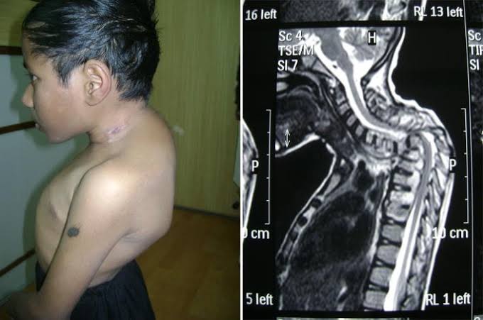
- Scoliosis: A lateral curvature of the spine that can occur during growth spurts in adolescence. It may require monitoring or intervention depending on severity.
- Fractures: Children are prone to various types of fractures due to their high activity levels and developing bones. Common types include greenstick fractures, which are incomplete breaks typical in younger children.
- Limb Length Discrepancy: This condition occurs when one limb is shorter than the other, which can be congenital or acquired due to trauma or infection.
- Patellar Instability: Often seen in active adolescents, this condition involves dislocation or subluxation of the kneecap, leading to pain and functional impairment.
- Perthes Disease: A childhood hip disorder resulting from temporary loss of blood supply to the femoral head, leading to bone death and subsequent regeneration.
- Torticollis: A condition characterized by a twisting of the neck that causes the head to tilt downwards towards one shoulder; it can be congenital or acquired.
Clinical Approach
The clinical approach for evaluating pediatric orthopedic conditions typically begins with a thorough history and physical examination:
- History Taking:
- Assess for symptoms such as pain, swelling, deformity, or functional limitations.
- Inquire about any previous injuries or family history of orthopedic conditions.
- Evaluate activity levels and any recent changes in physical activity.
- Physical Examination:
- Inspect for asymmetry, range of motion limitations, tenderness, and signs of inflammation.
- Perform specific tests relevant to suspected conditions (e.g., Ortolani test for DDH).
- Functional Assessment:
- Observe how the child walks or runs.
- Assess gait patterns and any compensatory movements.
Radiological Evaluation
After clinical assessment suggests an orthopedic issue, radiological evaluation is often necessary for diagnosis:
- X-rays:
- Standard initial imaging modality for most pediatric orthopedic conditions.
- Useful for identifying fractures, dislocations, alignment issues (e.g., scoliosis), and joint abnormalities (e.g., DDH).
- Ultrasound:
- Particularly useful for assessing soft tissue structures around joints (e.g., hip ultrasound for DDH).
- Can help visualize fluid collections or guide injections in certain cases.
- MRI (Magnetic Resonance Imaging):
- Provides detailed images of soft tissues including cartilage, ligaments, muscles, and bone marrow.
- Indicated when there is suspicion of osteomyelitis or when soft tissue injuries are suspected but not visible on X-ray.
- CT Scans (Computed Tomography):
- Occasionally used for complex fractures where detailed bony anatomy needs clarification.
- Not routinely used due to higher radiation exposure compared to X-rays.
- Bone Scintigraphy (Bone Scan):
- May be utilized in cases where there is suspicion of infection or inflammatory processes affecting multiple sites but is less common due to specificity concerns compared with MRI.
In summary, a systematic approach combining clinical evaluation with appropriate radiological studies allows for accurate diagnosis and management planning for common pediatric orthopedic conditions.
Management Modalities of Developmental Dysplasia of the Hip (DDH) and Clubfoot
Developmental Dysplasia of the Hip (DDH) and clubfoot are two distinct orthopedic conditions that primarily affect infants and children. Both conditions require early diagnosis and appropriate management to ensure optimal outcomes. The management strategies for each condition differ significantly due to their unique pathophysiology.
Management of Developmental Dysplasia of the Hip (DDH)
- Diagnosis
- DDH is typically diagnosed through physical examination, which may include the Ortolani and Barlow maneuvers, as well as imaging studies such as ultrasound or X-rays, particularly in older infants.
- Non-Surgical Management
- Pavlik Harness: This is the most common initial treatment for infants diagnosed with DDH under six months old. The Pavlik harness holds the hip in a flexed and abducted position, allowing for proper joint development while preventing dislocation.
- Observation: In cases where there is mild dysplasia without dislocation, careful monitoring may be sufficient, especially if the child is younger than six months.
- Surgical Management
- If non-surgical methods fail or if DDH is diagnosed after six months of age, surgical intervention may be necessary. Options include:
- Closed Reduction: This procedure involves manipulating the hip back into its socket without making an incision.
- Open Reduction: In more severe cases or when closed reduction fails, an open surgical approach may be required to reposition the femoral head within the acetabulum.
- Pelvic Osteotomy: This procedure reorients the acetabulum to provide better coverage of the femoral head.
- If non-surgical methods fail or if DDH is diagnosed after six months of age, surgical intervention may be necessary. Options include:
- Postoperative Care
- After surgery, patients often require bracing or casting to maintain hip stability during recovery. Follow-up imaging is essential to assess healing and joint function.
Management of Clubfoot
- Diagnosis
- Clubfoot (talipes equinovarus) is characterized by a foot that appears twisted or turned inward at birth. Diagnosis is primarily clinical but can be confirmed via ultrasound in utero or X-ray postnatally.
- Non-Surgical Management
- Ponseti Method: This is a widely accepted non-surgical technique involving a series of gentle manipulations and casting over several weeks to gradually correct foot positioning.
- Initial manipulation involves stretching the foot into a corrected position followed by application of a cast.
- Casts are changed weekly until optimal correction is achieved, usually within 6-8 weeks.
- A percutaneous Achilles tendon tenotomy may be performed if tightness persists after casting.
- Ponseti Method: This is a widely accepted non-surgical technique involving a series of gentle manipulations and casting over several weeks to gradually correct foot positioning.
- Post-Correction Management
- After achieving correction with casting, it’s crucial to prevent relapse:
- Foot Abduction Brace (FAB): Children typically wear this brace for 23 hours a day for three months followed by nighttime use until age four to maintain correction.
- After achieving correction with casting, it’s crucial to prevent relapse:
- Surgical Management
- In cases where non-surgical methods fail or if there are residual deformities after initial treatment, surgical options may include:
- Soft tissue releases
- Osteotomies
- Fusion procedures depending on severity and age at presentation.
- In cases where non-surgical methods fail or if there are residual deformities after initial treatment, surgical options may include:
- Long-term Follow-up
- Regular follow-ups are necessary to monitor growth and development, as well as any potential recurrence of deformity.
Conclusion
In summary, both DDH and clubfoot require early identification and tailored management strategies that vary significantly between conditions. While DDH often starts with non-invasive methods like bracing before potentially progressing to surgical interventions if necessary, clubfoot management typically begins with manipulation and casting techniques followed by bracing post-correction.
Understanding the Basics of Orthopedic Surgical Approach to Pediatric Patients
Pediatric orthopedic surgery is a specialized field that addresses musculoskeletal issues in children, who have unique anatomical and physiological characteristics compared to adults. The surgical approach in pediatric patients requires careful consideration of their growing bodies, as children’s bones and joints are still developing. This necessitates a tailored approach to ensure optimal outcomes while minimizing potential complications.
Key Considerations in Pediatric Orthopedic Surgery
- Growth Plates and Developmental Factors: One of the most critical aspects of pediatric orthopedic surgery is the understanding of growth plates (epiphyseal plates), which are areas of developing cartilage tissue near the ends of long bones. Surgeries involving or near these growth plates must be performed with caution, as improper handling can lead to growth disturbances or deformities.
- Anesthesia and Pain Management: Pediatric patients often require different anesthesia techniques than adults due to their smaller size and varying physiological responses. Anesthesiologists specializing in pediatrics are trained to manage these differences effectively. Postoperative pain management also needs to be age-appropriate, utilizing both pharmacological and non-pharmacological methods.
- Minimally Invasive Techniques: Whenever possible, pediatric orthopedic surgeons prefer minimally invasive techniques, such as arthroscopy or small incisions, which can reduce recovery time and minimize trauma to surrounding tissues. These approaches help decrease hospital stays and allow for quicker rehabilitation.
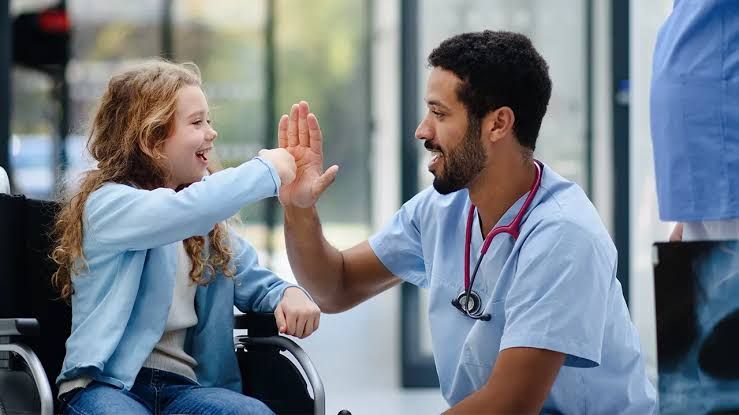
- Family-Centered Care: Engaging families in the surgical process is crucial. Surgeons often provide detailed explanations about the procedure, expected outcomes, and recovery processes not only to the patient but also to their parents or guardians. This helps alleviate anxiety and fosters trust between families and medical staff.
- Postoperative Care and Rehabilitation: After surgery, children may require specific rehabilitation protocols tailored to their age and developmental stage. Physical therapy plays a vital role in restoring function and strength while ensuring that activities are appropriate for their growth status.
- Long-Term Follow-Up: Continuous monitoring after surgery is essential due to the dynamic nature of a child’s growth. Regular follow-up appointments allow healthcare providers to assess healing progress, adjust treatment plans if necessary, and address any emerging concerns related to growth or function.
- Common Procedures: Some common orthopedic procedures performed on pediatric patients include:
- Fracture fixation
- Correction of scoliosis through spinal fusion
- Treatment of clubfoot using casting or surgical intervention
- Osteotomies for correcting bone deformities
- Communication with Young Patients: Effective communication strategies are vital when dealing with children undergoing surgery. Surgeons often use child-friendly language and visual aids to explain procedures in an understandable manner, helping reduce fear and anxiety associated with medical interventions.
Conclusion
The orthopedic surgical approach for pediatric patients is multifaceted, requiring specialized knowledge about children’s unique anatomical features, careful planning regarding growth considerations, effective communication with both young patients and their families, as well as a commitment to ongoing care throughout recovery.


