Morphology of Candida albicans
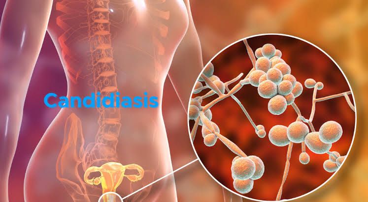
Candida albicans is a dimorphic fungus, meaning it can exist in two distinct forms: yeast and filamentous (hyphal) forms. The yeast form appears as oval to round cells, typically measuring 4-6 micrometers in diameter. These cells reproduce asexually through budding, where a new cell develops from the parent cell. In contrast, the hyphal form consists of elongated, tubular structures known as hyphae that can be several micrometers long. When C. albicans transitions to its filamentous form, it can produce pseudohyphae—characterized by constricted septa and irregular shapes—as well as true hyphae.
The morphology of C. albicans is influenced by environmental conditions such as temperature, pH, and nutrient availability. At body temperature (37°C), C. albicans predominantly exists in the hyphal form, which is associated with its pathogenicity. The ability to switch between these forms is crucial for its survival and virulence.
Pathogenesis of Candida albicans
C. albicans is an opportunistic pathogen that primarily affects immunocompromised individuals but can also cause infections in healthy hosts under certain conditions. Its pathogenesis involves several key factors:
- Adhesion: C. albicans adheres to host tissues using surface proteins known as adhesins. This adhesion is critical for colonization and biofilm formation on mucosal surfaces and medical devices.
- Invasion: The transition from yeast to hyphal form enhances its ability to invade epithelial tissues and evade host defenses. Hyphae can penetrate deeper into tissues compared to yeast cells.
- Immune Evasion: C. albicans has evolved mechanisms to evade the host immune system, including altering its surface antigens and producing enzymes that degrade immune components like antibodies and complement proteins.
- Biofilm Formation: C. albicans can form biofilms on various surfaces, including catheters and prosthetic devices, which protect it from both the immune response and antifungal treatments.
- Toxin Production: It produces various virulence factors such as proteases and phospholipases that contribute to tissue damage and inflammation during infection.
Infections caused by C. albicans range from superficial mucosal infections (such as oral thrush or vulvovaginal candidiasis) to systemic infections like candidemia, which can be life-threatening if not treated promptly.
Association Between the Immune System and Fungal Infections
The immune system plays a crucial role in defending against fungal infections, including those caused by C. albicans:
- Innate Immunity: The first line of defense includes physical barriers (skin and mucosa), phagocytic cells (neutrophils and macrophages), and pattern recognition receptors (PRRs) that detect fungal components like β-glucans in the cell wall of fungi.
- Adaptive Immunity: T lymphocytes (particularly Th17 cells) are vital for orchestrating an effective response against fungal pathogens by producing cytokines that recruit neutrophils and enhance their antifungal activity.
- Immunocompromised States: Individuals with weakened immune systems—due to conditions such as HIV/AIDS, cancer treatments, or organ transplants—are at increased risk for invasive candidiasis because their ability to mount an effective immune response is compromised.
- Cytokine Response: A balanced cytokine response is essential; excessive inflammation can lead to tissue damage while insufficient responses may fail to control fungal growth effectively.
- Vaccination Efforts: Research into vaccines targeting specific antigens of C. albicans aims to bolster the immune response in at-risk populations.
Understanding the interplay between Candida albicans morphology, its pathogenic mechanisms, and the host immune response is critical for developing effective prevention strategies and treatments for candidiasis.
Clinical Presentation and Nature of Vaginal Discharge in Candidiasis
Candidiasis, commonly known as a yeast infection, is primarily caused by the overgrowth of Candida species, most frequently Candida albicans. The clinical presentation of vaginal candidiasis typically includes:
- Symptoms: Patients often report intense itching and irritation in the vaginal area. This discomfort can be exacerbated by sexual intercourse or during urination. Other common symptoms include swelling and redness of the vulva and vagina.
- Vaginal Discharge: The nature of the discharge associated with candidiasis is characteristically thick, white, and curd-like, resembling cottage cheese. It is usually odorless but may have a slight yeasty smell. In contrast to bacterial vaginosis or trichomoniasis, which may produce a more watery or foul-smelling discharge, the discharge in candidiasis is distinctively thick and adherent.
- Physical Examination Findings: Upon examination, healthcare providers may observe erythema (redness) and edema (swelling) of the vulva and vagina. There may also be white patches on the vaginal walls or cervix.
- Risk Factors: Certain factors can predispose individuals to develop candidiasis, including antibiotic use, hormonal changes (such as those occurring during pregnancy or with oral contraceptive use), diabetes mellitus, immunosuppression (e.g., HIV/AIDS), and wearing tight-fitting clothing that retains moisture.
Laboratory Methods of Diagnosis
Diagnosis of vaginal candidiasis typically involves both clinical evaluation and laboratory testing:
- Clinical Diagnosis: A healthcare provider may initially diagnose candidiasis based on symptoms and physical examination findings.
- Microscopic Examination: A sample of vaginal discharge can be collected for microscopic examination. A wet mount preparation allows for direct visualization of yeast cells under a microscope.
- Culture Tests: If initial tests are inconclusive or if recurrent infections occur, a culture may be performed to identify the specific Candida species involved. This involves inoculating a sample onto a selective medium that supports yeast growth.
- Molecular Methods: Polymerase chain reaction (PCR) assays can also be utilized for rapid identification of Candida species from vaginal samples, providing higher sensitivity compared to traditional culture methods.
- pH Testing: The vaginal pH is typically normal (between 3.8 to 4.5) in cases of candidiasis; an elevated pH suggests other infections such as bacterial vaginosis or trichomoniasis.
Drugs Used for Treatment of Candidiasis
The treatment options for vaginal candidiasis include:
- Antifungal Medications:
- Topical Antifungals: Over-the-counter options such as clotrimazole (Lotrimin), miconazole (Monistat), and tioconazole (Vagistat) are commonly used topical treatments.
- Oral Antifungals: Fluconazole (Diflucan) is an effective oral antifungal medication often prescribed for more severe cases or recurrent infections.
- Duration of Treatment: Topical treatments are usually administered for 1 to 7 days depending on the formulation used, while fluconazole is typically given as a single dose but can be repeated after several days if symptoms persist.
- Considerations for Recurrent Cases: For women experiencing recurrent episodes (four or more per year), longer-term maintenance therapy with fluconazole may be recommended along with lifestyle modifications to reduce risk factors.
In summary, candidiasis presents with characteristic symptoms including itching and thick white discharge; diagnosis involves clinical assessment combined with laboratory tests such as microscopy and cultures; treatment primarily consists of antifungal medications either topically or orally.


