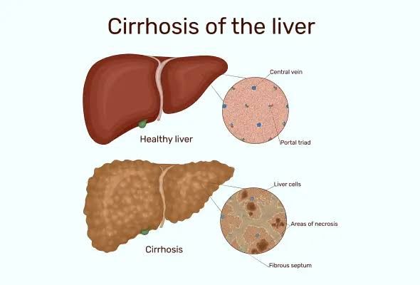
Liver cirrhosis is a late-stage scarring (fibrosis) of the liver caused by many forms of liver diseases and conditions, such as hepatitis and chronic alcoholism. In cirrhosis, healthy liver tissue is replaced with scar tissue, which impairs the liver’s ability to function properly. This condition can lead to severe complications, including liver failure, portal hypertension, and an increased risk of liver cancer.
Common Types of Liver Cirrhosis
- Alcoholic Cirrhosis: This type results from chronic alcohol abuse. The liver metabolizes alcohol, which can lead to inflammation and damage over time. Continued excessive drinking leads to the development of scar tissue.
- Hepatitis C Cirrhosis: Chronic infection with the hepatitis C virus (HCV) can cause long-term inflammation and damage to the liver, leading to cirrhosis. Hepatitis C is often asymptomatic in its early stages but can progress to severe liver disease.
- Nonalcoholic Fatty Liver Disease (NAFLD): This condition involves fat accumulation in the liver not due to alcohol consumption. It can progress to nonalcoholic steatohepatitis (NASH), which causes inflammation and fibrosis that may lead to cirrhosis.
- Biliary Cirrhosis: This type occurs when there is damage to the bile ducts within or outside the liver, leading to bile accumulation and subsequent liver damage. Primary biliary cholangitis (PBC) is an autoimmune disorder that can cause this type of cirrhosis.
- Hemochromatosis: This genetic disorder causes excessive iron accumulation in the body, particularly in the liver, leading to damage and cirrhosis over time.
- Wilson’s Disease: A rare genetic disorder that leads to copper buildup in the body, particularly affecting the liver and brain, resulting in cirrhosis if untreated.
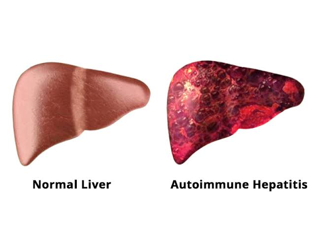
- Autoimmune Hepatitis: This condition occurs when the body’s immune system attacks liver cells, causing inflammation and potentially leading to cirrhosis if not managed effectively.
- Chronic Biliary Obstruction: Conditions that block bile flow from the liver can lead to bile accumulation and subsequent damage over time.
Clinical Manifestations of Liver Cirrhosis
The clinical manifestations can be categorized based on these two primary complications.
1. Clinical Manifestations of Liver Cell Failure
Liver cell failure occurs when the liver is unable to perform its normal functions due to extensive damage. The following are key manifestations:
- Jaundice: This is characterized by yellowing of the skin and eyes due to an accumulation of bilirubin in the blood, which occurs when the liver cannot adequately process this waste product.
- Coagulopathy: The liver produces many proteins necessary for blood clotting. In cirrhosis, decreased production leads to increased bleeding tendencies, easy bruising, and prolonged bleeding times.
- Hepatic Encephalopathy: This neurological condition arises from the accumulation of toxins (such as ammonia) that the damaged liver cannot clear. Symptoms range from mild confusion and altered sleep patterns to severe cognitive impairment and coma.
- Ascites: Fluid accumulation in the abdominal cavity occurs due to decreased albumin production (leading to low oncotic pressure) and increased portal pressure. Patients may present with abdominal distension and discomfort.
- Fatigue and Weakness: Generalized fatigue is common due to metabolic disturbances, malnutrition, and anemia associated with chronic disease.
- Malnutrition: Patients often experience weight loss and muscle wasting due to poor dietary intake, malabsorption, or increased metabolic demands.
- Spider Angiomas and Palmar Erythema: These vascular lesions are indicative of hormonal changes associated with liver dysfunction.
2. Clinical Manifestations of Portal Hypertension
Portal hypertension results from increased pressure in the portal venous system due to obstruction caused by cirrhosis. Key manifestations include:
- Esophageal Varices: These are dilated veins in the esophagus that develop as collateral circulation forms in response to elevated portal pressure. They pose a risk for life-threatening hemorrhage if they rupture.
- Gastric Varices: Similar to esophageal varices but located in the stomach; they can also lead to significant bleeding.
- Splenomegaly: Enlargement of the spleen occurs due to congestion in the portal system, leading to hypersplenism, which can cause further cytopenias (e.g., thrombocytopenia).
- Caput Medusae: This refers to visible distended veins around the umbilicus resulting from collateral circulation due to portal hypertension.
- Hemorrhoids: Increased pressure can lead to engorgement of rectal veins, causing hemorrhoids which may bleed or become painful.
In summary, patients with cirrhosis may exhibit a combination of symptoms stemming from both liver cell failure—such as jaundice, coagulopathy, hepatic encephalopathy, ascites—and complications arising from portal hypertension—such as varices, splenomegaly, caput medusae, and hemorrhoids. The interplay between these manifestations significantly impacts patient management and prognosis.
Features of Compensated and Decompensated Liver Cirrhosis
1. Compensated Cirrhosis
Compensated cirrhosis is the early stage of liver cirrhosis where the liver is still able to perform its essential functions despite the presence of scarring. This stage often does not present noticeable symptoms, which can make it difficult for individuals to recognize that they have a liver condition. Some key features include:
- Asymptomatic Nature: Many individuals with compensated cirrhosis do not exhibit any significant symptoms. They may feel generally well and may not look ill.
- Mild Symptoms: If symptoms do occur, they are typically mild and can include:
- Nausea
- Fatigue
- Loss of appetite
- Spider-like blood vessels on the skin (telangiectasia)
- Weight loss and muscle wasting
- Liver Function: The liver maintains adequate function in this stage, meaning that while there is damage, it has not yet progressed to a point where liver function is severely compromised.
- Survival Rate: Individuals with compensated cirrhosis generally have a better prognosis, with median survival times exceeding 12 years if they manage their condition effectively and avoid further liver damage.
- Risk Factors: Common risk factors include chronic alcohol consumption (more than 40 grams per day), viral hepatitis infections, non-alcoholic fatty liver disease (NAFLD), obesity, type 2 diabetes, and metabolic syndrome.
2. Decompensated Cirrhosis
Decompensated cirrhosis represents a more advanced stage of the disease where the liver can no longer compensate for its damage. This leads to significant complications and symptoms that indicate severe liver dysfunction. Key features include:
- Symptomatic Presentation: Patients typically experience overt symptoms due to complications arising from liver failure. These symptoms may include:
- Jaundice (yellowing of the skin and eyes)
- Ascites (fluid buildup in the abdomen)
- Variceal bleeding (bleeding from swollen veins in the esophagus)
- Hepatic encephalopathy (cognitive changes due to toxin buildup)
- Complications: Decompensated cirrhosis is characterized by serious complications such as kidney failure (hepatorenal syndrome), portal hypertension (high blood pressure in the veins leading to the liver), and increased risk of infections.
- Survival Rate: The prognosis for individuals with decompensated cirrhosis is significantly poorer, with median survival times around 2 years depending on various factors including treatment response and underlying causes.
- Potential for Reversibility: In some cases, if the underlying cause of decompensation is addressed—such as abstaining from alcohol or treating viral hepatitis—there may be potential for improvement back to a compensated state.
- Monitoring and Treatment Needs: Patients require close monitoring and may need interventions such as diuretics for ascites management or even consideration for liver transplantation if their condition continues to deteriorate.
In summary, compensated cirrhosis is marked by minimal symptoms and preserved liver function, while decompensated cirrhosis involves significant complications and impaired function requiring urgent medical attention.
Management of Liver Cirrhosis
The management of cirrhosis, particularly in its decompensated form, involves addressing complications such as bleeding varices, ascites, and hepatic encephalopathy. Below is a detailed outline of the management strategies for these major features.
1. Management of Bleeding Varices
Bleeding from esophageal varices is a life-threatening complication of cirrhosis that requires immediate intervention.
- Initial Assessment and Stabilization:
- Assess the patient’s hemodynamic status (blood pressure, heart rate).
- Establish intravenous access and initiate fluid resuscitation with crystalloids.
- Monitor vital signs closely.
- Pharmacological Treatment:
- Administer vasoactive drugs such as octreotide or vasopressin to reduce portal pressure and control bleeding.
- Endoscopic Intervention:
- Urgent endoscopy should be performed within 12 hours to manage variceal bleeding through band ligation or sclerotherapy.
- Antibiotic Prophylaxis:
- Start prophylactic antibiotics (e.g., intravenous cefotaxime) to prevent infections like spontaneous bacterial peritonitis (SBP), which can complicate variceal hemorrhage.
- Transjugular Intrahepatic Portosystemic Shunt (TIPS):
- In cases where endoscopic treatment fails or if there are recurrent bleeds, TIPS may be indicated to decompress the portal system.
2. Management of Ascites
Ascites is the accumulation of fluid in the abdominal cavity and is a common complication in patients with cirrhosis.
- Dietary Modifications:
- Implement a low-sodium diet (typically <2 grams/day) to help reduce fluid retention.
- Diuretics:
- Use diuretics such as spironolactone and furosemide to promote fluid excretion. The typical starting dose for spironolactone is 100 mg daily, adjusted based on response.
- Paracentesis:
- Perform therapeutic paracentesis for symptomatic relief or when there is tense ascites. This procedure helps remove excess fluid and can provide diagnostic information regarding infection or malignancy.
- Management of Refractory Ascites:
- For patients with refractory ascites who do not respond adequately to diuretics or paracentesis, consider TIPS or surgical options like peritoneovenous shunt placement.
3. Management of Hepatic Encephalopathy
Hepatic encephalopathy (HE) results from the accumulation of neurotoxins due to impaired liver function and can manifest as confusion, altered consciousness, and coma.
- Identification and Treatment of Precipitating Factors:
- Common triggers include gastrointestinal bleeding, infections (especially SBP), electrolyte imbalances, renal failure, and excessive dietary protein intake. Addressing these factors is crucial in managing HE.
- Pharmacological Therapy:
- Lactulose is the first-line treatment; it works by reducing ammonia levels through osmotic diarrhea and altering gut flora.
- Typical dosing starts at 15–30 mL orally every hour until bowel movements are achieved.
- Rifaximin may be added for patients who do not respond adequately to lactulose alone; it reduces ammonia-producing bacteria in the gut.
- Lactulose is the first-line treatment; it works by reducing ammonia levels through osmotic diarrhea and altering gut flora.
- Nutritional Support:
- Provide adequate nutrition while monitoring protein intake; moderate protein restriction may be necessary during acute episodes but should be individualized based on patient tolerance.
In summary, effective management of liver cirrhosis focuses on preventing complications associated with decompensation through timely interventions tailored to each patient’s needs. Regular follow-up and monitoring are essential components in managing this chronic condition effectively.
Understanding the Role of MELD and CTP Scores in Surgery and Transplantation
What is MELD Score?
The Model for End-Stage Liver Disease (MELD) score is a numerical scale used to assess the severity of chronic liver disease and prioritize patients for liver transplantation. The MELD score ranges from 6 to 40, with higher scores indicating more severe liver dysfunction and a greater urgency for transplantation. It is calculated based on four laboratory values:
- Creatinine: Reflects kidney function.
- Bilirubin: Indicates liver’s ability to clear bile.
- INR (International Normalized Ratio): Measures blood clotting ability, reflecting liver function.
- Serum Sodium: Assesses fluid balance in the body.
The MELD score is crucial in determining a patient’s place on the transplant waiting list managed by the Organ Procurement and Transplantation Network (OPTN). Patients with higher MELD scores are prioritized for receiving livers from deceased donors, as they are deemed to have a higher risk of mortality without transplantation.
Role of MELD Score in Surgery
In addition to its role in transplantation, the MELD score can influence surgical decisions for patients with liver disease. A high MELD score may indicate that a patient is too ill to undergo certain elective surgeries due to the increased risk of complications or mortality associated with their underlying liver condition. Surgeons often consider the MELD score when evaluating whether a patient can safely tolerate surgery, especially procedures that could further stress the liver.
What is Child-Pugh Score?
The Child-Pugh score (also known as the Child-Turcotte-Pugh score) assesses the prognosis of chronic liver disease and helps guide treatment decisions. This scoring system evaluates five clinical parameters:
- Total Bilirubin
- Serum Albumin
- Prothrombin Time (or INR)
- Ascites
- Encephalopathy
Each parameter receives points based on severity, leading to classification into three categories: Class A (least severe), Class B, and Class C (most severe). The Child-Pugh score helps predict outcomes after surgery and can inform decisions regarding interventions such as transjugular intrahepatic portosystemic shunt (TIPS) placement or other surgical options.
Role of Child-Pugh Score in Surgery
The Child-Pugh score plays a significant role in assessing surgical risk for patients with liver disease. For instance, patients classified as Class C may face higher risks during surgery due to poor liver function, which can lead to complications such as bleeding or infection post-operatively. Therefore, surgeons often use this scoring system alongside other assessments when determining whether a patient should proceed with surgery or if alternative treatments should be considered.
Conclusion
Both the MELD and Child-Pugh scores are essential tools in managing patients with liver disease, particularly concerning transplantation eligibility and surgical risk assessment. The MELD score primarily focuses on prioritizing patients for organ allocation based on their immediate need for a transplant, while the Child-Pugh score provides insight into long-term prognosis and surgical candidacy.


