Parameters Used to Detect Hemolysis
Detecting hemolysis involves several laboratory parameters that assess the breakdown of red blood cells (RBCs) and the subsequent release of hemoglobin into the plasma. The following are key parameters used in the detection of hemolysis:
1. Plasma Free Hemoglobin Levels
Plasma free hemoglobin is a direct indicator of hemolysis. When RBCs lyse, hemoglobin is released into the bloodstream, which can be measured quantitatively. Elevated levels of plasma free hemoglobin indicate significant hemolysis. Studies have shown a wide range in the thresholds for defining clinically relevant hemolysis, with some studies using a Haemolysis Index (HI) that averages around 846 mg/L.
2. Lactate Dehydrogenase (LDH)
LDH is an enzyme found in many tissues, including RBCs. When these cells are destroyed, LDH is released into the bloodstream, leading to elevated serum levels. High LDH levels are indicative of hemolytic processes and can help differentiate between various causes of anemia.
3. Unconjugated Bilirubin
Bilirubin is a byproduct of heme breakdown from lysed RBCs. In cases of hemolysis, there is an increase in unconjugated bilirubin due to the rapid destruction of RBCs outpacing the liver’s ability to conjugate it for excretion. Elevated levels of unconjugated bilirubin can suggest ongoing hemolytic activity.
4. Haptoglobin
Haptoglobin is a protein that binds free hemoglobin in circulation. In cases of hemolysis, haptoglobin levels typically decrease because it binds to excess free hemoglobin released from lysed RBCs. Low haptoglobin levels are therefore indicative of hemolytic anemia.
5. Reticulocyte Count
Reticulocytes are immature red blood cells released from the bone marrow into circulation as a response to anemia or increased destruction of RBCs. An elevated reticulocyte count indicates that the bone marrow is responding appropriately to compensate for the loss of red blood cells due to hemolysis.
6. Peripheral Blood Smear
A peripheral blood smear allows for visual examination of blood cells under a microscope and can provide clues about potential causes of hemolytic anemia based on cell morphology (e.g., spherocytes in hereditary spherocytosis). While not definitive on its own, it complements other tests by providing additional context regarding cell shape and presence.
7. Coombs Test
The Coombs test (direct and indirect) helps determine if there is an autoimmune component to the hemolysis by detecting antibodies bound to RBCs or circulating antibodies against RBC antigens.
These parameters collectively contribute to diagnosing and understanding the underlying causes and severity of hemolytic conditions.
Classification of Hemolytic Anemias
Hemolytic anemias can be classified into two main categories: Inherited Hemolytic Anemias and Acquired Hemolytic Anemias. Each category encompasses various specific conditions based on their underlying causes and mechanisms.
1. Inherited Hemolytic Anemias
Inherited hemolytic anemias are genetic disorders that result from faulty genes affecting red blood cell production, structure, or lifespan. The primary types include:
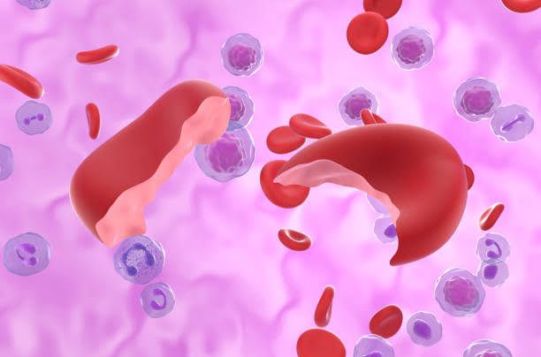
- Sickle Cell Anemia: This condition is characterized by the production of abnormal hemoglobin (hemoglobin S), leading to the distortion of red blood cells into a sickle shape. These sickle cells have a significantly reduced lifespan, typically dying within 10 to 20 days.
- Thalassemias: Thalassemias are a group of inherited blood disorders where the body produces insufficient amounts of certain types of hemoglobin, resulting in fewer healthy red blood cells. This condition is prevalent among individuals of Mediterranean, Southeast Asian, Indian, Chinese, and African descent.
- Hereditary Spherocytosis: This disorder involves defects in the red blood cell membrane that cause the cells to become spherical rather than disc-shaped. These spherocytes are more prone to destruction in the spleen.
- Hereditary Elliptocytosis (Ovalocytosis): Similar to hereditary spherocytosis but characterized by elliptical-shaped red blood cells that lack flexibility and have a shorter lifespan.
- Glucose-6-Phosphate Dehydrogenase (G6PD) Deficiency: This enzymatic deficiency leads to increased susceptibility of red blood cells to oxidative stress, causing them to rupture under certain conditions such as exposure to specific drugs or foods.
- Pyruvate Kinase Deficiency: A rare genetic disorder where a deficiency in the enzyme pyruvate kinase leads to premature breakdown of red blood cells. It is more commonly found among the Amish population.
2. Acquired Hemolytic Anemias
Acquired hemolytic anemias occur when normal red blood cells are destroyed due to external factors or diseases rather than inherited genetic defects. Key types include:
- Immune Hemolytic Anemia (AIHA): In this condition, the immune system mistakenly produces antibodies that attack and destroy red blood cells. AIHA can be further divided into:
- Autoimmune Hemolytic Anemia: The body’s immune system creates antibodies against its own red blood cells.
- Alloimmune Hemolytic Anemia: Occurs when antibodies are formed against transfused red blood cells.
- Drug-Induced Hemolytic Anemia: Certain medications can trigger an immune response leading to hemolysis.
- Nonimmune Causes: These include various factors such as:
- Thrombotic Microangiopathies: Conditions that cause small clots in vessels leading to mechanical destruction of red blood cells.
- Direct Trauma: Physical damage from injuries or medical procedures.
- Infections: Certain infections can lead to hemolysis through direct invasion or toxin production.
- Systemic Diseases: Conditions like liver disease or malignancies may also contribute to acquired hemolysis.
- Oxidative Insults: Exposure to oxidizing agents can damage red blood cell membranes leading to their destruction.
In summary, hemolytic anemias can be broadly classified into inherited and acquired forms, each with distinct pathophysiological mechanisms and clinical implications.
Immune Processes Leading to Hemolysis
Hemolysis refers to the destruction of red blood cells (RBCs), which can occur through various immune mechanisms. Understanding these processes is crucial for diagnosing and managing diseases associated with hemolysis. The immune-mediated hemolysis can be broadly categorized into two types: autoimmune hemolytic anemia (AIHA) and alloimmune hemolytic anemia.
1. Autoimmune Hemolytic Anemia (AIHA)
In AIHA, the body’s immune system mistakenly targets its own RBCs as foreign. This process involves several steps:
- Antibody Production: The immune system produces antibodies against RBC antigens. These antibodies can be of two types: warm-reacting (IgG) or cold-reacting (IgM). Warm AIHA typically occurs at body temperature, while cold agglutinin disease occurs at lower temperatures.
- Complement Activation: In some cases, particularly with IgM antibodies, complement proteins are activated upon binding to the antibody-coated RBCs. This leads to the formation of the membrane attack complex, resulting in lysis of the RBCs.
- Phagocytosis: Macrophages in the spleen and liver recognize and engulf opsonized (antibody-coated) RBCs. This process is facilitated by Fc receptors on macrophages that bind to the Fc region of antibodies attached to RBCs.
- Destruction in Spleen: The spleen plays a significant role in filtering out damaged or antibody-coated RBCs, leading to a reduction in overall red cell mass and subsequent anemia.
2. Alloimmune Hemolytic Anemia
Alloimmune hemolytic anemia occurs when an individual’s immune system reacts against transfused blood cells or fetal cells during pregnancy due to incompatibility:
- Transfusion Reactions: When a patient receives a blood transfusion containing incompatible blood type antigens, their immune system recognizes these foreign antigens as threats. This triggers an immune response where antibodies are produced against the transfused RBCs.
- Hemolytic Disease of the Newborn (HDN): In cases where an Rh-negative mother carries an Rh-positive fetus, maternal antibodies may cross the placenta and attack fetal RBCs, leading to hemolysis. This condition is primarily due to maternal sensitization during previous pregnancies or transfusions.
Other Immune-Mediated Mechanisms
Apart from AIHA and alloimmunity, other conditions can also lead to hemolysis through immune mechanisms:
- Infections: Certain infections can trigger hemolysis through immune responses. For example, malaria infects RBCs directly but also induces an immune response that contributes to their destruction.
- Drug-Induced Hemolysis: Some drugs can induce hemolysis by modifying RBC surface antigens or by eliciting an immune response against drug-modified erythrocytes. Examples include penicillin and methyldopa.
Diseases Associated with Hemolysis
Several diseases are characterized by hemolytic processes:
- Autoimmune Hemolytic Anemia (AIHA): As described above, this condition results from autoantibodies targeting RBCs.
- Sickle Cell Disease: In sickle cell disease, abnormal hemoglobin leads to distorted red blood cells that are prone to premature destruction.
- Thalassemia: Thalassemia involves genetic defects in hemoglobin production leading to ineffective erythropoiesis and increased destruction of abnormal red blood cells.
- Hereditary Spherocytosis: A genetic disorder affecting red blood cell membrane proteins causes spherically shaped cells that are more susceptible to destruction by splenic macrophages.
- G6PD Deficiency: Glucose-6-phosphate dehydrogenase deficiency leads to oxidative stress on red blood cells under certain conditions (e.g., infections or certain foods), resulting in hemolysis.
Conclusion
The processes leading to hemolysis involve complex interactions between antibodies, complement systems, and phagocytic activity within various contexts such as autoimmune disorders or reactions following transfusions. Understanding these mechanisms is essential for diagnosing related diseases effectively.
Enzyme Defects Leading to Hemolysis: Clinical and Laboratory Findings
Hemolytic anemia is a condition characterized by the premature destruction of red blood cells (RBCs), which can be caused by various factors, including enzyme defects. The most frequent enzyme defects associated with hemolysis include glucose-6-phosphate dehydrogenase (G6PD) deficiency, pyruvate kinase (PK) deficiency, and hexokinase deficiency. Each of these conditions has distinct clinical and laboratory findings.
1. Glucose-6-Phosphate Dehydrogenase (G6PD) Deficiency
Clinical Findings: G6PD deficiency is one of the most common enzymatic defects leading to hemolytic anemia, particularly in males due to its X-linked inheritance pattern. Patients may remain asymptomatic until exposed to oxidative stress from certain medications (e.g., sulfa drugs, antimalarials), infections, or foods (e.g., fava beans). Symptoms of hemolysis can include:
- Fatigue
- Jaundice
- Dark urine
- Abdominal pain
Laboratory Findings: Laboratory tests typically reveal:
- Reticulocytosis: An increased number of reticulocytes as the bone marrow responds to anemia.
- Hemoglobinuria: Presence of hemoglobin in urine due to hemolysis.
- Elevated indirect bilirubin levels: Resulting from increased breakdown of heme.
- Low haptoglobin levels: Haptoglobin binds free hemoglobin; low levels indicate hemolysis.
- Peripheral blood smear: May show bite cells and blister cells indicative of oxidative damage.
2. Pyruvate Kinase (PK) Deficiency
Clinical Findings: PK deficiency is an autosomal recessive disorder that leads to chronic hemolytic anemia. Symptoms often manifest in infancy or early childhood but can vary widely among individuals. Common clinical features include:
- Pallor
- Fatigue
- Jaundice
- Splenomegaly
Severe cases may lead to complications such as gallstones due to increased bilirubin production.
Laboratory Findings: Key laboratory findings for PK deficiency include:
- Reticulocytosis: Similar to G6PD deficiency, indicating active erythropoiesis.
- Elevated indirect bilirubin levels: Due to increased RBC breakdown.
- Low haptoglobin levels: Reflecting ongoing hemolysis.
- Peripheral blood smear: May show echinocytes (spiky red blood cells).
Additionally, specific enzyme assays can confirm the diagnosis by demonstrating reduced PK activity in red blood cells.
3. Hexokinase Deficiency
Clinical Findings: Hexokinase deficiency is a rare cause of hereditary non-spherocytic hemolytic anemia. It is also inherited in an autosomal recessive manner. Clinical manifestations are less common but may include:
- Mild to moderate anemia
- Jaundice
Symptoms are generally milder compared to G6PD or PK deficiencies.
Laboratory Findings: The laboratory findings associated with hexokinase deficiency include:
- Reticulocytosis: As seen in other enzyme deficiencies.
- Elevated indirect bilirubin levels: Indicative of hemolysis.
A peripheral blood smear may not show distinctive features; however, enzyme assays will demonstrate decreased hexokinase activity.
Conclusion
In summary, enzyme defects leading to hemolysis primarily involve G6PD and PK deficiencies, with hexokinase deficiency being less common. Each condition presents with characteristic clinical symptoms and specific laboratory findings that aid in diagnosis. Understanding these defects is crucial for appropriate management and treatment strategies for affected individuals.
Identification of Blood Cell Abnormalities
In hematology, various abnormalities in red blood cells (RBCs) can indicate different pathological conditions. Below is a detailed identification of specific RBC abnormalities: spherocytes, schistocytes, nucleated RBCs, Heinz bodies, elliptocytes, and Howell-Jolly bodies.
1. Spherocyte
Spherocytes are abnormally shaped red blood cells that appear as small, round cells lacking the typical biconcave disc shape. They are often associated with conditions such as hereditary spherocytosis and autoimmune hemolytic anemia. The lack of central pallor in spherocytes distinguishes them from normal RBCs. These cells result from membrane defects that lead to increased rigidity and decreased surface area relative to volume.
2. Schistocyte
Schistocytes are fragmented red blood cells that appear irregularly shaped and are typically smaller than normal RBCs. They are often seen in conditions involving microangiopathic hemolytic anemia, such as thrombotic thrombocytopenic purpura (TTP) or disseminated intravascular coagulation (DIC). The presence of schistocytes indicates mechanical destruction of RBCs due to turbulent blood flow or damage from fibrin strands.
3. Nucleated RBCs
Nucleated red blood cells (nRBCs) are immature forms of red blood cells that still contain a nucleus. Their presence in the peripheral blood is abnormal in adults and usually indicates severe stress on the bone marrow or increased erythropoiesis due to conditions like severe anemia or hypoxia. In newborns, nRBCs can be present normally but should not be found in significant numbers in older children or adults.
4. Heinz Bodies
Heinz bodies are aggregates of denatured hemoglobin that form within red blood cells due to oxidative stress. They can be visualized using special staining techniques such as the supravital stain. The presence of Heinz bodies is commonly associated with conditions like glucose-6-phosphate dehydrogenase (G6PD) deficiency and certain types of hemolytic anemia. These inclusions can lead to increased fragility of the RBC membrane and subsequent hemolysis.
5. Elliptocyte
Elliptocytes, also known as ovalocytes, are elongated red blood cells that resemble an ellipse rather than the typical round shape. They can be seen in various conditions including hereditary elliptocytosis and certain types of anemias. The presence of elliptocytes may indicate a defect in the cytoskeletal proteins that maintain the normal biconcave shape of RBCs.
6. Howell-Jolly Bodies
Howell-Jolly bodies are small nuclear remnants found within red blood cells, appearing as dark purple granules on a stained slide. They indicate a failure of splenic function or absence (asplenia), as the spleen normally removes these nuclear fragments from circulating RBCs. Howell-Jolly bodies can be observed in patients who have undergone splenectomy or have functional hyposplenism due to various causes.
In summary:
- Spherocyte: Small, round RBC lacking central pallor; associated with hereditary spherocytosis.
- Schistocyte: Fragmented RBC; indicative of mechanical destruction.
- Nucleated RBCs: Immature RBC with a nucleus; suggests increased erythropoiesis.
- Heinz Bodies: Denatured hemoglobin aggregates; linked to oxidative stress.
- Elliptocyte: Elongated RBC; associated with hereditary elliptocytosis.
- Howell-Jolly Bodies: Nuclear remnants in RBC; indicate splenic dysfunction.
RBC Membrane Cytoskeleton and Hereditary Spherocytosis
Introduction to RBC Membrane Cytoskeleton
Red blood cells (RBCs) have a unique structure that allows them to efficiently transport oxygen throughout the body. The membrane cytoskeleton of RBCs is a complex network that provides mechanical stability, flexibility, and resilience to the cell. This cytoskeletal framework is primarily composed of spectrin, actin, and various associated proteins that work together to maintain the biconcave shape of the erythrocyte.
Components of the RBC Membrane Cytoskeleton
- Spectrin: Spectrin is a key component of the RBC cytoskeleton, forming a mesh-like structure beneath the plasma membrane. It consists of alpha and beta chains that assemble into tetramers, which then polymerize into long filaments. These spectrin filaments are interconnected by actin filaments and other proteins, creating a supportive scaffold.
- Actin: Actin filaments are crucial for maintaining the shape and deformability of RBCs. They interact with spectrin to form a dynamic network that can quickly respond to mechanical stress during circulation.
- Ankyrin: Ankyrin is an important adaptor protein that links spectrin to integral membrane proteins such as band 3 (anion exchanger) and glycophorin C. This connection helps anchor the cytoskeletal network to the plasma membrane.
- Band 3 Protein: Band 3 serves not only as an ion transporter but also as a structural component that interacts with ankyrin and spectrin, playing a vital role in maintaining membrane integrity.
- Other Proteins: Additional proteins such as protein 4.1, tropomyosin, and adducin contribute to the stability and organization of the cytoskeletal network.
Role in Maintaining Cell Shape and Function
The RBC membrane cytoskeleton is essential for preserving the characteristic biconcave shape of erythrocytes, which maximizes surface area for gas exchange while allowing flexibility as they traverse narrow capillaries. The integrity of this cytoskeletal structure ensures proper deformability under shear stress encountered during circulation.
Hereditary Spherocytosis Overview
Hereditary spherocytosis (HS) is a genetic disorder characterized by defects in the RBC membrane cytoskeleton leading to spherical-shaped red blood cells instead of their normal biconcave shape. This condition results from mutations in genes encoding components of the cytoskeleton, particularly those involved in spectrin or ankyrin function.
- Genetic Basis: HS is typically inherited in an autosomal dominant manner but can also arise from recessive mutations. Commonly affected genes include ANK1 (ankyrin), SPTB (beta-spectrin), SPTA1 (alpha-spectrin), and EPB42 (protein 4.1). Mutations disrupt normal interactions within the cytoskeletal network, compromising cell stability.
- Pathophysiology: The altered shape of RBCs leads to increased rigidity and decreased deformability, making them more prone to hemolysis—destruction due to mechanical stress or splenic filtration. The spleen recognizes these abnormally shaped cells as defective and removes them from circulation prematurely.
- Clinical Manifestations: Patients with HS often present with anemia due to hemolysis, jaundice from elevated bilirubin levels, splenomegaly due to increased workload on the spleen, and potential complications like gallstones resulting from increased bilirubin production.
- Diagnosis and Management: Diagnosis typically involves blood tests showing spherocytes on peripheral smear along with elevated reticulocyte counts indicating compensatory erythropoiesis. Confirmatory tests may include osmotic fragility testing or genetic testing for specific mutations associated with HS. Management may involve folic acid supplementation for anemia support or splenectomy in severe cases to reduce hemolysis.
- Prognosis: With appropriate management, individuals with hereditary spherocytosis can lead relatively normal lives; however, they require regular monitoring for complications related to hemolytic anemia.
In summary, hereditary spherocytosis exemplifies how defects in the RBC membrane cytoskeleton can lead to significant clinical consequences through alterations in red blood cell shape and function.


