Organs Responsible for Hematopoiesis in the Fetus
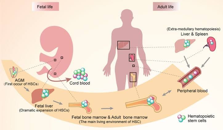
Hematopoiesis, the process of blood cell production, begins early in embryonic development and involves several key organs throughout different stages of fetal growth. The primary organs responsible for hematopoiesis in the fetus include:
1. Yolk Sac
During the earliest weeks of embryonic development, hematopoiesis occurs in the yolk sac. This structure is crucial as it provides the first site for blood cell formation before other organs are fully developed. The yolk sac produces primitive red blood cells that are essential for supplying oxygen to the developing embryo.
2. Liver
As development progresses, particularly around the sixth week of gestation, the liver becomes a major site of hematopoiesis. It takes over from the yolk sac and significantly contributes to the production of various blood cells, including red blood cells and some types of white blood cells. The liver remains an important hematopoietic organ until shortly before birth.
3. Spleen
The spleen also plays a role in fetal hematopoiesis, particularly during mid-gestation. While its contribution is less significant than that of the liver, it assists in producing lymphocytes and other immune-related cells that are vital for establishing a functional immune system.
4. Bone Marrow
Towards the end of gestation, specifically around the third trimester, bone marrow begins to take over as the primary site for hematopoiesis. Although it starts developing earlier, its function becomes more prominent as birth approaches. After birth, bone marrow becomes the main organ responsible for ongoing blood cell production throughout life.
In summary, the organs responsible for hematopoiesis in the fetus include: 1) Yolk Sac; 2) Liver; 3) Spleen; and 4) Bone Marrow, with each playing a critical role at different stages of fetal development.
Developmental Stages of Hematopoiesis Prenatally and Postnatally
Hematopoiesis is the process through which blood cells are formed. This complex process occurs in different stages throughout development, both prenatally and postnatally. Understanding these stages is crucial for comprehending how the body produces its essential components of blood, including red blood cells, white blood cells, and platelets.
Prenatal Hematopoiesis
Prenatal hematopoiesis can be divided into three main phases based on the location and type of hematopoietic stem cells involved:
- Yolk Sac Phase (Mesoblastic Phase):
- Timeframe: Begins around the 3rd week of gestation.
- Location: The yolk sac, an extra-embryonic structure.
- Description: In this initial phase, primitive erythrocytes (red blood cells) are produced from mesodermal progenitor cells. These primitive erythrocytes are large and contain embryonic hemoglobin (HbE). This phase primarily supports the developing embryo until more definitive sources of hematopoiesis are established.
- Liver Phase (Hepatic Phase):
- Timeframe: Starts around the 6th week of gestation and continues until birth.
- Location: The fetal liver becomes the primary site of hematopoiesis.
- Description: During this stage, definitive hematopoietic stem cells migrate to the liver from the yolk sac. The liver produces not only red blood cells but also other lineages such as granulocytes and megakaryocytes (which produce platelets). This phase is characterized by a transition from embryonic to fetal hemoglobin (HbF).
- Bone Marrow Phase (Medullary Phase):
- Timeframe: Begins around the 5th month of gestation and continues after birth.
- Location: The bone marrow becomes the primary site for hematopoiesis.
- Description: As gestation progresses, hematopoietic stem cells migrate to the bone marrow where they establish a stable environment for producing all types of blood cells. By birth, the bone marrow has taken over as the main site for hematopoiesis, continuing to produce red blood cells, white blood cells, and platelets throughout life.
Postnatal Hematopoiesis
Postnatal hematopoiesis primarily occurs in the bone marrow but can also involve other organs under certain conditions:
- Bone Marrow Hematopoiesis:
- Location: Primarily in the red bone marrow found in flat bones (e.g., pelvis, sternum) and proximal ends of long bones.
- Description: After birth, all types of blood cell lineages arise from multipotent hematopoietic stem cells located in the bone marrow. These stem cells differentiate into various progenitor cells that give rise to myeloid lineage (including erythrocytes, granulocytes, monocytes) and lymphoid lineage (including T-cells and B-cells). The process is regulated by various growth factors such as erythropoietin for red cell production.
- Extramedullary Hematopoiesis:
- Conditions Leading to Extramedullary Hematopoiesis: Under stress conditions such as severe anemia or certain diseases like leukemia or myelofibrosis.
- Locations Involved: Spleen and liver may resume their roles in producing blood cells when bone marrow function is compromised.
- Description: Although not typical in healthy adults, extramedullary hematopoiesis can occur when there is an increased demand for blood cell production or when normal bone marrow function is impaired.
In summary, prenatal hematopoiesis transitions from yolk sac-derived primitive erythropoiesis to liver-based definitive hematopoiesis before establishing a lifelong reliance on bone marrow postnatally.
Post-Natal Development of Blood Formed Elements
The post-natal development of blood formed elements involves several key processes, including erythropoiesis (the formation of red blood cells), granulopoiesis (the formation of granulocytes), monocytopoiesis (the formation of monocytes), and megakaryopoiesis (the formation of platelets). Each process occurs in specific stages and is characterized by distinct cellular features.
1. Erythropoiesis
Erythropoiesis is the process through which red blood cells (erythrocytes) are produced. This process primarily occurs in the bone marrow after birth.
- Stages:
- Proerythroblast Stage: The earliest precursor, large cells with a prominent nucleus and basophilic cytoplasm.
- Basophilic Erythroblast: Characterized by intense basophilia due to ribosomal RNA; the nucleus is still large.
- Polychromatic Erythroblast: Cells begin to synthesize hemoglobin, leading to a change in cytoplasmic color from blue to a mix of blue and pink.
- Orthochromatic Erythroblast: The nucleus condenses and is eventually extruded; the cell becomes more pink due to increased hemoglobin content.
- Reticulocyte: An immature red blood cell that still contains some organelles; released into circulation where it matures into an erythrocyte within a day or two.
- Characteristic Features:
- Mature erythrocytes lack nuclei and organelles, have a biconcave shape, and contain hemoglobin for oxygen transport.
2. Granulopoiesis
Granulopoiesis refers to the production of granulocytes, which include neutrophils, eosinophils, and basophils.
- Stages:
- Myeloblast: The earliest precursor with a large nucleus and scant cytoplasm.
- Promyelocyte: Characterized by the appearance of azurophilic granules; still has a large nucleus.
- Myelocyte: The first stage where specific granules appear depending on the type of granulocyte being formed (neutrophilic, eosinophilic, or basophilic).
- Metamyelocyte: Nucleus begins to indent; granules become more abundant.
- Band Cell: An immature form with a band-shaped nucleus; this stage precedes maturation into fully functional granulocytes.
- Characteristic Features:
- Neutrophils have multi-lobed nuclei and fine granules; eosinophils have bi-lobed nuclei with larger granules that stain red; basophils have bi-lobed nuclei with dark-staining granules.
3. Monocytopoiesis
Monocytopoiesis is the process through which monocytes are formed from progenitor cells in the bone marrow.
- Stages:
- Monoblast: A large precursor cell with a round nucleus and abundant cytoplasm.
- Promonocyte: A transitional stage where the cell size decreases slightly, and the nucleus becomes more irregularly shaped.
- Characteristic Features:
- Monocytes are larger than other white blood cells, have kidney-shaped nuclei, and differentiate into macrophages or dendritic cells upon entering tissues.
4. Megakaryopoiesis
Megakaryopoiesis is the process responsible for producing megakaryocytes, which are large cells that give rise to platelets.
- Stages:
- Megakaryoblast: A large precursor cell that undergoes endomitosis (nuclear division without cytoplasmic division) leading to polyploidy.
- Megakaryocyte: A mature cell characterized by a lobulated nucleus and extensive cytoplasmic processes that extend into sinusoids in the bone marrow where platelets are released.
- Characteristic Features:
- Megakaryocytes are very large compared to other hematopoietic cells; they have multiple nuclei due to endomitosis and produce platelets through fragmentation of their cytoplasm.
In summary, post-natal development of blood formed elements involves complex processes characterized by distinct stages and cellular features essential for maintaining homeostasis in the body through effective oxygen transport, immune response, and hemostasis.


