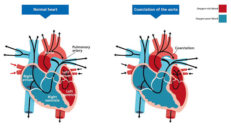
Coarctation of the Aorta (CoA) is a congenital cardiovascular anomaly characterized by a localized narrowing of the descending thoracic aorta, typically occurring distal to the origin of the left subclavian artery near the insertion of the ductus arteriosus (the juxtaductal region). This obstruction creates a pressure barrier, resulting in hypertension proximal to the narrowing and hypoperfusion distal to it. CoA accounts for approximately 5-8% of all congenital heart defects and necessitates systematic diagnosis and timely intervention to prevent life-threatening complications.
Types of Coarctation of the Aorta
CoA is primarily classified based on its anatomical location relative to the ductus arteriosus/ligamentum arteriosum, which dictates the severity of presentation and associated lesions.
A. Anatomical Classification
- Juxtaductal Coarctation: This is the most common form in both infants and adults, where the narrowing (usually a shelf-like inward projection) is positioned directly opposite the ductal ostium.
- Preductal (or Infantile) Coarctation: The narrowing occurs proximal to the ductus arteriosus. This form is often severe and dependent on a patent ductus arteriosus (PDA) for perfusion to the descending aorta and lower body. When the PDA closes, the infant rapidly decompensates, leading to circulatory shock. This type is frequently associated with other left-sided obstructive lesions, such as a hypoplastic left heart or mitral stenosis.
- Postductal (or Adult) Coarctation: The narrowing is located distal to the insertion point of the ductus arteriosus (or ligamentum arteriosum). This type often manifests later in life, as collateral circulation has typically developed, softening the acute hemodynamic consequence, though systemic hypertension remains a key feature.
B. Associated Cardiac Anomalies
CoA rarely occurs in isolation. The most common associated defect is a bicuspid aortic valve (BAV), present in over 80% of CoA patients. Other associated lesions include ventricular septal defects (VSD), patent ductus arteriosus (PDA), and mitral valve abnormalities. CoA is also frequently observed in genetic syndromes, particularly Turner Syndrome (45,XO).
Clinical Features and Presentation
The clinical manifestation of CoA is highly dependent on the patient’s age, the severity of the obstruction, and the functional status of the ductus arteriosus.
A. Critical Presentation in Neonates and Infants
When the ductus arteriosus closes (typically within the first week of life), infants with critical CoA experience rapid hemodynamic deterioration due to insufficient systemic blood flow.
- Symptoms: Signs of congestive heart failure (CHF) and cardiogenic shock, including lethargy, poor feeding, respiratory distress (tachypnea), and irritability.
- Physical Examination: Weak or absent lower extremity pulses (femoral, popliteal, dorsalis pedis). Differential cyanosis may be present if the coarctation is preductal and the PDA is supplying desaturated blood to the lower body. Evidence of metabolic acidosis and renal impairment is common in severe cases.
B. Presentation in Older Children and Adults
In patients with adequate collateral circulation, symptoms may be subtle or absent for years, with discovery often resulting from routine screening for hypertension.
- Symptoms:
- Hypertension-related: Headaches, epistaxis, dizziness, tinnitus.
- Hypoperfusion-related: Lower extremity claudication (pain, weakness, or fatigue in the legs during exercise) due to insufficient blood flow.
- Physical Examination (Hallmark Findings):
- Differential Blood Pressure: Systemic blood pressure measured in the upper extremities (brachial artery) is significantly higher than that in the lower extremities (popliteal artery). A difference exceeding 20 mmHg is highly suggestive of CoA.
- Radio-Femoral Delay: The femoral pulse is weaker and palpably delayed compared to the radial or brachial pulse due to the distance the blood must travel through the high-resistance collateral vessels.
- Auscultation: A systolic ejection murmur may be heard over the left scapula or upper back, reflecting flow through the narrowed segment. Continuous murmurs may arise from turbulent flow through large collateral arteries (e.g., intercostal arteries).
Investigations and Diagnosis
A combination of physical examination and specialized imaging is crucial for confirming the diagnosis, assessing severity, and planning intervention.
A. Physical Examination
The measurement of blood pressure in all four limbs and the assessment of pulse quality (radio-femoral delay) remain the cornerstone of initial diagnosis.
B. Electrocardiography (ECG)
- Neonates/Infants: May show signs of right ventricular (RV) volume or pressure overload, reflecting fetal circulation patterns or pulmonary hypertension.
- Older Patients: Typically demonstrates left ventricular hypertrophy (LVH) and left atrial enlargement, resulting from chronic systemic hypertension proximal to the narrowing.
C. Chest X-ray (CXR)
CXR findings are often diagnostic in older children and adults but may lag in young infants.
- Rib Notching: Pathognomonic finding caused by pressure erosion of the inferior borders of the posterior ribs (ribs 3-8) from enlarged, pulsating intercostal collateral arteries. This finding is usually not visible until after 5–10 years of age.
- Figure-3 Sign: An indentation visible in the aortic contour on a posteroanterior view. The upper arc represents the dilated ascending aorta and left subclavian artery, the middle constriction represents the coarctation site, and the lower arc represents post-stenotic dilation.
D. Echocardiography (ECHO)
Echocardiography is the initial non-invasive imaging modality of choice.
- Visual Confirmation: Allows direct visualization of the transverse aortic arch, the narrowing segment, and the presence of associated lesions (e.g., bicuspid aortic valve, VSD).
- Hemodynamic Assessment: Doppler techniques are used to measure the peak systolic pressure gradient across the coarctation site. A peak-to-peak systolic gradient exceeding 20 mmHg is generally considered hemodynamically significant, necessitating intervention.
E. Advanced Imaging (CT Angiography and Magnetic Resonance Imaging – MRI)
CT and MRI—particularly Magnetic Resonance Angiography (MRA)—are the gold standards for definitive anatomical assessment, especially prior to intervention.
- Pre-Intervention Mapping: These modalities provide precise 3D delineation of the coarctation length, diameter, and the extent of collateral circulation.
- Post-Repair Surveillance: They are vital for long-term follow-up to detect recoarctation, pseudoaneurysm formation, or aneurysmal dilation of the ascending aorta, which is a common late complication.
Complications of Coarctation of the Aorta
Complications arise from chronic upper-body hypertension and end-organ damage, or acute circulatory failure in infancy.
A. Acute Complications (Untreated Infants)
The closure of the PDA in newborns with critical CoA leads to:
- Cardiovascular collapse, shock, and death.
- Severe refractory congestive heart failure.
- Metabolic acidosis and renal failure due to poor distal perfusion.
B. Chronic Complications (Older Children and Adults)
- Systemic Hypertension: This is the most prevalent long-term issue. Even after successful repair, a significant percentage of patients develop residual or recurrent hypertension, often related to structural changes in the great arteries or persistent renin-angiotensin system activation.
- Aortic Dissection and Rupture: Chronic high pressure and increased shear stress in the ascending aorta and proximal arch can lead to medial degeneration, increasing the risk of aneurysm formation or catastrophic dissection, especially during strenuous activity.
- Cerebrovascular Events: High BP in the cerebral vasculature increases the risk of stroke and is strongly linked to the formation of Berry aneurysms in the Circle of Willis. Ruptured cerebral aneurysms are a major cause of death in unrepaired adults.
- Infective Endarteritis/Endocarditis: The turbulent flow across the narrowed segment or an associated bicuspid aortic valve predisposes the patient to bacterial colonization (infective endarteritis at the coarctation site, or endocarditis on the aortic valve).
5. Management of Coarctation of the Aorta
Management aims to relieve the obstruction, normalize blood pressure, and prevent irreversible cardiovascular and cerebral damage.
A. Immediate Medical Stabilization (Neonates)
For critically ill infants, stabilization is paramount:
- Prostaglandin E1 (PGE1) Infusion: This is essential to ensure survival. PGE1 maintains the patency of the ductus arteriosus, restoring blood flow to the descending aorta, mitigating shock, and stabilizing metabolic acidosis.
- Supportive Care: Diuretics (for pulmonary edema) and inotropic agents (to improve myocardial function) are used to manage severe CHF secondary to pressure overload.
- Blood Pressure Control: Gentle BP management may be necessary, although aggressive lowering must be avoided until the obstruction is relieved, as this can compromise vital organ perfusion.
B. Definitive Intervention
Intervention is indicated for a peak-to-peak gradient >20 mmHg, or if there is clinical evidence of hypertension or end-organ damage, regardless of the measured gradient.
1. Surgical Repair
Surgical intervention is generally the preferred approach for infants and young children, as it carries a lower risk of late aneurysm formation and recoarctation compared to balloon angioplasty in this age group.
- Resection and Primary End-to-End Anastomosis: Considered the procedure of choice. The constricted segment is removed, and the cut ends of the aorta are sewn together. This technique minimizes the risk of recoarctation and late aneurysm formation.
- Subclavian Flap Aortoplasty: Uses the left subclavian artery tissue to widen the coarctation segment. While effective, it sacrifices the subclavian artery, potentially causing growth restriction or weakness in the left arm.
2. Catheter-Based Intervention
Catheterization techniques are increasingly favored in older children, adolescents, and adults.
- Balloon Angioplasty: Involves inflating a specialized balloon catheter across the stenotic segment to tear the stricture. While effective for initial relief, it carries a higher risk of aneurysm formation or recoil if used in native CoA of infants. It is a highly effective treatment for recoarctation (recurrence after surgical repair).
- Stent Implantation: Currently the preferred method for CoA in older children, adolescents, and adults. Stents provide immediate relief, offer long-term scaffolding, and significantly reduce the risk of acute recoil and aneurysm formation compared to ballooning alone. They are typically oversized to allow for growth in younger patients.
C. Lifelong Follow-up
Post-intervention, patients require mandatory lifelong surveillance by a cardiologist specializing in Adult Congenital Heart Disease (ACHD). The focus of follow-up includes:
- Hypertension Management: Up to 30-50% of patients remain hypertensive, even years after successful repair, requiring pharmacological treatment.
- Surveillance for Recoarctation: Recurrence risk is highest in the first few years, but periodic imaging is required indefinitely.
- Aortic Surveillance: Regular MRI or CT angiography is necessary to monitor for ascending aortic dilation, aneurysm formation, or dissection, particularly given the high prevalence of associated bicuspid aortic valve disease.
References
- Doshi AR, Shirali GS. Coarctation of the aorta. Pediatric Clinics of North America. 2014;61(3):515-534.
- Moore, P. Coarctation of the aorta and interrupted aortic arch. In: Allen HD, Shaddy RE, Penny DJ, et al., eds. Moss and Adams’ Heart Disease in Infants, Children, and Adolescents: Including the Fetus and Young Adult. 9th ed. Wolters Kluwer; 2019:808-831.
- Valdes, AM, de la Cruz, MV. Coarctation of the aorta: Anatomical types and haemodynamic correlation. Archives of Disease in Childhood. 1971;46(248):495–506.
- Forbes TJ. Stent implantation for coarctation of the aorta. Catheterization and Cardiovascular Interventions. 2005;65(3):364-372.
- Vahanian A, Alfieri O, Andreotti F, et al. Guidelines on the management of valvular heart disease (version 2012): The Joint Task Force on the Management of Valvular Heart Disease of the European Society of Cardiology (ESC) and the European Association for Cardio-Thoracic Surgery (EACTS). European Heart Journal. 2012;33(19):2451–2496.


