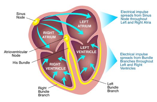
The human heart, a marvel of biological engineering, functions as a highly efficient pump, tirelessly circulating blood throughout the body. This relentless, rhythmic contraction is orchestrated by a sophisticated intrinsic electrical system, enabling the heart to generate its own impulses and propagate them in a precise, coordinated manner. Understanding the electrical activity of the heart is fundamental to comprehending its normal function and the various pathologies that can arise from electrical dysregulation.
Electrical Activity of the Heart: An Overview
The heart’s electrical activity originates in specialized pacemaker cells and propagates through a dedicated conduction system, culminating in the contraction of cardiac muscle cells (myocytes). This intricate system ensures that atrial contraction precedes ventricular contraction, optimizing blood flow. The primary components of this system include:
- Sinoatrial (SA) Node: Located in the right atrium, it is the heart’s natural pacemaker, possessing the highest intrinsic rate of spontaneous depolarization.
- Atrioventricular (AV) Node: Situated between the atria and ventricles, it delays the electrical impulse, allowing for complete ventricular filling before contraction.
- Bundle of His (AV Bundle): Extends from the AV node into the interventricular septum.
- Bundle Branches: Divide into right and left branches, conducting impulses to the respective ventricles.
- Purkinje Fibers: A network of specialized conducting fibers that rapidly distribute the electrical signal throughout the ventricular myocardium, ensuring synchronized contraction.
The electrical signals that drive contraction are known as action potentials, which are transient changes in the membrane potential of excitable cells. In the heart, these action potentials vary significantly depending on the cell type, dictating their unique roles in impulse generation and conduction.
Two Types of Action Potential in the Heart Muscle
Cardiac muscle cells exhibit two primary types of action potentials: the fast response action potential and the slow response action potential. These differ in their ionic mechanisms, morphology, and physiological roles.
1. Fast Response Action Potential
The fast response action potential is characteristic of ventricular and atrial myocytes, as well as Purkinje fibers. These cells have a stable resting membrane potential and are responsible for rapid, robust conduction and contraction. The fast action potential is typically divided into five phases (Phase 4, 0, 1, 2, 3):
- Phase 4: Resting Membrane Potential (-90 mV): This is a stable phase in which the cell maintains a negative charge across its membrane. It is primarily determined by the high permeability of the membrane to potassium ions (K+) through inward rectifying K+ channels (IK1), allowing K+ to leak out of the cell. The Na+/K+ ATPase pump actively maintains the ionic gradients by extruding 3 Na+ ions for every 2 K+ ions brought into the cell.
- Phase 0: Rapid Depolarization (Upstroke): Upon reaching a threshold potential (around -70 mV), voltage-gated fast sodium channels (Nav1.5) open rapidly, leading to a massive influx of Na+ ions into the cell. This causes a swift and steep depolarization, pushing the membrane potential to positive values (+20 to +30 mV). This phase is crucial for rapid signal propagation.
- Phase 1: Initial Repolarization: Shortly after depolarization, the fast Na+ channels inactivate, halting Na+ influx. Simultaneously, transient outward K+ channels (Ito) open, allowing a brief efflux of K+ ions. This combination leads to a transient, partial repolarization of the membrane.
- Phase 2: Plateau Phase: This is a unique and prolonged phase in cardiac action potentials, critical for extending the refractory period and preventing tetanic contractions. It is maintained by a delicate balance between the inward current of calcium ions (Ca2+) through L-type voltage-gated calcium channels (ICa,L) and the outward current of K+ ions through delayed rectifier K+ channels (IKr, IKs). The influx of Ca2+ also triggers calcium-induced calcium release from the sarcoplasmic reticulum, initiating muscle contraction.
- Phase 3: Rapid Repolarization: The L-type Ca2+ channels gradually inactivate, reducing the inward Ca2+ current. Concurrently, the delayed rectifier K+ channels (IKr, IKs) become fully active, leading to a dominant efflux of K+ ions. This causes the membrane potential to rapidly return to its resting negative state (Phase 4), preparing the cell for the next excitation.
The prolonged plateau phase (Phase 2) results in an extended absolute refractory period, during which the cardiac muscle cannot be re-excited, preventing sustained, tetanic contractions that would impair the heart’s pumping ability.
2. Slow Response Action Potential
The slow response action potential is characteristic of the pacemaker cells in the SA node and AV node. Unlike fast response cells, these cells do not have a stable resting membrane potential; instead, they exhibit spontaneous, gradual depolarization, known as the pacemaker potential. The slow action potential is typically described with three main phases:
- Phase 4: Pacemaker Potential (Slow Depolarization): This is the most crucial phase, where the membrane potential gradually depolarizes from its maximum diastolic potential (around -60 mV). This spontaneous depolarization is due to a net inward current primarily generated by three mechanisms:
- Funny Current (If): A unique, mixed Na+/K+ inward current activated by hyperpolarization at the end of the preceding action potential.
- T-type Calcium Channels (ICa,T): Transient, voltage-gated Ca2+ channels that open as the membrane potential approaches the threshold, contributing to the final depolarization.
- Decreased Potassium Efflux: As the cell repolarizes, the outward K+ current gradually decreases, further contributing to the net inward current.
- Phase 0: Depolarization (Upstroke): When the pacemaker potential reaches the threshold (around -40 mV), voltage-gated L-type Ca2+ channels (ICa,L) open, leading to an influx of Ca2+ ions. This causes the depolarization of the cell. Notably, this upstroke is slower and less steep than in fast response cells because it is carried by Ca2+ rather than the faster Na+ influx.
- Phase 3: Repolarization: This phase is primarily mediated by the efflux of K+ ions through voltage-gated delayed rectifier K+ channels (IKr, IKs) that open during the upstroke. As K+ leaves the cell, the membrane potential returns to its most negative value (maximum diastolic potential), which then triggers the activation of the funny current, initiating the next pacemaker potential.
The slow response action potential is responsible for the heart’s automaticity and rhythmic beating. Its slower conduction velocity in the AV node is vital for ensuring ventricular filling.
Genesis of Pacemaker Potential at the SA Node
The SA node is the heart’s natural pacemaker due to its unique ability to spontaneously depolarize and generate action potentials at the fastest rate. The genesis of this pacemaker potential (Phase 4 of the slow response action potential) is a complex interplay of several ion currents, collectively resulting in a gradual rise of the membrane potential towards a threshold.
The primary mechanisms contributing to the spontaneous depolarization of SA nodal cells are:
- Activation of the “Funny Current” (If): This is the most important and unique current contributing to pacemaker activity. The “funny” channels, also known as hyperpolarization-activated cyclic nucleotide-gated (HCN) channels, are permeable to both Na+ and K+ ions. They are activated when the membrane potential hyperpolarizes to its maximum diastolic potential (around -60 mV) at the end of repolarization. As the channels open, there is a net inward current, predominantly of Na+, which slowly depolarizes the cell. The name “funny” comes from its unusual activation by hyperpolarization and its mixed Na+/K+ permeability.
- Deactivation of Voltage-Gated Potassium Channels: As the SA nodal cell repolarizes, voltage-gated K+ channels that were open during Phase 3 begin to close. This reduction in the outward K+ current further contributes to the net inward current, allowing the membrane potential to rise. The decrease in K+ permeability prevents the cell from maintaining a stable resting potential.
- Opening of T-type Calcium Channels (ICa,T): As the membrane potential slowly depolarizes due to the funny current and decreasing K+ efflux, it eventually reaches a point (around -50 mV) where transient, voltage-gated T-type Ca2+ channels open. The influx of Ca2+ through these channels provides an additional depolarizing current, accelerating the rise of the membrane potential towards the threshold for the L-type Ca2+ channels.
- Opening of L-type Calcium Channels (ICa,L): Once the membrane potential reaches the threshold (around -40 mV), the long-lasting, voltage-gated L-type Ca2+ channels open. The large influx of Ca2+ through these channels rapidly depolarizes the cell, initiating Phase 0 of the action potential.
The rhythmic interplay of these currents ensures that once an action potential has repolarized, the SA nodal cell immediately begins to depolarize again, setting the pace for the entire heart.
Effects of Vagal and Sympathetic Stimulations on the Pacemaker Potential
The heart’s intrinsic rate, dictated by the SA node, is constantly modulated by the autonomic nervous system to meet the body’s changing physiological demands. The two branches of the autonomic nervous system—parasympathetic (vagal) and sympathetic—exert opposing effects on the pacemaker potential, thereby regulating heart rate.
1. Vagal (Parasympathetic) Stimulation
The parasympathetic nervous system primarily influences the heart via the vagus nerve, which releases the neurotransmitter acetylcholine (ACh). ACh binds to muscarinic M2 receptors on SA nodal cells, leading to a cascade of events that decrease heart rate (negative chronotropy):
- Increased K+ Conductance (Hyperpolarization): Binding of ACh to M2 receptors activates a G-protein (Gi) that opens acetylcholine-sensitive inwardly rectifying K+ channels (IK,ACh). This increases the efflux of K+ ions, leading to hyperpolarization of the SA nodal cell membrane (making it more negative). This means the pacemaker potential must start from a more negative maximum diastolic potential, requiring a longer time to reach threshold.
- Decreased Rate of Phase 4 Depolarization: The Gi protein activated by M2 receptors also inhibits adenylyl cyclase, reducing the production of cyclic AMP (cAMP). Since the funny current (If) is modulated by cAMP (cAMP directly binds to HCN channels, increasing their opening probability), a decrease in cAMP reduces the activity of If channels. This slows down the rate of the initial spontaneous depolarization in Phase 4, prolonging the time to reach threshold.
- Decreased L-type Ca2+ Current: ACh also directly or indirectly reduces the activity of L-type Ca2+ channels, which raises the threshold potential that the pacemaker potential needs to reach to trigger an action potential.
The combined effect of these actions is a reduced heart rate. By hyperpolarizing the cell, slowing the rate of depolarization, and increasing the threshold, vagal stimulation prolongs the interval between successive action potentials.
2. Sympathetic Stimulation
The sympathetic nervous system augments heart rate and contractility, primarily via the release of norepinephrine (NE) from sympathetic nerve terminals and epinephrine (Epi) from the adrenal medulla. NE and Epi bind to beta-1 (β1) adrenergic receptors on SA nodal cells, initiating a signaling pathway that increases heart rate (positive chronotropy):
- Increased Rate of Phase 4 Depolarization: Binding of NE/Epi to β1 receptors activates a G-protein (Gs), which stimulates adenylyl cyclase. This increases intracellular cAMP levels. cAMP directly binds to and activates the funny channels (HCN channels), increasing the inward funny current (If). This accelerates the rate of spontaneous depolarization in Phase 4, causing the membrane potential to reach threshold more quickly.
- Increased L-type Ca2+ Current (Lowered Threshold and Faster Upstroke): Increased cAMP also enhances the activity of L-type Ca2+ channels. This has two effects: firstly, it lowers the threshold potential for action potential generation, meaning the cell reaches threshold sooner. Secondly, it contributes to a faster and more robust Phase 0 depolarization.
- Increased T-type Ca2+ Current: Sympathetic stimulation also enhances the activity of T-type Ca2+ channels, further contributing to the rapid depolarization towards the action potential threshold.
The combined effect of these actions is an increased heart rate. By making the maximum diastolic potential slightly less negative, speeding up the rate of depolarization in Phase 4, and lowering the threshold potential, sympathetic stimulation shortens the interval between successive action potentials.
Conclusion
The electrical activity of the heart is a sophisticated and highly regulated process, foundational to its role as an effective pump. The distinct ionic mechanisms underlying fast and slow action potentials allow cardiac cells to perform specialized functions—from rapid conduction and powerful contraction (fast response) to spontaneous rhythm generation (slow response). The SA node’s inherent automaticity, driven by the unique funny current and other ion channels, establishes the heart’s intrinsic rhythm. This rhythm is, in turn, exquisitely modulated by the autonomic nervous system. Vagal stimulation slows the heart by hyperpolarizing cells and decreasing depolarization rates, while sympathetic stimulation accelerates the heart by enhancing depolarizing currents. A comprehensive understanding of these electrophysiological principles is indispensable for diagnosing and treating cardiac rhythm disorders and for appreciating the intricate regulatory mechanisms that maintain cardiovascular homeostasis.
References
- Boron, W. F., & Boulpaep, E. L. (2017). Medical Physiology (3rd ed.). Elsevier. (This is a comprehensive textbook covering cellular and organ system physiology, including detailed sections on cardiac electrophysiology.)
- Guyton, A. C., & Hall, J. E. (2021). Textbook of Medical Physiology (14th ed.). Elsevier. (Another foundational medical physiology textbook with extensive coverage of cardiac muscle and electrophysiology.)
- Katz, A. M. (2011). Physiology of the Heart (5th ed.). Lippincott Williams & Wilkins. (A specialized textbook focused entirely on cardiovascular physiology, providing in-depth explanations of action potentials and their regulation.)
- Rhoades, R. A., & Bell, D. R. (2017). Medical Physiology: Principles for Clinical Medicine (5th ed.). Elsevier. (Offers a clinically oriented perspective on physiological principles, including cardiac electrical activity.)
- Braunwald, E., & Zipes, D. P. (Eds.). (2019). Braunwald’s Heart Disease: A Textbook of Cardiovascular Medicine (11th ed.). Elsevier. (While a clinical cardiology textbook, it contains robust foundational chapters on basic cardiac physiology and electrophysiology.)


