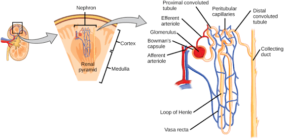
The kidneys play a pivotal role in maintaining the body’s internal environment through a complex interplay of filtration, reabsorption, and secretion. While glomerular filtration produces a large volume of filtrate, the vast majority of its useful components are reclaimed, and waste products are actively removed, ensuring precise control over fluid volume, electrolyte balance, and acid-base homeostasis. This intricate process is primarily orchestrated within the renal tubules, where finely tuned mechanisms regulate the movement of substances between the tubular lumen and the peritubular capillaries.
Regulation of Tubular Reabsorption and Secretion
The regulation of tubular reabsorption and secretion is a sophisticated process involving hormonal, neural, and intrinsic mechanisms, allowing the kidneys to adapt rapidly to changing physiological demands.
1. Hormonal Regulation:
- Aldosterone: Released from the adrenal cortex in response to angiotensin II or high plasma potassium, aldosterone primarily acts on the principal cells of the collecting ducts. It upregulates the synthesis and activity of epithelial sodium channels (ENaC) on the apical membrane and Na+/K+ ATPase pumps on the basolateral membrane, leading to increased sodium reabsorption and concomitant potassium secretion. This action is crucial for maintaining blood volume and blood pressure.
- Antidiuretic Hormone (ADH) / Vasopressin: Produced by the hypothalamus and released by the posterior pituitary, ADH regulates water reabsorption in the collecting ducts and distal tubules. It binds to V2 receptors on the basolateral membrane of principal cells, triggering the insertion of aquaporin-2 (AQP2) water channels into the apical membrane. This makes these segments permeable to water, allowing water to be reabsorbed down its osmotic gradient into the hypertonic medullary interstitium, thereby concentrating the urine and conserving body water.
- Parathyroid Hormone (PTH): Secreted by the parathyroid glands in response to low plasma calcium, PTH enhances calcium reabsorption in the distal tubules and collecting ducts. It also inhibits phosphate reabsorption in the proximal tubule, promoting phosphate excretion. This dual action helps restore normal calcium levels while preventing calcium-phosphate precipitation.
- Angiotensin II: A potent vasoconstrictor, Angiotensin II is formed in response to renin release from the juxtaglomerular apparatus. Beyond its systemic effects, it directly stimulates sodium and water reabsorption in the proximal tubule, loop of Henle, and collecting duct. It also triggers aldosterone release, further enhancing sodium reabsorption. Its primary role is to increase blood volume and pressure.
- Atrial Natriuretic Peptide (ANP): Released by atrial myocytes in response to atrial distension (high blood volume), ANP antagonizes the effects of Angiotensin II and aldosterone. It inhibits sodium reabsorption in the collecting duct, increases glomerular filtration rate (GFR), and reduces renin and ADH secretion, leading to increased sodium and water excretion (natriuresis and diuresis) and a reduction in blood volume and pressure.
2. Autoregulation / Intrinsic Mechanisms:
- Glomerulotubular Balance: This intrinsic mechanism ensures that a relatively constant fraction (about 65-70%) of the filtered sodium and water is reabsorbed in the proximal tubule, regardless of variations in GFR. If GFR increases, the amount of filtered fluid increases, but the reabsorption rate in the proximal tubule also proportionately increases, preventing excessive load on the downstream segments.
- Tubuloglomerular Feedback (TGF): The macula densa cells in the juxtaglomerular apparatus sense the sodium chloride concentration in the tubular fluid at the end of the thick ascending limb. If NaCl delivery to the macula densa increases (indicating a high GFR), the macula densa releases vasoconstrictors (e.g., adenosine), leading to afferent arteriolar constriction and a decrease in GFR, thereby protecting downstream tubules from excessive flow.
3. Neural Regulation:
- Sympathetic Nervous System: Activation of renal sympathetic nerves primarily leads to vasoconstriction of afferent and efferent arterioles, reducing GFR. However, moderate sympathetic stimulation also directly enhances sodium reabsorption in the proximal tubule, thick ascending limb, and collecting duct, likely through alpha-1 receptors on tubular epithelial cells, contributing to fluid conservation in situations like hypovolemia.
Transport Maximum (Tm), Renal Plasma Threshold, and Splay
These concepts are critical for understanding how the kidneys handle substances that are reabsorbed or secreted via carrier-mediated transport systems.
- Transport Maximum (Tm): The transport maximum refers to the maximum rate at which a substance can be actively reabsorbed from the tubular lumen or secreted into it, once all the transport proteins responsible for that substance are fully saturated. This occurs because there is a finite number of carrier proteins available. For example, glucose is reabsorbed by specific transporters (SGLTs) in the proximal tubule. If the filtered load of glucose (plasma glucose concentration × GFR) exceeds the Tm for glucose, the excess glucose cannot be reabsorbed and will appear in the urine.
- Renal Plasma Threshold: The renal plasma threshold is the plasma concentration of a substance at which that substance first begins to appear in the urine. For substances like glucose, which are normally completely reabsorbed, the renal plasma threshold is the plasma concentration at which the filtered load just exceeds the combined reabsorptive capacity of the nephrons, leading to its excretion. In a healthy individual, the glucose threshold is typically around 180-200 mg/dL (10-11.1 mmol/L). Below this threshold, all filtered glucose is reabsorbed; above it, glucose spills into the urine (glucosuria).
- Splay: Splay describes the phenomenon where a substance, such as glucose, begins to appear in the urine even before the overall renal plasma threshold is reached, or before the theoretical Tm of all nephrons is saturated. This “spread” or “splay” in the reabsorption curve is primarily due to several factors:
- Nephron Heterogeneity: Not all nephrons have identical GFRs or the same number of specific transporters, meaning their individual reabsorptive capacities vary slightly. Some nephrons will reach their Tm earlier than others.
- Uneven Delivery: The distribution of filtered load to different nephrons can be slightly uneven.
- Kinetic Factors: It’s a continuous process, not a sharp switch, so some transporters may leak or be less efficient at very high concentrations. This means there’s a gradual increase in urinary glucose excretion as plasma glucose rises above a certain level, rather than an abrupt appearance once a single, sharp threshold is crossed. This “splay” is clinically relevant as it explains why some glucosuria might be observed at plasma glucose levels slightly below the theoretical Tm.
Mode of Reabsorption of Different Substances
Reabsorption mechanisms vary widely, employing both active and passive transport, often coupled in complex ways. Key substances include:
- Sodium (Na+): Sodium reabsorption is paramount, driving the reabsorption of many other solutes and water.
- Proximal Tubule: Approximately 65-70% of filtered Na+ is reabsorbed. On the apical membrane, Na+ enters the cell downhill via secondary active transport, often coupled with H+ (Na+/H+ antiporter, NHE3) or with glucose and amino acids (SGLTs). On the basolateral membrane, Na+ is actively pumped out of the cell into the interstitial fluid by the Na+/K+ ATPase, creating a low intracellular Na+ concentration that drives apical entry.
- Loop of Henle (Thick Ascending Limb): About 25% of filtered Na+ is reabsorbed. Here, the Na+-K+-2Cl- cotransporter (NKCC2) on the apical membrane moves Na+, K+, and 2Cl- into the cell. Na+ is then actively pumped out basolaterally by Na+/K+ ATPase.
- Distal Tubule (Early): Approximately 5% of filtered Na+ is reabsorbed via the Na+-Cl- cotransporter (NCC) on the apical membrane, with Na+ exiting via Na+/K+ ATPase basolaterally.
- Collecting Duct (Principal Cells): The remaining 1-3% of Na+ is reabsorbed through epithelial Na+ channels (ENaC) on the apical membrane, exiting via Na+/K+ ATPase basolaterally. This segment is under tight hormonal control by aldosterone.
- Potassium (K+): K+ handling is complex, involving both reabsorption and secretion.
- Proximal Tubule: Roughly 65-70% of filtered K+ is passively reabsorbed, primarily paracellularly, driven by solvent drag and K+ concentration gradients.
- Loop of Henle (Thick Ascending Limb): Another 20-25% of filtered K+ is reabsorbed via the NKCC2 cotransporter on the apical membrane. Some K+ also recycles back into the lumen via ROMK channels, creating a positive lumen potential that drives paracellular cation reabsorption.
- Distal Tubule/Collecting Duct: These segments are the primary sites of K+ secretion, but also have mechanisms for reabsorption. In alpha-intercalated cells, K+ can be reabsorbed via an H+/K+ ATPase on the apical membrane, particularly in states of K+ depletion.
- Chloride (Cl-): Chloride reabsorption largely follows sodium reabsorption to maintain electroneutrality.
- Proximal Tubule: Primarily reabsorbed paracellularly, driven by the electrochemical gradient created by Na+ reabsorption and solvent drag. Some transcellular reabsorption occurs via Cl-/anion antiporters and Cl- channels.
- Loop of Henle (Thick Ascending Limb): Reabsorbed via the NKCC2 cotransporter and paracellularly.
- Distal Tubule (Early): Reabsorbed via the NCC cotransporter.
- Glucose: Normally, 100% of filtered glucose is reabsorbed in the proximal tubule.
- Proximal Tubule (S1 and S2 segments): Glucose is transported from the lumen into the tubular cells against its concentration gradient by secondary active transport, coupled with Na+. Two main sodium-glucose cotransporters (SGLTs) are involved: SGLT2 (low affinity, high capacity) in the early proximal tubule, and SGLT1 (high affinity, low capacity) in the later segments. Once inside the cell, glucose exits across the basolateral membrane into the interstitial fluid by facilitated diffusion via glucose transporters (GLUT2).
- Urea: Urea reabsorption is crucial for establishing the medullary osmotic gradient.
- Proximal Tubule: Approximately 50% of filtered urea is passively reabsorbed down its concentration gradient as water is reabsorbed.
- Loop of Henle (Descending Thin Limb, Inner Medullary Collecting Duct): Urea is secreted into the descending thin limb and then reabsorbed in the inner medullary collecting duct (IMCD) via specific urea transporters (UT-A1 and UT-A3). This recycling contributes significantly to the hypertonicity of the renal medulla, which is essential for concentrated urine formation.
- Water: Water reabsorption is either obligatory or facultative.
- Proximal Tubule: Approximately 65-70% of filtered water is obligatorily reabsorbed via aquaporin-1 (AQP1) channels, following the osmotic gradient created by solute (primarily Na+) reabsorption.
- Loop of Henle (Descending Limb): Highly permeable to water via AQP1, allowing water to move into the hypertonic medulla. The ascending limb is virtually impermeable to water.
- Distal Tubule and Collecting Duct: Water reabsorption here is facultative and regulated by ADH. In the presence of ADH, aquaporin-2 (AQP2) channels are inserted into the apical membrane, making these segments permeable to water, allowing water to be reabsorbed and urine to be concentrated.
Mode of Secretion of Different Substances
Tubular secretion allows the kidneys to add substances to the tubular fluid, playing a vital role in eliminating waste products and regulating ion balance.
- Potassium (K+): The primary site of K+ secretion is the principal cells of the distal tubule and collecting duct.
- Mechanism: On the basolateral membrane, the Na+/K+ ATPase actively pumps K+ into the cell, creating a high intracellular K+ concentration. On the apical membrane, K+ then passively diffuses out of the cell into the tubular lumen through potassium channels, primarily ROMK (Renal Outer Medullary K+) channels and, to a lesser extent, BK (Big Conductance K+) channels. This process is stimulated by aldosterone, which increases the activity of both Na+/K+ ATPase and apical K+ channels, and by high tubular flow rates.
- Hydrogen Ions (H+): H+ secretion is critical for acid-base balance.
- Proximal Tubule: H+ is secreted primarily via the Na+/H+ antiporter (NHE3) on the apical membrane, exchanging intracellular H+ for luminal Na+. This H+ combines with filtered bicarbonate to form carbonic acid, which then dissociates, allowing for bicarbonate reabsorption.
- Distal Tubule and Collecting Duct (Alpha-Intercalated Cells): These cells are the main sites for direct H+ secretion and acidification of the urine. They possess an H+ ATPase pump and an H+/K+ ATPase pump on their apical membrane, which actively transport H+ into the lumen. Simultaneously, a Cl-/HCO3- exchanger on the basolateral membrane moves bicarbonate into the blood, reabsorbing it.
- Organic Ions (Drugs, Toxins): The proximal tubule is a major site for the secretion of a wide variety of organic anions and cations, including many drugs, metabolites, and toxins.
- Organic Anions: Organic Anion Transporters (OATs), located on the basolateral membrane, mediate the uptake of organic anions (e.g., penicillin, uric acid, bile salts, salicylates, para-aminohippurate [PAH]) from the peritubular capillaries into the tubular cells. From the cell, these anions are then secreted into the lumen across the apical membrane, often via secondary active transport mechanisms or specific efflux pumps (e.g., MRPs – Multidrug Resistance-associated Proteins).
- Organic Cations: Organic Cation Transporters (OCTs), also located on the basolateral membrane, facilitate the uptake of organic cations (e.g., creatinine, dopamine, quinine, atropine) from the blood into the tubular cells. These are then secreted into the lumen across the apical membrane, often by H+/organic cation antiporters. These systems are non-specific and can transport a broad range of structurally diverse compounds, making them crucial for drug elimination.
In conclusion, the renal tubules employ a remarkable array of transport mechanisms and regulatory pathways to precisely control the composition of the body’s internal fluids. This sophisticated integration of reabsorption and secretion, governed by hormones, nerves, and intrinsic controls, ensures the maintenance of homeostasis, highlighting the kidneys’ indispensable role in physiological function and overall health.
References:
- Guyton, A. C., & Hall, J. E. (2021). Guyton and Hall Textbook of Medical Physiology (14th ed.). Elsevier.
- Boron, W. F., & Boulpaep, E. L. (2017). Medical Physiology (3rd ed.). Elsevier.
- Ganong, W. F. (2019). Ganong’s Review of Medical Physiology (26th ed.). McGraw-Hill Education.


