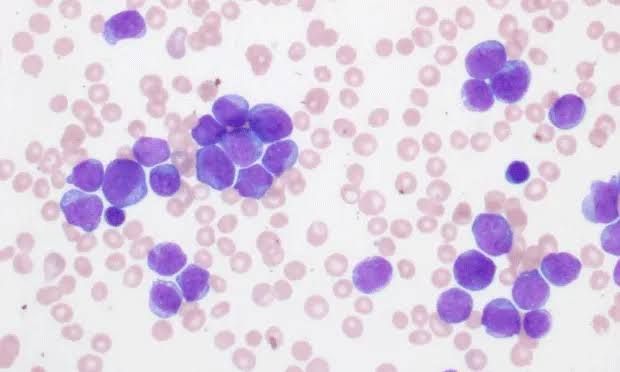
Acute Lymphoblastic Leukemia (ALL) is an aggressive hematologic malignancy characterized by the uncontrolled proliferation and accumulation of immature lymphoid cells, known as lymphoblasts, in the bone marrow and peripheral blood. A critical understanding of its pathogenesis, diverse clinical features, and comprehensive laboratory diagnostic approach is paramount for timely intervention and effective management.
Understanding Acute Lymphoblastic Leukemia: Pathogenesis, Clinical Features, and Laboratory Diagnosis
Acute Lymphoblastic Leukemia (ALL) represents a heterogeneous group of cancers originating from the malignant transformation of lymphoid progenitor cells. Predominantly affecting children, ALL also occurs in adults, exhibiting distinct biological and clinical characteristics across age groups. Its rapid progression necessitates prompt diagnosis and aggressive treatment.
1. Pathogenesis of Acute Lymphoblastic Leukemia
The pathogenesis of ALL is complex, stemming from a multi-step process involving specific genetic and epigenetic alterations within a lymphoid progenitor cell, leading to its uncontrolled proliferation, impaired differentiation, and survival advantage. This transformation results in the accumulation of malignant lymphoblasts in the bone marrow, peripheral blood, and frequently in extramedullary sites.
1.1. Cellular Origin and Malignant Transformation: ALL originates from an aberrant lymphoid progenitor cell (lymphoblast) arrested at an early stage of differentiation. Depending on the lineage of the transformed cell, ALL is broadly classified into B-cell ALL (B-ALL), accounting for approximately 85% of cases, and T-cell ALL (T-ALL), comprising about 15%. This malignant transformation is initiated by somatic mutations or chromosomal rearrangements that confer a proliferative advantage and block normal lymphoid maturation.
1.2. Genetic Alterations and Molecular Lesions: The hallmark of ALL pathogenesis is the presence of diverse chromosomal abnormalities and gene mutations that drive leukemogenesis. These genetic lesions are crucial for classification, risk stratification, and therapeutic targeting.
- Chromosomal Translocations: These are common and often prognostically significant.
- t(12;21)(p13;q22) / ETV6-RUNX1 (TEL-AML1): The most common translocation in pediatric B-ALL. It is associated with a good prognosis.
- t(9;22)(q34;q11) / BCR-ABL1: Philadelphia chromosome (Ph+) ALL. More common in adults, associated with high-risk disease and requires targeted therapy with tyrosine kinase inhibitors (TKIs).
- t(4;11)(q21;q23) / KMT2A-AFF1 (MLL-AF4): Observed in infants and high-risk adult B-ALL, associated with a very poor prognosis.
- t(1;19)(q23;p13) / TCF3-PBX1 (E2A-PBX1): Found in both pediatric and adult B-ALL, confers an intermediate to adverse prognosis.
- Intrachromosomal amplification of chromosome 21 (iAMP21): Associated with a poor prognosis, particularly in children.
- Aneuploidies:
- Hyperdiploidy: Presence of more than 50 chromosomes (e.g., trisomies 4, 10, 17) is a favorable prognostic factor, especially in children.
- Hypodiploidy: Less than 44 chromosomes, associated with a very poor prognosis.
- Gene Mutations: Next-generation sequencing has identified numerous recurrent mutations.
- IKZF1 deletions: Associated with Ph-like ALL and poor prognosis, especially in BCR-ABL1 positive ALL.
- NOTCH1 mutations: Frequent in T-ALL, often associated with a better response to therapy.
- TP53 mutations: Generally associated with high-risk disease and resistance to treatment.
- RAS pathway mutations (KRAS, NRAS, PTPN11): Common across various ALL subtypes, contributing to cell proliferation.
- JAK-STAT pathway mutations (JAK1, JAK2, STAT5B): Particularly in Ph-like ALL and T-ALL, leading to constitutive activation of signaling pathways.
1.3. Epigenetic Modifications: Beyond genetic mutations, epigenetic alterations such as DNA methylation and histone modifications play a significant role. Abnormal methylation patterns can silence tumor suppressor genes or activate oncogenes, further driving leukemic transformation and progression.
1.4. Microenvironment and Predisposing Factors: The bone marrow microenvironment, including stromal cells, cytokines, and growth factors, can support the survival and proliferation of leukemic blasts. Additionally, certain inherited conditions increase the risk of ALL:
- Down syndrome (Trisomy 21): Children with Down syndrome have a 10-20 times higher risk of developing ALL.
- Li-Fraumeni syndrome: Associated with TP53 germline mutations.
- Ataxia-telangiectasia: Due to ATM gene mutations.
- Fanconi anemia: A bone marrow failure syndrome. Environmental factors, though less clearly defined, are also hypothesized to play a role (e.g., exposure to certain chemicals or radiation, viral infections).
In essence, ALL pathogenesis is a culmination of initial genetic insults leading to a clonal expansion of immature lymphocytes, which then accumulate further genetic and epigenetic changes, ultimately overwhelming normal hematopoiesis.
2. Clinical Features of Acute Lymphoblastic Leukemia
The clinical presentation of ALL is diverse, often reflecting the consequences of bone marrow failure and infiltration of leukemic cells into various organs. Symptoms typically have a rapid onset, progressing over days to weeks.
2.1. Symptoms due to Bone Marrow Failure: The rapid proliferation of lymphoblasts in the bone marrow suppresses normal hematopoiesis, leading to:
- Anemia (Lack of Red Blood Cells):
- Fatigue, weakness, pallor: Due to reduced oxygen-carrying capacity.
- Dyspnea (shortness of breath): Especially on exertion.
- Tachycardia: Compensatory increase in heart rate.
- Thrombocytopenia (Low Platelet Count):
- Petechiae and Purpura: Small pinpoint and larger bruise-like hemorrhages on the skin and mucous membranes.
- Easy bruising: Minor trauma leading to significant bruising.
- Epistaxis (nosebleeds), gingival bleeding: Spontaneous bleeding from mucous membranes.
- Menorrhagia: Heavy or prolonged menstrual bleeding in females.
- Neutropenia (Low Neutrophil Count):
- Fever of unknown origin: Often the first sign, due to impaired immune response.
- Recurrent or severe infections: Bacterial, fungal, or viral infections, often in unusual sites or with atypical severity.
2.2. Symptoms due to Extramedullary Infiltration: Leukemic cells can infiltrate various organs beyond the bone marrow, causing specific symptoms:
- Bone and Joint Pain:
- Approximately 25% of patients, especially children, experience bone or joint pain (arthralgia, ostealgia). This is due to direct leukemic infiltration into the periosteum or expanding bone marrow space, particularly in long bones, vertebrae, and joints. It can mimic juvenile idiopathic arthritis.
- Lymphadenopathy:
- Enlarged, non-tender lymph nodes are common, often in the cervical, axillary, and inguinal regions.
- Hepatosplenomegaly:
- Enlargement of the liver (hepatomegaly) and spleen (splenomegaly) is frequent, leading to abdominal fullness or discomfort.
- Central Nervous System (CNS) Involvement:
- Occurs in up to 5-10% of patients at diagnosis, more common in T-ALL and specific genetic subtypes (e.g., KMT2A-rearranged ALL).
- Symptoms include headache, nausea, vomiting, lethargy, irritability, nuchal rigidity (stiff neck), and cranial nerve palsies (e.g., facial nerve palsy, visual disturbances due to optic nerve infiltration).
- Can also manifest as seizures or altered mental status.
- Mediastinal Mass (More common in T-ALL):
- Enlarged thymus or lymph nodes in the mediastinum can lead to cough, dyspnea, chest pain, and superior vena cava (SVC) syndrome (facial swelling, plethora, engorged neck veins) due to compression of the SVC.
- Testicular Involvement (More common in B-ALL):
- Painless, firm enlargement of one or both testicles. This can be a site of relapse if not adequately treated.
- Skin Lesions (Leukemia Cutis):
- Less common, presenting as nodules, papules, or plaques, often reddish-brown or purple.
- Renal Infiltration:
- Rarely causes renal dysfunction but can lead to kidney enlargement.
2.3. General Symptoms:
- Weight loss, anorexia, and malaise: Non-specific symptoms reflecting the systemic nature of the disease and high metabolic turnover.
2.4. Age-Specific Presentations:
- Children: Peak incidence between 2-5 years. Often present with fever, pallor, fatigue, bruising, and bone pain.
- Adults: Bimodal distribution with another peak in older adults. Adults tend to have more aggressive subtypes and a higher incidence of Ph+ ALL.
The rapid onset and systemic nature of symptoms often necessitate prompt medical attention, leading to diagnosis.
3. Laboratory Diagnosis of Acute Lymphoblastic Leukemia
The diagnosis of ALL requires a multi-faceted approach, integrating findings from peripheral blood, bone marrow, and specialized molecular and cytogenetic studies. The goal is not only to confirm the diagnosis but also to classify the subtype, assess prognostic risk factors, and guide therapeutic decisions.
3.1. Initial Peripheral Blood Work:
- Complete Blood Count (CBC) with Differential:
- White Blood Cell (WBC) Count: Can be low, normal, or markedly elevated (leukocytosis, often >50,000/µL, sometimes >100,000/µL), but the key finding is the presence of immature cells.
- Anemia: Typically normocytic, normochromic.
- Thrombocytopenia: Universally present.
- Neutropenia: Often present, increasing infection risk.
- Peripheral Blood Smear:
- Crucial for initial suspicion. Reveals circulating blasts (lymphoblasts), which are immature lymphoid cells. These blasts are typically larger than mature lymphocytes, have a high nuclear-to-cytoplasmic ratio, fine chromatin, and often prominent nucleoli. They generally lack granules. Distinguishing blasts from mature lymphocytes or myeloid blasts is fundamental.
- Blood Chemistry:
- Lactate Dehydrogenase (LDH): Often elevated due to high cell turnover.
- Uric Acid: May be elevated (hyperuricemia) due to rapid purine catabolism from dying leukemic cells, risking tumor lysis syndrome.
- Electrolytes: May show imbalances (e.g., hyperkalemia, hyperphosphatemia, hypocalcemia) in cases of spontaneous tumor lysis syndrome.
- Liver and Renal Function tests: To assess organ involvement or baseline function.
3.2. Bone Marrow Aspiration and Biopsy (Gold Standard):
This is the definitive diagnostic procedure.
- Morphology:
- Bone marrow is typically hypercellular (packed with cells) and extensively infiltrated by lymphoblasts, often >20% of all nucleated cells (WHO criterion for acute leukemia).
- The blasts display characteristic features: high nuclear-to-cytoplasmic ratio, immature chromatin, and prominent nucleoli.
- The FAB (French-American-British) classification (L1, L2, L3) is historically used but has been largely supplanted by immunophenotyping for more precise classification.
- Histology (Biopsy):
- Confirms the extent of marrow replacement by blasts and assesses marrow cellularity and fibrosis.
3.3. Immunophenotyping (Flow Cytometry):
This is essential for lineage assignment (B-ALL vs. T-ALL) and subclassification, which dictates treatment protocols. It involves detecting specific cell surface and intracellular antigens using fluorescently labeled antibodies.
- B-ALL Markers:
- Positive: CD19 (pan-B cell marker), cytoplasmic CD79a, CD22, TdT (terminal deoxynucleotidyl transferase), CD34 (hematopoietic stem cell marker), HLA-DR.
- Common ALL (cALL): Expresses CD10 (CALLA).
- Pro-B ALL: Lacks CD10.
- Pre-B ALL: Expresses cytoplasmic immunoglobulin µ chain.
- T-ALL Markers:
- Positive: Cytoplasmic CD3 (most specific T-cell marker), surface CD3 (variable), CD2, CD5, CD7 (pan-T cell markers), TdT, CD34, CD1a (cortical T-cell marker), CD4, CD8 (can be single-positive, double-positive, or double-negative).
- Often expresses myeloid markers aberrantly (e.g., CD13, CD33) in a small subset, which does not indicate biphenotypic leukemia unless lineage-specific blasts are co-expressed.
3.4. Cytogenetics and Molecular Genetics:
These tests are critical for identifying specific chromosomal abnormalities and gene mutations that guide risk stratification and targeted therapy.
- Conventional Karyotyping:
- Identifies gross chromosomal changes (translocations, aneuploidies like hyperdiploidy or hypodiploidy). Provides a global view of the genome.
- Fluorescence In Situ Hybridization (FISH):
- Uses fluorescent probes to detect specific gene fusions (e.g., BCR-ABL1, KMT2A rearrangements, ETV6-RUNX1) or copy number alterations (e.g., iAMP21). It’s faster than karyotyping for specific targets.
- Reverse Transcription Polymerase Chain Reaction (RT-PCR):
- Detects specific fusion transcripts (e.g., BCR-ABL1, ETV6-RUNX1) at a very high sensitivity. It’s also used for monitoring Minimal Residual Disease (MRD) during and after treatment.
- Next-Generation Sequencing (NGS):
- Provides a comprehensive analysis of gene mutations and copy number alterations, identifying a broader range of driver mutations and prognostic markers (e.g., IKZF1 deletions, NOTCH1 mutations, RAS pathway mutations).
3.5. Lumbar Puncture (LP) with Cerebrospinal Fluid (CSF) Analysis:
Performed at diagnosis to assess for CNS involvement.
- CSF Cytology: Examination for the presence of leukemic blasts.
- CSF Cell Count and Protein Levels: Elevated protein and cell count can be indicative of inflammation or infiltration.
- Even in the absence of neurological symptoms, CNS assessment is crucial for staging and determining the need for intrathecal chemotherapy.
3.6. Imaging Studies:
- Chest X-ray: To evaluate for a mediastinal mass, particularly in T-ALL, which can cause respiratory compromise.
- Ultrasound or CT Scans: May be used to assess for lymphadenopathy, hepatosplenomegaly, or testicular involvement.
3.7. Other Tests:
- Coagulation Profile: To assess for coagulopathies, especially if disseminated intravascular coagulation (DIC) is suspected in very high WBC counts.
- Viral Serologies: Screening for infections that may complicate chemotherapy (e.g., CMV, EBV, Hepatitis B/C).
The integration of these laboratory findings provides a comprehensive diagnostic picture, enabling clinicians to accurately classify ALL, stratify risk, and tailor an individualized treatment plan for optimal patient outcomes.
Conclusion
Acute Lymphoblastic Leukemia is a formidable hematologic malignancy whose understanding is built upon a detailed grasp of its underlying molecular pathogenesis, the varied clinical manifestations it presents, and the rigorous diagnostic steps necessary for its precise characterization. The evolution of diagnostic techniques, from conventional morphology to sophisticated molecular assays, has revolutionized our ability to identify specific ALL subtypes, predict prognosis, and ultimately guide targeted, life-saving therapies. Continued research into the genomic landscape of ALL promises further refinements in diagnosis and the development of novel therapeutic strategies, moving closer to curative outcomes for all patients.
References
- Arber, D. A., Orazi, A., Hasserjian, R., Thiele, J., Borowitz, M. J., Le Beau, M. M., … & Campo, E. (2016). The 2016 revision to the World Health Organization classification of myeloid neoplasms and acute leukemia. Blood, 127(20), 2391-2405. (For diagnostic criteria and classification)
- Pui, C. H., & Robison, L. L. (2017). Childhood acute lymphoblastic leukemia. New England Journal of Medicine, 377(8), 754-767. (Comprehensive review covering pathogenesis, clinical features, and diagnosis, especially in pediatric ALL)
- Terwilliger, T., & Abdul-Hay, M. (2017). Acute lymphoblastic leukemia: a comprehensive review and 2017 update. Blood Cancer Journal, 7(6), e577. (Excellent overview for both pediatric and adult ALL, focusing on molecular aspects and clinical management)
- Inaba, H., & Pui, C. H. (2017). Molecular pathogenesis and therapeutic targets in pediatric acute lymphoblastic leukemia. Cancer Cell, 32(1), 17-30. (Focuses on genetic drivers and their therapeutic implications)
- Jabbour, E., & Kantarjian, H. (2016). Optimizing therapy and management of acute lymphoblastic leukemia in adults. Blood, 127(18), 2214-2224. (Specifically addresses adult ALL, which often has different genetic profiles and treatment considerations).


