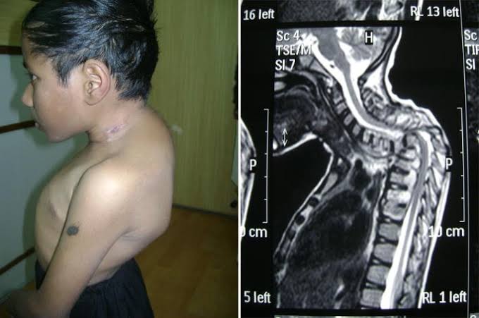
Kyphoscoliosis is a complex three-dimensional spinal deformity characterized by an abnormal curvature in both the sagittal (kyphosis, or excessive forward rounding of the back) and coronal (scoliosis, or lateral curvature) planes, often accompanied by vertebral rotation. This intricate anatomical deviation can lead to a myriad of physiological impairments and clinical challenges. Understanding its multifactorial etiology, diverse clinical manifestations, potential complications, and comprehensive management strategies is paramount for effective patient care.
Etiology of Kyphoscoliosis
The origins of kyphoscoliosis are diverse, ranging from congenital abnormalities to acquired conditions, and often defying a single identifiable cause. Understanding these etiologies is crucial for accurate diagnosis and tailored intervention.
- Idiopathic Kyphoscoliosis: This is the most common form, accounting for approximately 80% of all cases of scoliosis and often coexisting with kyphotic tendencies. The term “idiopathic” signifies that the exact cause remains unknown, though genetic predispositions and biomechanical factors are suspected. It is further categorized by the age of onset:
- Infantile (0-3 years): Rare, often resolving spontaneously but can be severe.
- Juvenile (4-9 years): Less common, but has a higher risk of progression.
- Adolescent (10-18 years): Most prevalent, typically presenting during growth spurts.
- Congenital Kyphoscoliosis: Resulting from malformations of the vertebrae that occur during fetal development, typically within the first six weeks of gestation. These can include:
- Failed Formation: Hemivertebrae (wedge-shaped vertebrae), causing a sharp angulation.
- Failed Segmentation: Block vertebrae or fused ribs, limiting spinal growth on one side.
- These often require early intervention due to a high potential for severe progression and complications.
- Neuromuscular Kyphoscoliosis: This type arises from underlying neurological or muscular disorders that weaken the muscles supporting the spine, leading to progressive curvature. Examples include:
- Cerebral Palsy: Muscle spasticity and imbalances.
- Muscular Dystrophies (e.g., Duchenne, Becker): Progressive muscle weakness.
- Spinal Muscular Atrophy: Degeneration of motor neurons.
- Polio: Paralysis of spinal muscles.
- Spinal Cord Trauma or Tumors: Can disrupt neurological control of spinal stability. These conditions often lead to long, C-shaped curves that progress rapidly, particularly in non-ambulatory patients.
- Syndromic Kyphoscoliosis: Associated with specific genetic syndromes that affect connective tissue, bone growth, and overall musculoskeletal integrity. Notable examples include:
- Marfan Syndrome: Connective tissue disorder, leading to lax ligaments and skeletal abnormalities.
- Ehlers-Danlos Syndrome: Hyperelasticity of connective tissues.
- Neurofibromatosis (Type 1): Nerve sheath tumors and bone dysplasias.
- Prader-Willi Syndrome, Rett Syndrome, Charcot-Marie-Tooth Disease: Various genetic conditions with musculoskeletal manifestations.
- Scheuermann’s Kyphosis (often with associated scoliosis): A structural increase in thoracic kyphosis due to wedging of three or more consecutive vertebrae. While primarily a kyphosis, it can be accompanied by a compensatory scoliosis.
- Post-Traumatic Kyphoscoliosis: Occurs following spinal fractures, especially if left unstable or improperly treated, leading to segmental collapse and deformity.
- Post-Infectious Kyphoscoliosis: Conditions like spinal tuberculosis (Pott’s disease) can destroy vertebral bodies, leading to kyphotic collapse and often an accompanying scoliosis.
- Degenerative Kyphoscoliosis (Adult De Novo): Develops in adulthood, typically after age 40, due to age-related degeneration of intervertebral discs, facet joints, and spinal ligaments. This often results in a loss of sagittal balance and a progressive lateral curve, commonly seen in the lumbar spine.
- Metabolic Kyphoscoliosis: Conditions like osteoporosis can weaken vertebral bodies, predisposing them to compression fractures and subsequent kyphotic deformity, which may evolve into kyphoscoliosis over time.
Clinical Features of Kyphoscoliosis
The clinical presentation of kyphoscoliosis varies widely depending on the age of onset, severity, location of the curve, and underlying etiology. It often manifests through observable physical signs and patient-reported symptoms.
- Visible Deformity: This is often the most noticeable and concerning feature for patients and their families.
- Uneven Shoulders: One shoulder may appear higher than the other.
- Prominent Shoulder Blade (Scapula): One scapula may protrude more than the other, often on the convex side of the scoliotic curve.
- Head Not Centered: The head may appear off-center, not directly over the pelvis.
- Rib Hump (Rotational Deformity): During Adam’s forward bend test, a prominent hump may be visible on one side of the back, particularly in the thoracic region, indicating vertebral rotation.
- Uneven Waistline: One side of the waist may appear flatter or higher.
- Pelvic Tilt/Leg Length Discrepancy: While not a direct feature of the spine, severe curves can lead to compensatory pelvic tilt, making one leg appear shorter.
- Increased Thoracic Kyphosis: An exaggerated forward rounding of the upper back.
- Pain:
- Back Pain: Common, especially in adults with degenerative or progressing curves. It can range from dull aches to sharp, localized pain caused by muscle fatigue, facet joint arthritis, or nerve compression.
- Radiating Pain: If nerve roots are compressed, pain, numbness, or tingling can radiate into the limbs.
- Respiratory Symptoms: In severe cases, particularly with significant thoracic curves (Cobb angles > 60-70 degrees) and rotation, the chest cavity can be compromised.
- Dyspnea (Shortness of Breath): Especially during exertion.
- Reduced Exercise Tolerance: Inability to perform physical activities due to breathlessness.
- Recurrent Respiratory Infections: Due to inefficient lung expansion and decreased cough effectiveness.
- Neurological Symptoms: While less common, severe spinal deformity can lead to spinal cord or nerve root compression, particularly in congenital or rapidly progressing forms.
- Weakness, Numbness, or Tingling: In the trunk or extremities.
- Gait Disturbances: Difficulty walking or imbalance.
- Bowel or Bladder Dysfunction: In very severe cases, indicative of significant spinal cord compromise.
- Fatigue: Chronic pain and the increased effort required to maintain posture can lead to generalized fatigue.
- Functional Limitations: Difficulty with daily activities, such as dressing, bathing, or sleeping, can occur depending on the severity and location of the curve.
- Psychosocial Impact: Visible deformities can lead to body image issues, low self-esteem, anxiety, and depression, especially in adolescents.
Diagnostic Evaluation:
- Physical Examination: Includes Adam’s forward bend test (for rib hump), plumb line assessment (for spinal balance), neurological examination, and assessment of global posture.
- Imaging Studies:
- Standing Full-Length Spine X-rays (AP and Lateral): Essential for diagnosis, measuring Cobb angles (for magnitude of scoliosis and kyphosis), assessing vertebral rotation, and evaluating sagittal balance.
- MRI (Magnetic Resonance Imaging): Indicated for atypical curves, rapid progression, neurological symptoms, congenital anomalies, or to rule out intraspinal pathologies (e.g., syrinx, tumor).
- CT Scan (Computed Tomography): Provides detailed bone anatomy, useful for surgical planning or complex congenital cases.
- Pulmonary Function Tests (PFTs): To assess lung capacity and function in severe thoracic curves.
Complications of Kyphoscoliosis
The progressive nature and structural impact of kyphoscoliosis can lead to a range of significant complications affecting various bodily systems, particularly if left untreated or unmanaged.
- Pulmonary Complications: These are among the most serious, especially with severe thoracic curves (Cobb angle > 70 degrees).
- Restrictive Lung Disease: The deformed rib cage restricts lung expansion, leading to reduced vital capacity and total lung capacity.
- Dyspnea and Reduced Exercise Tolerance: Due to compromised lung function.
- Chronic Hypoventilation: Insufficient breathing, particularly during sleep, leading to hypoxemia and hypercapnia.
- Recurrent Respiratory Infections: Impaired lung mechanics and cough effectiveness increase susceptibility to pneumonia and bronchitis.
- Pulmonary Hypertension and Cor Pulmonale: Chronic hypoxemia can lead to increased pressure in the pulmonary arteries, eventually causing right-sided heart failure (cor pulmonale), which is life-threatening.
- Neurological Complications: While rare, they are severe when they occur.
- Spinal Cord Compression: Progressive deformity, especially with sharp angular kyphosis, can compress the spinal cord, leading to myelopathy.
- Radiculopathy: Nerve root compression causing pain, numbness, weakness, or tingling in the distribution of the affected nerve.
- Paralysis: In extreme cases of spinal cord injury.
- Chronic Pain:
- Persistent Back Pain: The most common complication, resulting from muscular fatigue, facet joint arthritis, degenerative disc disease, and mechanical stress on the spine. It can significantly impact quality of life.
- Sciatica (Lumbar Radiculopathy): If lumbar nerve roots are compressed.
- Functional Limitations and Disability:
- Limited Mobility: Reduced range of motion in the spine and difficulty with activities of daily living (ADLs) such as bending, lifting, or prolonged sitting/standing.
- Gait Abnormalities: Imbalance and difficulty walking, especially in severe cases or those with neurological involvement.
- Reduced Quality of Life: Chronic pain, physical limitations, and negative body image can significantly diminish overall well-being.
- Psychosocial Issues:
- Body Image Distortion: Visible deformity can lead to significant psychological distress, low self-esteem, social anxiety, and depression, particularly in adolescents.
- Social Stigma: Patients may face challenges in social interactions and recreational activities.
- Progression of Deformity: Without intervention, curves can progressively worsen, especially during growth spurts or in adulthood due to degenerative changes. This further exacerbates all other complications.
- Degenerative Arthritis: The abnormal loading on spinal joints and discs can accelerate degenerative changes, leading to premature arthritis.
Management of Kyphoscoliosis
The management of kyphoscoliosis is highly individualized, depending on the patient’s age, curve magnitude, flexibility, potential for progression, symptoms, and underlying etiology. A multidisciplinary approach involving orthopedists, neurologists, pulmonologists, physical therapists, and pain specialists is often necessary.
Goals of Management:
- Prevent curve progression.
- Alleviate pain.
- Improve spinal balance and function.
- Prevent or mitigate cardiopulmonary complications.
- Improve cosmetic appearance.
A. Observation:
- Indication: Mild curves (Cobb angle < 20-25 degrees) in growing children or non-progressive curves in adults without significant symptoms.
- Approach: Regular clinical check-ups (every 4-6 months) and standing X-rays (every 6-12 months) to monitor for progression. Patient education on posture and general fitness.
B. Conservative Management:
- Physical Therapy and Exercise:
- Indication: All patients, regardless of curve severity, can benefit. Essential for pain management, improving muscle strength, flexibility, and posture.
- Approach: Core strengthening exercises, stretching, specific scoliosis-specific exercises (e.g., Schroth method), and postural retraining. These aim to improve spinal stability, reduce muscle imbalances, and enhance body awareness. While not proven to correct structural curves, they can alleviate symptoms and improve function.
- Bracing:
- Indication: Moderate curves (Cobb angle 20-45 degrees) in growing adolescents (Risser sign 0-2, pre- or peri-pubertal) to prevent progression and potentially reduce curve magnitude. Less effective in adults for structural correction but may be used for pain relief or to prevent further collapse in specific cases.
- Approach: Braces (e.g., Thoraco-Lumbo-Sacral Orthosis/TLSO, Boston brace, Charleston bending brace) are worn for a prescribed number of hours daily (typically 18-23 hours) until skeletal maturity. The brace applies corrective pressures to the spine to prevent the curve from worsening. Compliance is critical for success.
- Pain Management:
- Indication: For patients experiencing chronic back pain.
- Approach: Over-the-counter analgesics (NSAIDs), muscle relaxants, heat/cold therapy, transcutaneous electrical nerve stimulation (TENS), and massage. Injections (epidural, facet joint) may be considered for severe localized pain, though less common for the kyphoscoliotic deformity itself.
C. Surgical Management:
- Indication:
- Severe curves: Typically Cobb angle > 45-50 degrees in adolescents or > 50-60 degrees in adults with documented progression.
- Progression despite bracing: When conservative measures fail to halt curve progression.
- Significant pain: Unresponsive to conservative treatment.
- Neurological deficits: Evidence of spinal cord or nerve root compression.
- Cardiopulmonary compromise: Severe curves interfering with lung or heart function.
- Unacceptable cosmetic deformity: Significantly impacting quality of life.
- Objectives: Correct the deformity, stabilize the spine, alleviate pain, decompress neural elements, and improve global spinal balance.
- Procedures:
- Spinal Fusion with Instrumentation: The most common surgical approach. It involves correcting the curve as much as safely possible and fusing the affected vertebrae together to create a solid, stable segment. Modern instrumentation (rods, screws, hooks) provides powerful three-dimensional correction and stabilization.
- Posterior Spinal Fusion: The most common approach, involving an incision in the back.
- Anterior Spinal Fusion: Less common for kyphoscoliosis, typically used for specific lumbar or thoracolumbar curves, often combined with a posterior approach.
- Combined Anterior-Posterior Fusion: For very rigid or severe deformities.
- Growing Rods: For very young children with progressive severe curves, where fusion would significantly stunt spinal growth. These rods are surgically implanted and periodically lengthened without major surgery until the child approaches skeletal maturity, at which point a definitive fusion is performed.
- Vertebral Column Resection (VCR) or Osteotomies: For very severe, rigid, or complex deformities, including congenital kyphoscoliosis, where bone removal is necessary to achieve adequate correction.
- Spinal Fusion with Instrumentation: The most common surgical approach. It involves correcting the curve as much as safely possible and fusing the affected vertebrae together to create a solid, stable segment. Modern instrumentation (rods, screws, hooks) provides powerful three-dimensional correction and stabilization.
- Surgical Risks: Significant surgery carries risks including bleeding, infection, neurological injury (e.g., paralysis), pseudoarthrosis (failure of fusion), implant failure, persistent pain, and junctional kyphosis/scoliosis (new deformity above or below the fused segment). Neuromonitoring is routinely used during surgery to reduce neurological risks.
- Post-operative Care: Includes pain management, mobilization as tolerated, physical therapy, and activity restrictions for several months while the fusion consolidates. Regular follow-up X-rays are crucial.
D. Rehabilitation:
- Pre- and Post-operative Rehabilitation: Essential for optimizing patient outcomes. It involves strengthening exercises, flexibility training, gait training, and education on proper body mechanics.
In conclusion, kyphoscoliosis is a challenging spinal condition requiring a thorough understanding of its varied etiologies, recognition of its diverse clinical features, awareness of its potentially severe complications, and the implementation of a tailored, often multidisciplinary, management plan. Early diagnosis, vigilant monitoring, and appropriate intervention — whether conservative or surgical — are key to preventing progression, alleviating symptoms, and improving the long-term quality of life for affected individuals.
References:
- Weinstein, S. L., Dolan, L. A., Cheng, J. C. Y., Danielsson, A., & Nachemson, A. L. (2009). Adolescent idiopathic scoliosis: natural history and long-term follow-up. Spine, 34(16), 1735-1740.
- Lonstein, J. E., & Bradford, D. S. (2007). Moe’s textbook of scoliosis and other spinal deformities. Saunders Elsevier.
- Newton, P. O., & Shufflebarger, H. L. (2014). Scoliosis: A practical approach to classification, diagnosis, and management. Thieme.
- Cahill, P. J., & Abel, M. F. (2019). Neuromuscular Scoliosis. Journal of the American Academy of Orthopaedic Surgeons, 27(1), e1-e15.
- Bridwell, K. H., & DeWald, R. L. (2011). The Textbook of Spinal Surgery (3rd ed.). Lippincott Williams & Wilkins.
- Kim, Y. J., & Lenke, L. G. (2012). Surgical treatment options for kyphosis. Spine, 37(13), E802-E806.
- Negrini, S., Donzelli, S., Aulisa, A. G., Czaprowski, D., Schreiber, S., de Mauroy, J. C., … & Zaina, F. (2018). 2016 SOSORT guidelines: orthopaedic and rehabilitation treatment of idiopathic scoliosis during growth. Scoliosis and Spinal Disorders, 13(1), 3.
- Schwab, F., Blondel, B., Bess, S., Hostin, L., Lafage, V., Smith, J. S., … & International Spine Study Group. (2018). Adult spinal deformity-postoperative complications: a review of the SRS database. Spine Deformity, 6(1), 1-10.


