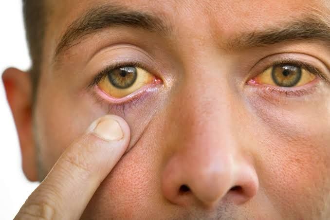
Obstructive jaundice is a clinical syndrome characterized by yellowish discoloration of the skin and sclera due to the accumulation of conjugated bilirubin, resulting from a blockage in the biliary drainage system. It is a common and serious presentation that demands a systematic and efficient diagnostic and therapeutic approach. One of the most concerning causes of obstructive jaundice, particularly in older adults, is carcinoma of the head of the pancreas.
A Systematic Approach to Obstructive Jaundice
The management of a patient presenting with obstructive jaundice hinges on a logical progression from clinical suspicion to definitive diagnosis and intervention. The primary goals are to confirm the presence of obstruction, identify its level and cause, relieve the blockage to prevent complications like cholangitis and liver failure, and ultimately treat the underlying pathology.
(a) Initial Clinical Assessment
A thorough history and physical examination are foundational. The clinician must differentiate obstructive (post-hepatic) jaundice from pre-hepatic (hemolytic) and hepatocellular causes.
- History: Key features suggesting obstruction include painless, progressive jaundice, which is a classic, ominous sign of malignancy, particularly pancreatic cancer. In contrast, painful jaundice may suggest a benign cause like choledocholithiasis (gallstones in the common bile duct). Associated symptoms provide vital clues:
- Constitutional Symptoms: Significant weight loss, anorexia, and deep-seated abdominal or back pain are red flags for malignancy.
- Biliary Obstruction Signs: Pruritus (itching) from bile salt deposition in the skin, dark-colored urine (from renal excretion of conjugated bilirubin), and acholic (pale, clay-colored) stools (from lack of bilirubin reaching the gut) are pathognomonic for biliary obstruction.
- Past Medical History: A history of gallstones, pancreatitis, recent biliary instrumentation (e.g., ERCP), or primary sclerosing cholangitis can point toward a specific etiology.
- Physical Examination: The examination should focus on confirming jaundice (best seen in the sclera) and searching for the underlying cause.
- Abdominal Examination: Palpation may reveal a mass in the right upper quadrant. Courvoisier’s law states that in the presence of jaundice, a palpable, non-tender gallbladder suggests the cause is unlikely to be gallstones (as chronic cholecystitis would lead to a fibrosed, non-distensible gallbladder) and more likely due to a malignant obstruction of the distal common bile duct, such as pancreatic cancer.
- General Examination: Look for signs of malignancy like cachexia (muscle wasting), a left supraclavicular lymph node (Virchow’s node), or a periumbilical nodule (Sister Mary Joseph’s nodule), which indicate metastatic disease.
(b) Laboratory Investigations
Blood tests are essential to confirm the biochemical pattern of cholestasis and assess hepatic function.
- Liver Function Tests (LFTs): The hallmark of obstructive jaundice is a cholestatic pattern:
- A disproportionately high elevation in Alkaline Phosphatase (ALP) and Gamma-Glutamyl Transferase (GGT).
- An elevation in Total and Direct (Conjugated) Bilirubin.
- Only a mild to moderate elevation in aminotransferases (AST and ALT), unless there is a concurrent ascending cholangitis or prolonged, severe obstruction causing hepatocellular damage.
- Coagulation Profile: An elevated Prothrombin Time (PT) or International Normalized Ratio (INR) can occur due to malabsorption of fat-soluble vitamin K, which is essential for the synthesis of clotting factors II, VII, IX, and X. This is correctable with parenteral vitamin K administration.
- Tumor Markers: Carcinoembryonic antigen (CEA) and Carbohydrate Antigen 19-9 (CA 19-9) may be elevated in pancreatobiliary malignancies. However, CA 19-9 can also be elevated in benign cholestasis and is therefore not a diagnostic tool, but rather used for prognostic purposes and monitoring treatment response.
(c) Imaging to Confirm and Localize Obstruction
Imaging is the cornerstone of diagnosis, used to confirm ductal dilation and identify the site and nature of the obstruction.
- First-Line Imaging (Abdominal Ultrasound – US): This is the initial imaging modality of choice as it is non-invasive, widely available, and inexpensive. It can readily identify intrahepatic and extrahepatic biliary ductal dilation, confirm the presence of gallstones, and often visualize a mass in the head of the pancreas or distal common bile duct.
- Second-Line/Definitive Imaging:
- Multidetector Computed Tomography (MDCT): A triple-phase, pancreatic protocol CT scan of the abdomen and pelvis is the gold standard for evaluating a suspected pancreatic mass. It is crucial for staging cancer by defining the tumor’s size, its relationship to critical blood vessels (e.g., superior mesenteric artery, portal vein), and detecting regional or distant metastases.
- Magnetic Resonance Cholangiopancreatography (MRCP): This non-invasive MRI technique provides excellent, detailed images of the biliary tree and pancreatic duct, making it superior to CT for defining the anatomy of the stricture and identifying non-obstructing stones.
(d) Advanced Diagnostics and Therapeutic Intervention
Once a likely obstruction is localized, further procedures are often needed for tissue diagnosis and biliary decompression.
- Endoscopic Retrograde Cholangiopancreatography (ERCP): ERCP is a dual-purpose endoscopic procedure. Diagnostically, it allows for direct visualization of the ampulla of Vater, cholangiography to delineate the stricture, and tissue sampling via brush cytology or biopsy. Therapeutically, it is the primary method for relieving the obstruction by placing a plastic or self-expanding metal stent across the stricture, allowing bile to drain into the duodenum.
- Endoscopic Ultrasound (EUS): EUS provides high-resolution imaging of the pancreas and bile duct. Its main advantage is its ability to guide Fine-Needle Aspiration (FNA) to obtain a cytological diagnosis from a pancreatic mass or suspicious lymph nodes with high accuracy and a low risk of tumor seeding.
- Percutaneous Transhepatic Cholangiography (PTC): This is an alternative to ERCP, used when ERCP is technically unsuccessful. A needle is passed through the skin and liver into a dilated bile duct under radiological guidance, followed by placement of a drainage catheter, which can be external or internal-external.
(e) Definitive Management
Management is tailored to the underlying cause. Biliary stenting via ERCP is performed to relieve jaundice, manage pruritus, and prevent cholangitis, especially in patients who are surgical candidates or require neoadjuvant chemotherapy. Definitive treatment for pancreatic cancer, if resectable, is surgical resection (pancreaticoduodenectomy or “Whipple procedure”), often combined with adjuvant or neoadjuvant chemotherapy. For benign causes like gallstones, stone extraction via ERCP is curative.
In other words, management of obstructive jaundice is multi-faceted, focusing on immediate stabilization, biliary decompression, definitive treatment of the underlying cause, and management of complications.
- Initial Stabilization and Supportive Care:
- Fluid Resuscitation: Correct dehydration.
- Coagulopathy Correction: Administer vitamin K (intramuscular or intravenous) to correct prolonged PT/INR secondary to fat-soluble vitamin malabsorption, especially before any invasive procedure.
- Antibiotics: Crucial if cholangitis is suspected (fever with jaundice). Broad-spectrum antibiotics covering gram-negative bacteria and anaerobes are initiated empirically (e.g., piperacillin-tazobactam, meropenem).
- Pain Management: Administer analgesics as needed.
- Nutritional Support: Address malnutrition, which is common in chronic obstruction or malignancy.
- Biliary Decompression: This is often the most urgent step once obstructive jaundice is confirmed, especially in patients with cholangitis or rapidly deteriorating liver function.
- Endoscopic Decompression (ERCP):
- Stent Placement: The most common method. Plastic stents are used for temporary drainage (e.g., benign strictures, bridging to surgery for malignancy). Self-expanding metal stents (SEMS) are preferred for long-term palliation in malignant obstruction due to their larger diameter and longer patency.
- Stone Extraction: Sphincterotomy followed by balloon trawling or basket retrieval for CBD stones.
- Percutaneous Decompression (PTC with External/Internal-External Drain): Performed when ERCP fails or is not technically feasible (e.g., tight proximal strictures, altered anatomy post-surgery). This involves placing a catheter through the liver parenchyma into the bile duct to drain bile externally or internally.
- Surgical Decompression: Rarely the primary initial decompression method due to its invasiveness, but may be performed in certain circumstances (e.g., immediate surgical bypass for complete obstruction with concurrent surgery).
- Endoscopic Decompression (ERCP):
- Definitive Treatment of the Underlying Cause: Once the patient is stabilized and biliary drainage is established, the focus shifts to treating the specific etiology.
- Choledocholithiasis: ERCP with stone extraction, followed by laparoscopic cholecystectomy (if gallbladder in situ) to prevent recurrence.
- Benign Biliary Strictures: Endoscopic balloon dilation and/or long-term placement of multiple plastic stents, or sometimes surgical bypass.
- Malignant Obstruction (e.g., Pancreatic Cancer, Cholangiocarcinoma):
- Resectable Disease: Surgical resection (e.g., Whipple procedure for pancreatic head/ampullary/distal CBD tumors, liver resection for cholangiocarcinoma). This may be preceded by neoadjuvant chemotherapy or radiation.
- Unresectable Disease: Palliative management involves ongoing biliary stenting (ERCP or PTC), chemotherapy, radiation therapy, and aggressive symptom management (pain, malabsorption, gastric outlet obstruction).
- Management of Complications:
- Cholangitis: Prompt antibiotics and urgent biliary decompression.
- Pancreatitis: Supportive care, pain management, fluid resuscitation.
- Coagulopathy: Vitamin K administration.
- Pruritus: Cholestyramine, rifampicin, or ursodeoxycholic acid may be used.
- Malabsorption: Pancreatic enzyme replacement, fat-soluble vitamin supplementation.
- Renal Impairment: Careful fluid and electrolyte management.
Post-Management Care: Regular follow-up is essential to monitor for stent patency (if applicable), disease progression, and manage long-term complications. Nutritional support, pain control, and psychological support are critical components of ongoing care, especially for patients with malignancy.
Counseling a Patient with Newly Diagnosed Carcinoma of the Head of the Pancreas
Delivering a diagnosis of pancreatic cancer is one of the most challenging communication tasks in medicine. It requires a structured approach that prioritizes empathy, clarity, and shared decision-making. The SPIKES protocol offers a reliable framework.
Step 1: Setting up the Interview (S)
- Arrange for privacy: Choose a quiet, private room. Ensure there will be no interruptions.
- Involve significant others: Ask the patient if they would like a family member or friend present.
- Sit down and connect: Sit at the same level as the patient, make eye contact, and give them your full attention. This non-verbal communication establishes rapport and shows respect.
Step 2: Assess the Patient’s Perception (P)
- Start with open-ended questions: Before delivering the news, gauge the patient’s understanding of their situation. Ask, “Before we talk about the test results, can you tell me what you understand so far about why we did the scans and the biopsy?” This helps you identify any misconceptions and assesses their emotional readiness.
Step 3: Obtain the Patient’s Invitation (I)
- Ask for permission to share information: Most patients want to know their diagnosis, but it is respectful to ask. “Would it be okay if we discuss the results of your tests now?” You can also ask about their preference for detail: “Some people prefer to know all the details, while others prefer a summary. What would you prefer?”
Step 4: Give Knowledge and Information (K)
- Give a warning shot: Prepare the patient for bad news. A phrase like, “I’m afraid the results are not what we had hoped for,” gives them a moment to brace themselves.
- Be direct, clear, and avoid jargon: Use the word “cancer.” State clearly, “The biopsy of the mass in your pancreas showed that it is a cancer.” Pause after delivering this information. Silence allows the patient to process the initial shock.
- Provide information in small chunks: Avoid overwhelming the patient with complex details. Focus on the basics first.
Step 5: Address the Patient’s Emotions with Empathic Responses (E)
- Identify and acknowledge the emotion: This is the most critical step. Observe the patient’s reaction—shock, silence, tears, anger, denial. Acknowledge their feelings with an empathetic response. For example:
- “I can see this is a huge shock.”
- “This is obviously very upsetting news. Take all the time you need.”
- “Tell me what’s going through your mind right now.”
- Connect the emotion to the reason: Show you understand why they are feeling this way. “Anyone hearing this news would feel overwhelmed.” Your role here is not to fix the situation but to sit with the patient in their distress and show that you care.
Step 6: Strategy and Summary (S)
- Outline a clear plan: Once the patient is ready to discuss the next steps, transitioning to a plan can help restore a sense of control and hope.
- Confirm the Diagnosis and Staging: “We now know this is a pancreatic adenocarcinoma. Our next step is to complete the ‘staging’ process, which tells us precisely the extent of the cancer. This is crucial for deciding the best treatment.”
- Introduce the Multidisciplinary Team (MDT): “You will not be going through this alone. We have a dedicated team of experts—including surgeons, medical oncologists, and radiation oncologists—who will meet to review your case and recommend the best possible treatment plan for you.”
- Discuss Treatment Goals and Options Broadly: Frame the discussion around the primary goal. “Our goal is to treat this cancer as effectively as possible. The options depend on the staging. If the cancer is confined to the pancreas, the primary treatment is often surgery to remove it, an operation called a Whipple procedure. If it has spread, or is involved with major blood vessels, then treatments like chemotherapy become the main focus to control the cancer and its symptoms.”
- Address Symptom Control: “In the meantime, our immediate priority is your well-being. The stent we placed is already helping with the jaundice. We have excellent ways to manage pain and support your nutrition.”
- Summarize and check for understanding: Use the “teach-back” method. “I know this has been an overwhelming amount of information. To make sure I’ve explained things clearly, could you tell me in your own words what you understand the plan is moving forward?”
- Provide a concrete follow-up: Before they leave, ensure they have a scheduled follow-up appointment, contact information for a clinical nurse specialist, and reliable written information. Encourage them to write down questions and bring a family member to the next visit. End with a statement of support: “This is the beginning of a difficult journey, but we have a clear path forward, and our entire team will be with you every step of the way.”
References:
- Lidofsky, S.D. (2020). Jaundice. In J. L. Jameson, A. S. Fauci, D. L. Kasper, S. L. Hauser, D. L. Longo, & J. Loscalzo (Eds.), Harrison’s Principles of Internal Medicine (20th ed.). McGraw-Hill Education.
- Lillemoe, K.D., & Yeo, C.J. (2018). Tumors of the Pancreas. In C. M. Townsend Jr., R. D. Beauchamp, B. M. Evers, & K. L. Mattox (Eds.), Sabiston Textbook of Surgery (20th ed.). Elsevier.
- National Comprehensive Cancer Network. (2023). Pancreatic Adenocarcinoma (Version 2.2023). NCCN Clinical Practice Guidelines in Oncology. Retrieved from https://www.nccn.org.
- Buckman, R. (1992). How to Break Bad News: A Guide for Health Care Professionals. The Johns Hopkins University Press. (This book is the origin of the SPIKES protocol).
- Gollan, J.L. (2019). Pathogenesis of Obstructive Jaundice. UpToDate. Retrieved from https://www.uptodate.com.


