Introduction to Polysaccharides and Heteropolysaccharides
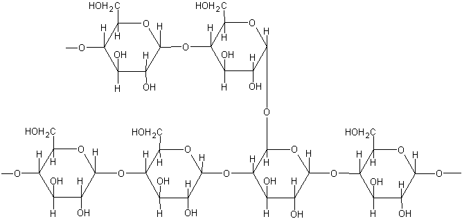
Polysaccharides are complex carbohydrates formed by the polymerization of multiple monosaccharide units linked together by glycosidic bonds. They are among the most abundant biomolecules, serving diverse roles ranging from energy storage (e.g., starch, glycogen) to structural support (e.g., cellulose, chitin) and cellular recognition. Polysaccharides can be broadly categorized into two main types based on their monosaccharide composition: homopolysaccharides and heteropolysaccharides.
Homopolysaccharides are composed of a single type of monosaccharide repeating unit, such as glucose in starch or glycogen. In contrast, heteropolysaccharides, also known as heteroglycans, are complex carbohydrates constructed from two or more different types of monosaccharide units. This structural diversity allows heteropolysaccharides to exhibit a vast array of intricate three-dimensional structures and perform highly specialized biological functions. They are often branched, frequently contain modified monosaccharides (e.g., amino sugars, uronic acids), and are commonly sulfated, conferring a significant negative charge. These characteristics are fundamental to their roles in hydration, lubrication, and mediating cell-cell and cell-matrix interactions.
Classification of Heteropolysaccharides
The most biologically significant class of heteropolysaccharides, especially in animal tissues, are the Glycosaminoglycans (GAGs). GAGs are linear anionic heteropolysaccharides composed of repeating disaccharide units, typically consisting of an amino sugar (D-glucosamine or D-galactosamine) and an uronic acid (D-glucuronic acid or L-iduronic acid) or, in the case of keratan sulfate, D-galactose. With the sole exception of hyaluronic acid, all GAGs are sulfated, which contributes significantly to their high negative charge density. This charge allows GAGs to attract large quantities of water, forming hydrated gels that are crucial for tissue resilience and function.
The principal GAGs found in mammals include:
- Hyaluronic Acid (HA) / Hyaluronan:
- Structure: Composed of repeating disaccharide units of D-glucuronic acid and N-acetyl-D-glucosamine.
- Unique Features: Unlike other GAGs, HA is non-sulfated, is not covalently linked to a core protein (exists as a free polysaccharide), and is synthesized at the plasma membrane rather than in the Golgi apparatus. It has a very high molecular weight, often reaching millions of Daltons.
- Location/Function: Abundant in synovial fluid, vitreous humor of the eye, umbilical cord, and loose connective tissues. HA is critical for joint lubrication, tissue hydration, space filling, cell migration (e.g., during embryogenesis and wound healing), and acts as a central backbone for proteoglycan aggregation.
- Chondroitin Sulfate (CS):
- Structure: Consists of repeating disaccharide units of D-glucuronic acid and N-acetyl-D-galactosamine, with sulfate groups most commonly at the 4- or 6-position of the N-acetyl-D-galactosamine (chondroitin-4-sulfate, chondroitin-6-sulfate).
- Unique Features: One of the most abundant GAGs. Always found covalently attached to core proteins, forming proteoglycans (e.g., aggrecan in cartilage).
- Location/Function: Predominant in cartilage, bone, heart valves, and the cornea. It provides structural integrity and resistance to compression, contributing to the elasticity and load-bearing capacity of these tissues.
- Dermatan Sulfate (DS):
- Structure: Similar to chondroitin sulfate but contains a high proportion of L-iduronic acid along with D-glucuronic acid, paired with N-acetyl-D-galactosamine, typically sulfated at the 4-position.
- Unique Features: Its iduronic acid content gives it more flexibility compared to CS. Always found as proteoglycans (e.g., decorin, biglycan).
- Location/Function: Found in skin, blood vessels, heart valves, and tendons. Involved in collagen fibril organization, wound healing, and regulating cell growth and differentiation.
- Keratan Sulfate (KS):
- Structure: Unique among GAGs as it contains D-galactose instead of an uronic acid in its repeating disaccharide unit (N-acetyl-D-glucosamine and D-galactose), with variable sulfation on both monosaccharides.
- Unique Features: The most heterogeneous GAG in terms of sulfation patterns. Always found as proteoglycans (e.g., aggrecan in cartilage, lumican in cornea).
- Location/Function: Primarily found in the cornea (maintains transparency), cartilage, and bone. Contributes to tissue hydration and transparency, and plays a role in cell adhesion.
- Heparan Sulfate (HS) and Heparin:
- Structure: Both are closely related GAGs composed of repeating disaccharide units of D-glucuronic acid or L-iduronic acid and N-sulfated D-glucosamine. They exhibit a highly variable and complex sulfation pattern, with sulfate groups on N-acetyl, 2-O-position of uronic acid, and 6-O-position of glucosamine. Heparin is generally more highly sulfated than HS.
- Unique Features: HS is ubiquitously expressed on cell surfaces and in the ECM, always as a proteoglycan (e.g., syndecans, glypicans). Heparin is primarily an intracellular component of mast cell granules and is released upon mast cell degranulation.
- Location/Function:
- Heparan Sulfate: Crucial for cell-surface interactions, acting as co-receptors for growth factors (e.g., FGF, VEGF), cytokines, and chemokines, modulating cell signaling. It plays roles in cell adhesion, migration, proliferation, and development. Found in basement membranes and on cell surfaces.
- Heparin: Best known as a potent natural anticoagulant, by binding to and activating antithrombin III, which inhibits various coagulation factors. Used therapeutically as an antithrombotic agent.
Beyond GAGs, other heteropolysaccharides include bacterial capsules (e.g., pneumococcal polysaccharides), certain plant gums, and some components of glycoconjugates (glycoproteins and glycolipids), though GAGs are the primary focus in the context of animal ECM.
Functions of Heteropolysaccharides
The diverse structures of heteropolysaccharides enable them to perform a wide array of critical biological functions:
- Hydration and Lubrication: Due to their highly anionic nature, GAGs attract and bind large amounts of water and positively charged ions (e.g., Na+). This creates a highly hydrated, gel-like matrix that can resist compressive forces, provide lubrication (e.g., HA in synovial fluid), and facilitate diffusion of nutrients and waste products.
- Structural Support and Resilience: In conjunction with fibrous proteins, GAGs, particularly in the form of proteoglycans, contribute significantly to the mechanical properties of tissues. They impart elasticity, tensile strength, and resistance to deformation, crucial for tissues like cartilage, skin, and blood vessels.
- Cell Adhesion and Migration: GAGs, especially HA and HS, interact with specific cell surface receptors (e.g., CD44 for HA, syndecans and glypicans for HS) to mediate cell adhesion, influence cell migration patterns during development, wound healing, and immune responses.
- Growth Factor and Cytokine Modulation: Heparan sulfate proteoglycans act as crucial co-receptors and reservoirs for many signaling molecules, including growth factors (e.g., FGF, VEGF, HGF), chemokines, and cytokines. They bind these ligands, present them to their cognate receptors, enhance receptor binding affinity, protect them from degradation, and create gradients, thereby regulating critical cellular processes like proliferation, differentiation, and morphogenesis.
- Molecular Filtration: The highly charged and porous nature of GAGs in structures like basement membranes (e.g., renal glomerulus) contributes to selective filtration, allowing passage of small molecules while restricting larger ones and negatively charged proteins.
- Anticoagulation: Heparin’s potent anticoagulant activity, through its interaction with antithrombin III, is a prime example of a highly specialized function.
- Development and Morphogenesis: The dynamic turnover and precise spatial-temporal distribution of GAGs are essential for guiding embryonic development, tissue patterning, and organogenesis.
Biochemical Significance of Heteropolysaccharides in Extracellular Matrix Formation
The extracellular matrix (ECM) is a complex and dynamic network of macromolecules secreted by cells, providing structural and biochemical support to surrounding cells. It is much more than mere scaffolding; it actively participates in regulating cell behavior, including adhesion, migration, proliferation, and differentiation. The ECM is primarily composed of two main classes of biomolecules: fibrous proteins (e.g., collagens, elastins, fibronectin, laminins) and highly hydrated, gel-forming polysaccharides, predominantly heteropolysaccharides in the form of GAGs and proteoglycans.
The integration of heteropolysaccharides into the ECM is of paramount biochemical significance:
- Formation of a Hydrated, Turgid Gel: The most defining contribution of GAGs to the ECM is their ability to form a highly hydrated, gel-like ground substance. Their numerous negative charges (from sulfate and carboxyl groups) strongly attract water molecules and counter-ions (like Na+). This creates an osmotic swelling pressure, rendering the ECM resistant to compressive forces. This turgidity is vital for tissues under mechanical stress, such as cartilage, where proteoglycan aggregates (e.g., aggrecan bound to HA) can occupy a volume 50 times greater than their isolated components, absorbing shock and distributing loads.
- Structural Scaffolding and Organization:
- Proteoglycan Aggregates: Hyaluronan often acts as a central backbone onto which numerous proteoglycans (like aggrecan in cartilage) non-covalently bind via link proteins. This forms massive, supramolecular aggregates that occupy vast volumes and organize the fibrous protein networks within the ECM.
- Interactions with Fibrous Proteins: Proteoglycans, particularly those containing chondroitin sulfate and dermatan sulfate (e.g., decorin, biglycan), directly interact with and modulate the assembly of collagen fibrils. Decorin, for instance, binds to collagen fibrils and influences their diameter and spacing, which is crucial for the tensile strength of tissues like skin and tendons.
- Regulation of Cell Behavior and Signaling:
- Cell Adhesion and Migration: Heparan sulfate proteoglycans (HSPGs) on the cell surface (e.g., syndecans, glypicans) and in the ECM (e.g., perlecan) serve as co-receptors for integrins and other adhesion molecules, facilitating cell attachment and spreading. HA interactions with CD44 on cell surfaces are critical for cell migration during development, inflammation, and wound healing.
- Growth Factor and Cytokine Reservoir: HSPGs within the ECM act as a crucial reservoir for various growth factors (e.g., FGFs, PDGF, TGF-β) and cytokines. They bind and sequester these signaling molecules, protecting them from degradation and creating local concentration gradients that guide cell migration and differentiation. This spatial presentation ensures that cells receive appropriate signals at the right time and place, profoundly influencing tissue development, homeostasis, and repair. For example, the precise binding of FGF to HS proteoglycans is essential for FGF receptor activation.
- Regulation of Protease Activity: Some GAGs can bind and modulate the activity of proteases and their inhibitors, influencing ECM turnover and tissue remodeling, which is vital during development, wound healing, and disease processes like cancer metastasis.
- Barrier and Filtration Properties: In structures like basement membranes, the highly anionic and dense network formed by HSPGs (e.g., perlecan) and other GAGs contributes to the selective permeability barrier. This is particularly evident in the kidney glomerulus, where the negatively charged GAGs repel negatively charged plasma proteins, preventing their leakage into the urine.
- Dynamic Remodeling: The ECM is not static but constantly remodeled through synthesis and degradation. Enzymes like hyaluronidases, heparanases, and various matrix metalloproteinases (MMPs) precisely break down GAGs and proteoglycans. This dynamic turnover is essential for tissue development, regeneration, and repair processes. Dysregulation of GAG synthesis or degradation is implicated in numerous pathologies, including osteoarthritis, fibrosis, and cancer progression.
In conclusion, heteropolysaccharides, primarily in the form of glycosaminoglycans and proteoglycans, are indispensable components of the extracellular matrix. Their unique structural features—diversity, high negative charge, and ability to bind large amounts of water—enable them to provide indispensable functions, from creating a hydrated, resilient tissue environment to actively orchestrating complex cellular behaviors and signaling pathways. Understanding their intricate biochemistry is crucial for comprehending tissue physiology, disease mechanisms, and for developing novel therapeutic strategies.
References
- Alberts, B., Johnson, A., Lewis, J., Raff, M., Roberts, K., & Walter, P. (2014). Molecular Biology of the Cell (6th ed.). Garland Science. (Chapters on ECM and Cell Signaling).
- K. M. Smith, M. S. C. (2018). Lehninger Principles of Biochemistry (7th ed.). W. H. Freeman. (Chapter on Carbohydrates and Polysaccharides).
- Voet, D., Voet, J. G., & Pratt, C. W. (2016). Fundamentals of Biochemistry: Life at the Molecular Level (5th ed.). Wiley. (Chapter on Carbohydrates).
- Capila, I., & Linhardt, R. J. (2002). Carbohydrate-protein interactions: biological recognition and its therapeutic implications. Current Opinion in Chemical Biology, 6(5), 639–644.
- Frantz, C., Stewart, P. K., & Weaver, V. M. (2010). The extracellular matrix at a glance. Journal of Cell Science, 123(24), 4195–4200.
- Iozzo, R. V., & Schaefer, L. (2016). Proteoglycan form and function: A comprehensive nomenclature of proteoglycans. Matrix Biology, 49, 12–21.
- Poncelet, A. C., & Van der Smissen, P. (2020). The Extracellular Matrix: A Fundamental Player in Tissue Organization and Function. International Journal of Molecular Sciences, 21(22), 8758.
- Toole, B. P. (2004). Hyaluronan: from extracellular glue to regulator of cellular processes. Journal of Internal Medicine, 256(5), 375–386.
- Sarrazin, S., Lamanna, V., & Esko, J. D. (2011). Heparan Sulfate Proteoglycans. Cold Spring Harbor Perspectives in Biology, 3(7), a004903.


