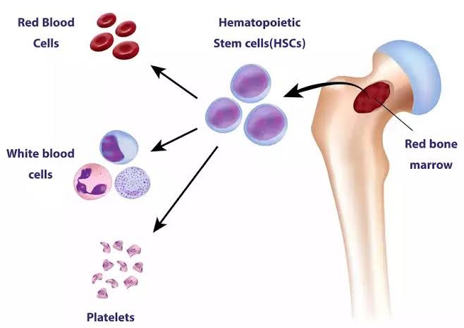
Hematopoiesis, the intricate and continuous process of blood cell formation, is fundamental to life, ensuring a constant supply of diverse cellular components for oxygen transport, immune defense, and hemostasis. This complex biological activity is meticulously regulated within specialized microenvironments, or niches, that evolve and shift their primary locations throughout an individual’s development, from embryonic stages to adulthood. Understanding the organization of hematopoietic tissue and the dynamic changes in hematopoietic sites is crucial for comprehending normal physiological function and the pathogenesis of various hematological disorders.
Organization of Hematopoietic Tissue: The Bone Marrow Niche
The primary site of definitive hematopoiesis in adults is the bone marrow, a highly vascularized, spongy tissue found within the medullary cavities of bones. It comprises two main types: red marrow, which is actively hematopoietic, and yellow marrow, predominantly composed of adipocytes (fat cells) and capable of reverting to red marrow under increased demand.
A. Cellular Components: The bone marrow is a dynamic ecosystem housing a diverse array of cells critical for blood production:
- Hematopoietic Stem Cells (HSCs): These are the cornerstone of hematopoiesis, characterized by their dual capacities:
- Self-renewal: The ability to produce identical daughter cells, maintaining the HSC pool throughout life.
- Multipotency: The capacity to differentiate into all types of mature blood cells, including myeloid (erythroid, granulocytic, monocytic, megakaryocytic) and lymphoid (B and T lymphocytes, NK cells) lineages. HSCs typically reside in a quiescent state, dividing only as needed.
- Hematopoietic Progenitor Cells (HPCs): These are descendants of HSCs that have committed to specific lineages (e.g., common myeloid progenitor, common lymphoid progenitor) and have lost some self-renewal capacity but possess high proliferative potential. They further differentiate into various blast forms before maturing.
- Mature Blood Cells: Erythrocytes, leukocytes, and platelets are produced and mature within the marrow, ready for release into the peripheral circulation.
- Stromal Cells: These non-hematopoietic cells form the structural and functional scaffold of the bone marrow, collectively contributing to the “hematopoietic niche.” Key stromal components include:
- Mesenchymal Stromal Cells (MSCs): Also known as multipotent stromal cells, these can differentiate into fibroblasts, adipocytes, chondrocytes, and osteoblasts, providing structural support and secreting regulatory factors.
- Adipocytes: Fat cells that can store energy and secrete cytokines influencing hematopoiesis.
- Endothelial Cells: Forming the lining of blood vessels (sinusoids) within the marrow, regulating cell trafficking and secreting growth factors.
- Macrophages and Osteoblasts: These cells also contribute to niche regulation, with macrophages clearing debris and osteoblasts influencing HSC quiescence and maintenance.
- Nerve Fibers: Autonomic nervous system innervation directly modulates HSC function.
B. The Hematopoietic Niche: The hematopoietic niche refers to the specialized microenvironment within the bone marrow that provides crucial signals for HSC maintenance, self-renewal, proliferation, and differentiation. It is not a static entity but a complex interplay of physical structures, cellular interactions, and soluble factors.
- Key Features: The niche provides a protective sanctuary for HSCs, protecting them from exhaustion or premature differentiation. It is characterized by specific concentrations of growth factors, cytokines (e.g., SCF, TPO, G-CSF, EPO), chemokines (e.g., CXCL12), and extracellular matrix components that regulate HSC fate decisions.
- Locations: Two primary niches are recognized:
- Endosteal Niche: Located near the bone surface, associated with osteoblasts, believed to promote HSC quiescence and self-renewal.
- Vascular Niche: Surrounding sinusoidal blood vessels, rich in endothelial cells and perivascular stromal cells, thought to be involved in HSC proliferation and mobilization.
- Extracellular Matrix (ECM): Components like collagen, fibronectin, and laminin provide structural support and act as reservoirs for growth factors, influencing cell adhesion and signaling.
Sites and Sources of Hematopoiesis Before Birth (Prenatal Hematopoiesis)
Fetal development witnesses a fascinating migratory pattern of hematopoietic activity, ensuring the continuous provision of blood cells as the organism grows and its physiological needs change.
A. Mesoblastic Phase (Yolk Sac Hematopoiesis):
- Timing: Approximately 2 to 8 weeks of gestation. This is the earliest site of blood formation.
- Location: Occurs in the mesoderm of the yolk sac wall.
- Output: Characterized by the formation of primitive erythroid cells (often nucleated) that produce embryonic hemoglobins (Gower-1, Gower-2, Portland). These early cells are crucial for oxygen transport in the developing embryo. Early macrophages and megakaryocytes are also produced. Importantly, the yolk sac is also considered the primary site for the de novo generation of definitive HSCs, which will then migrate to subsequent hematopoietic organs.
B. Hepatic Phase (Liver and Spleen Hematopoiesis):
- Timing: Begins around 6 weeks of gestation, becoming the dominant site by 3 months, and peaks around 3-4 months. It gradually declines after 6-7 months but can persist until shortly after birth.
- Location: The fetal liver becomes the primary hematopoietic organ, while the spleen also contributes significantly, especially to lymphopoiesis and erythrocyte destruction.
- Output: This phase marks the beginning of definitive hematopoiesis, producing enucleated red blood cells containing fetal hemoglobin (HbF), which has a higher affinity for oxygen. All myeloid lineages (granulocytes, monocytes, megakaryocytes) are produced, along with early B lymphocytes. HSCs generated in the yolk sac colonize the fetal liver, establishing a more complex hematopoietic environment.
C. Medullary Phase (Bone Marrow Hematopoiesis):
- Timing: Initiates around 4-5 months of gestation and gradually increases in activity, eventually becoming the sole primary site of definitive hematopoiesis.
- Location: Hematopoiesis begins in the developing bone marrow cavities, starting with the clavicle, then long bones (femur, tibia), and subsequently flat bones (vertebrae, ribs, sternum, pelvis, skull).
- Output: The bone marrow takes over the full spectrum of definitive hematopoiesis, producing all mature blood cell types (erythrocytes, granulocytes, monocytes, platelets, B and T lymphocytes). HSCs migrate from the fetal liver to colonize the developing bone marrow, establishing the adult hematopoietic system.
- Thymus: Concurrently, the thymus becomes a crucial site for T-lymphocyte maturation, starting from mid-gestation.
Sites and Sources of Hematopoiesis After Birth (Postnatal Hematopoiesis)
After birth, the bone marrow consolidates its role as the primary site of hematopoiesis, with specific locations changing over the lifespan.
A. Infancy and Childhood:
- Location: During infancy and childhood, virtually all bone marrow cavities throughout the skeleton are actively hematopoietic, filled with red marrow. This includes the long bones (femur, tibia, humerus) as well as the axial skeleton (vertebrae, sternum, ribs, pelvis, skull).
- Output: The high demand for blood cell production to support rapid growth and development necessitates widespread hematopoietic activity. All myeloid and lymphoid cell lineages are continuously produced.
B. Adulthood:
- Location: As an individual matures, hematopoietic activity gradually retracts from the appendicular skeleton (long bones of limbs) and becomes concentrated primarily in the axial skeleton. The red marrow in the shafts of long bones is progressively replaced by yellow marrow (adipose tissue) in a process called fatty infiltration. In adults, active red marrow is predominantly found in the vertebrae, sternum, ribs, pelvis, skull, and the proximal epiphyses of the femurs and humeri.
- Output: The adult bone marrow maintains a steady state of blood cell production, replacing senescent or damaged cells and responding to increased demand during infection, inflammation, or hemorrhage. HSCs remain the ultimate source of all blood cells, residing within the specialized bone marrow niche.
C. Extramedullary Hematopoiesis (EMH):
- Condition: Under conditions of severe or chronic hematopoietic stress, such as severe anemia (e.g., thalassemia, sickle cell disease), myelofibrosis, or certain leukemias, the adult bone marrow may be unable to meet the body’s demands. In such pathological situations, hematopoietic activity can reactivate in sites that were active during fetal development.
- Location: The most common sites for EMH are the liver and spleen, which can undergo significant enlargement (hepatosplenomegaly) due to their renewed hematopoietic function. Other less common sites include lymph nodes, adrenal glands, kidneys, and even the skin.
- Significance: While EMH represents an adaptive response to insufficient bone marrow function, it is often less efficient than normal medullary hematopoiesis and can lead to complications related to organ dysfunction or mass effects.
The Central Role of Hematopoietic Stem Cells (HSCs)
Throughout all stages of development, from the mesoblastic yolk sac to the adult bone marrow, Hematopoietic Stem Cells (HSCs) are the foundational element. Their remarkable capacity for self-renewal ensures the lifelong maintenance of the hematopoietic system, while their multipotency guarantees the generation of all necessary blood cell types. The journey of HSCs from their origin in the yolk sac, through transient residence in the fetal liver, and ultimately to their permanent home in the bone marrow, underscores the dynamic and adaptive nature of hematopoiesis, meticulously orchestrated to meet the evolving physiological demands of the organism.
Conclusion
Hematopoiesis is a quintessential example of biological adaptability and precision. From its earliest embryonic origins in the yolk sac, through its flourishing in the fetal liver, to its refined localization in the adult bone marrow, the sites and mechanisms of blood cell production undergo significant transformations. The sophisticated organization of hematopoietic tissue, particularly the intricate bone marrow niche, provides the essential microenvironment for the self-renewal and differentiation of hematopoietic stem cells, ensuring a continuous and robust supply of blood cells crucial for life. This dynamic system, capable of adapting to various physiological demands and even reactivating fetal sites in pathological conditions, is a cornerstone of human health.
References:
- Orkin, S. H., & Zon, L. I. (2008). Hematopoiesis: an evolving paradigm for stem cell biology. Cell, 132(4), 631-644.
- Morrison, S. J., & Scadden, D. T. (2014). The hematopoietic stem cell niche. Nature, 507(7493), 76-82.
- Dzierzak, E., & Speck, N. A. (2008). Of zebrafish and mice: developmental hematopoiesis. Immunity, 29(6), 863-874.
- Zhao, M., Li, L., & Scadden, D. T. (2014). Hematopoietic stem cells and the bone marrow niche. Advances in Experimental Medicine and Biology, 871, 13-21.
- Frenette, P. S., Pinho, S., Lucas, D., & Scheiermann, C. (2013). Myeloid control of hematopoietic stem cell trafficking. Cell Stem Cell, 12(6), 673-683.
- Robin, C., & Dzierzak, E. (2011). Developmental origins of hematopoietic stem cell fate. Current Opinion in Cell Biology, 23(6), 661-665.
- Bianco, P., Robey, P. G., & Simmons, P. J. (2008). Mesenchymal stem cells: revisitting history, concepts, and assays. Cell Stem Cell, 2(4), 313-319.


