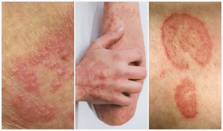
Fungi are a diverse group of eukaryotic organisms that can cause a wide spectrum of diseases in humans, collectively known as mycoses. These infections range from superficial, self-limiting conditions to life-threatening systemic diseases. Understanding the classification, properties, transmission, pathogenesis, clinical manifestations, and laboratory diagnosis of fungal infections is crucial for effective management.
Cutaneous Fungi (Dermatophytes)
Cutaneous fungi, primarily dermatophytes, are a specialized group of molds that cause infections of the skin, hair, and nails. They are unique in their ability to colonize and invade keratinized tissues.
- Classification and Important Properties: Dermatophytes are classified into three main genera: Trichophyton, Microsporum, and Epidermophyton. They are characterized by their keratinophilic nature, meaning they possess enzymes (keratinases) that allow them to digest keratin, the main protein in skin, hair, and nails. Unlike many other pathogenic fungi, dermatophytes typically do not exhibit thermal dimorphism; they grow as molds at both environmental and host temperatures. They are generally restricted to the superficial, non-living keratinized layers of the epidermis, hair, and nails, rarely invading deeper tissues or becoming systemic. Their growth often leads to a characteristic inflammatory response.
- Transmission: Transmission of dermatophytes occurs through direct contact with infected individuals (anthropophilic species), infected animals (zoophilic species), or contaminated soil (geophilic species). Indirect transmission is also common via fomites such as contaminated towels, combs, clothing, shower floors, or shared personal items. Factors like warmth, humidity, minor skin trauma, and occlusive footwear facilitate transmission and infection.
- Pathogenesis: The pathogenesis begins with the adherence of arthroconidia (spores) or hyphal fragments to the stratum corneum. These fungi then germinate and grow radially, forming hyphae that extend into the keratinized tissue. The production of keratinases, proteases, and elastases allows them to break down and utilize keratin as a nutrient source. The host’s immune response, primarily cell-mediated immunity, results in the characteristic inflammatory reaction, including erythema, scaling, pruritus, and vesicle formation, often with central clearing (hence “ringworm”). The infection is typically confined to the epidermis because host factors and serum inhibitory factors prevent deeper invasion.
- Clinical Findings: Dermatophyte infections are collectively known as tinea, followed by the Latin term for the body site. Common clinical presentations include:
- Tinea corporis: Ringworm of the body, characterized by annular, erythematous, scaling lesions with raised active borders.
- Tinea pedis: Athlete’s foot, affecting the spaces between toes (interdigital), soles, or dorsal foot, presenting with scaling, maceration, itching, or blistering.
- Tinea cruris: Jock itch, involving the groin area, typically pruritic, erythematous, and scaling.
- Tinea capitis: Ringworm of the scalp and hair, common in children, causing patchy hair loss (alopecia), scaling, and inflammation. Can present as black dot ringworm (broken hairs) or kerion (highly inflammatory, boggy lesion).
- Tinea unguium (Onychomycosis): Fungal infection of the nails, leading to discoloration, thickening, and crumbling of the nail plate.
- Tinea manuum/barbae: Ringworm of the hand/beard area, respectively.
- Lab Diagnosis:
- Direct Microscopy: The most common and rapid method. Skin scrapings, hair shafts, or nail clippings are treated with 10-20% potassium hydroxide (KOH) to dissolve keratin and visualize fungal elements (hyphae, arthroconidia). Calcofluor white stain can enhance visualization.
- Culture: Samples are inoculated onto Sabouraud Dextrose Agar (SDA) often supplemented with antibiotics (chloramphenicol) to inhibit bacterial growth and cycloheximide to inhibit saprophytic fungi. Cultures are incubated at room temperature for 2-4 weeks. Identification is based on macroscopic colony morphology (color, texture, topography) and microscopic morphology (conidia, hyphal structures).
- Wood’s Lamp: Ultraviolet light can cause certain Microsporum species (e.g., M. canis, M. audouinii) to fluoresce (green-yellow) on infected hair, aiding in screening for tinea capitis.
- PCR: Molecular methods are increasingly available for rapid and accurate species identification, especially for onychomycosis.
Systemic Fungi (Endemic Mycoses)
Systemic fungi, or endemic mycoses, are true pathogenic fungi capable of causing disease in immunocompetent individuals. They are geographically restricted and are acquired through inhalation of spores from environmental sources. A defining characteristic is their thermal dimorphism.
- Classification and Important Properties: The major systemic pathogenic fungi include Histoplasma capsulatum, Blastomyces dermatitidis, Coccidioides immitis/posadasii, and Paracoccidioides brasiliensis. Their most important property is thermal dimorphism: they exist as molds with filamentous hyphae and spores (conidia) in the environment (at ambient temperatures, often 25-30°C) and transform into yeast forms (or spherules for Coccidioides) within the host at body temperature (37°C). This dimorphic transition is crucial for their pathogenicity. They are found in specific environmental niches and are not typically transmitted person-to-person.
- Transmission: All systemic fungal infections are acquired by inhalation of airborne conidia or spores from their environmental reservoirs.
- Histoplasma capsulatum: Found in soil enriched with bird or bat droppings (e.g., caves, old chicken coops, construction sites).
- Blastomyces dermatitidis: Associated with moist soil, decaying wood, and riparian areas (riverbanks, lakeshores).
- Coccidioides immitis/posadasii: Found in arid and semi-arid regions of the Americas (Southwestern US, parts of Mexico, Central and South America) in disturbed soil.
- Paracoccidioides brasiliensis: Endemic to Latin America, associated with soil disturbances in warm, humid climates.
- Pathogenesis: Upon inhalation, the conidia/spores reach the alveoli in the lungs. At body temperature, they transform into their pathogenic yeast (or spherule) forms. These forms resist phagocytosis or, as in the case of Histoplasma and Coccidioides, survive and replicate within macrophages. The initial infection is often pulmonary and can be asymptomatic or cause a mild, self-limiting flu-like illness. In some individuals, particularly those with compromised immunity or a high inoculum, the organisms can disseminate hematogenously or lymphatically from the lungs to other organs. The host mounts a granulomatous inflammatory response to contain the infection, forming granulomas and sometimes calcifications.
- Clinical Findings: Clinical presentations vary, ranging from asymptomatic to severe disseminated disease:
- Histoplasmosis: Most commonly asymptomatic. Can cause acute pulmonary histoplasmosis (flu-like illness, cough, fever), chronic pulmonary histoplasmosis (mimicking tuberculosis), or progressive disseminated histoplasmosis (PDH) affecting multiple organs (liver, spleen, lymph nodes, bone marrow, CNS, skin) in immunocompromised individuals.
- Blastomycosis: Primarily affects the lungs (pneumonia-like illness). Can disseminate to the skin (verrucous or ulcerative lesions), bones, joints, and genitourinary tract.
- Coccidioidomycosis (Valley Fever): Majority are asymptomatic or cause a mild respiratory illness. Can lead to chronic progressive pulmonary disease, nodular lesions, or disseminated disease affecting the skin, bones, joints, and meninges (coccidioidal meningitis is very serious).
- Paracoccidioidomycosis: Primarily affects the lungs, but often disseminates to mucocutaneous lesions (especially oral and nasal mucosa), lymph nodes, and adrenal glands.
- Lab Diagnosis:
- Direct Microscopy: Examination of clinical specimens (sputum, bronchoalveolar lavage (BAL) fluid, tissue biopsies) using KOH, India ink, or specialized stains (GMS, PAS) to visualize yeast forms (Histoplasma, Blastomyces, *Paracoccidioides) or spherules (Coccidioides).
- Culture: Considered the gold standard. Samples are cultured on SDA, BHI, and other specialized media at both room temperature (mold phase) and 37°C (yeast phase) to demonstrate dimorphism. Caution: Handling mold cultures poses a serious biohazard risk due to airborne conidia.
- Serology: Detection of antibodies (IgM, IgG) via immunodiffusion, complement fixation, or ELISA. Useful for diagnosis of chronic or disseminated disease.
- Antigen Detection: Particularly useful for Histoplasma (urine, serum, BAL, CSF) and Coccidioides (urine, serum) in disseminated disease and for monitoring treatment response.
- Molecular Methods: PCR assays are increasingly used for rapid identification in clinical samples.
- Histopathology: Biopsy of affected tissue often reveals characteristic granulomas and fungal elements.
Opportunistic Fungi
Opportunistic fungi are ubiquitous organisms that typically cause disease only in individuals with compromised immune systems or altered host defenses. As the population of immunocompromised patients grows (e.g., transplant recipients, HIV/AIDS patients, cancer patients undergoing chemotherapy), opportunistic fungal infections have become a significant cause of morbidity and mortality.
- Classification and Important Properties: Key opportunistic fungi include:
- Candida species (e.g., C. albicans): Yeasts that are part of the normal human microbiota (oral cavity, GI tract, skin, vagina). Can cause mucocutaneous and invasive infections. Some Candida species are dimorphic (yeast and pseudohyphae/hyphae).
- Aspergillus species (e.g., A. fumigatus): Molds found widely in the environment (soil, decaying vegetation, air). Characterized by septate hyphae and conidia. They are primarily acquired by inhalation.
- Cryptococcus neoformans/gatti: Encapsulated yeasts found in the environment (C. neoformans in soil with bird droppings; C. gatti associated with trees). Primary infection is pulmonary, but dissemination to the central nervous system (CNS) is common.
- Mucorales (e.g., Rhizopus, Mucor, Lichtheimia): Rapidly growing molds with broad, aseptate or sparsely septate hyphae. Ubiquitous in soil and decaying organic matter. Cause mucormycosis, a highly aggressive and often fatal infection.
- Pneumocystis jirovecii: Atypical fungus (once classified as a protozoan) that causes pneumonia exclusively in immunocompromised hosts, especially those with AIDS. Lacks ergosterol in its cell membrane.
- Transmission:
- Candida: Predominantly endogenous (overgrowth of normal flora due to disruption of microbial balance or host immunity) but can also be exogenous (healthcare-associated transmission).
- Aspergillus, Cryptococcus, Mucorales: Acquired by inhalation of airborne spores/conidia from the environment.
- Pneumocystis: Inhalation of airborne cysts. Not typically considered person-to-person.
- Pathogenesis: Opportunistic fungi exploit breaches in host defenses.
- Candida: Factors like broad-spectrum antibiotics (disrupting normal flora), indwelling catheters, surgery, diabetes, and immunosuppression allow Candida to overgrow, adhere to epithelial surfaces, form biofilms, and invade tissues.
- Aspergillus: In immunocompromised hosts (e.g., severe neutropenia, corticosteroid use), inhaled conidia are not effectively cleared by alveolar macrophages. They germinate into hyphae, which then invade blood vessels (angioinvasion), leading to thrombosis, infarction, and dissemination.
- Cryptococcus: Inhaled yeasts are usually cleared by the immune system. In immunocompromised individuals, the polysaccharide capsule of Cryptococcus is a major virulence factor, enabling evasion of phagocytosis and facilitating dissemination, particularly to the CNS.
- Mucorales: Spores are inhaled or gain entry through skin trauma. In patients with conditions like diabetic ketoacidosis, deferoxamine therapy, or severe neutropenia, these fungi exhibit rapid growth and profound angioinvasion, causing tissue necrosis and rapid dissemination.
- Pneumocystis: In AIDS patients, depletion of CD4+ T cells leads to inability to clear the organism, resulting in pneumonia.
- Clinical Findings: Clinical manifestations are diverse and depend on the specific fungus and the extent of immunosuppression.
- Candida infections (Candidiasis):
- Mucocutaneous: Oral thrush, esophagitis (common in HIV), vulvovaginitis, diaper rash, intertrigo, chronic mucocutaneous candidiasis.
- Invasive: Candidemia (bloodstream infection), endocarditis, peritonitis, urinary tract infection, candidal meningitis, hepatosplenic candidiasis.
- Aspergillus infections (Aspergillosis):
- Allergic: Allergic bronchopulmonary aspergillosis (ABPA), severe asthma with fungal sensitization (SAFS).
- Colonizing: Aspergilloma (fungus ball in a pre-existing lung cavity).
- Invasive: Invasive pulmonary aspergillosis (IPA), often with angioinvasion and infarction, leading to hemoptysis, pleuritic pain. Can disseminate to CNS, skin, or other organs.
- Cryptococcus infections (Cryptococcosis):
- Most common manifestation is cryptococcal meningitis, especially in HIV/AIDS patients, presenting with headache, fever, altered mental status.
- Pulmonary cryptococcosis (cough, dyspnea), cutaneous lesions, and bone lesions can also occur.
- Mucormycosis:
- Rhinocerebral: Rapidly progressive, originating in the sinuses and invading orbits, palate, and brain, causing facial pain, black eschar on palate, proptosis, vision loss, and neurological deficits.
- Pulmonary: Severe pneumonia in immunocompromised hosts.
- Other forms: Cutaneous, gastrointestinal, disseminated.
- Pneumocystis pneumonia (PCP): Dry cough, dyspnea, fever, and hypoxemia, disproportionate to physical findings. Ground-glass opacities on chest imaging.
- Candida infections (Candidiasis):
- Lab Diagnosis:
- Direct Microscopy: Examination of clinical specimens (sputum, BAL, CSF, tissue biopsies, blood for Candida) using various stains:
- KOH, Gram stain (Candida yeast and pseudohyphae).
- India ink stain for Cryptococcus (demonstrates capsule as a clear halo).
- Calcofluor white, GMS (Grocott’s Methenamine Silver), PAS (Periodic Acid-Schiff) stains on tissue biopsies to visualize fungal elements (hyphae, yeast, sporangia).
- Culture: Inoculation of samples onto SDA and other enriched media. Identification based on macroscopic and microscopic morphology, biochemical tests, or molecular sequencing.
- Antigen Detection:
- Beta-D-glucan: A pan-fungal cell wall component, elevated in invasive candidiasis, aspergillosis, and Pneumocystis pneumonia (not Cryptococcus or Mucorales).
- Galactomannan: Cell wall component of Aspergillus, detected in serum, BAL for invasive aspergillosis.
- Cryptococcal Antigen (CrAg): Detected in CSF, serum, urine via latex agglutination or Lateral Flow Assay (LFA); highly sensitive for cryptococcosis.
- Mannan/Anti-Mannan: For Candida infections.
- Molecular Methods: PCR assays for specific fungal DNA (e.g., Aspergillus, Pneumocystis, Candida in blood) offer rapid and sensitive diagnosis, especially from sterile sites or when culture is slow/negative.
- Histopathology: Biopsy and microscopic examination of infected tissue remain critical for definitive diagnosis, especially for invasive diseases.
- Direct Microscopy: Examination of clinical specimens (sputum, BAL, CSF, tissue biopsies, blood for Candida) using various stains:
Conclusion
Fungal infections represent a complex and growing challenge in medicine. Cutaneous mycoses, primarily caused by dermatophytes, are superficial infections confined to keratinized tissues after direct contact. Systemic mycoses, caused by endemic dimorphic fungi, are acquired environmentally through inhalation and can cause severe disseminated disease in both healthy and immunocompromised individuals. Opportunistic mycoses, caused by ubiquitous fungi like Candida, Aspergillus, Cryptococcus, and Mucorales, primarily affect immunocompromised hosts, leading to a wide array of life-threatening invasive diseases. Each category has distinct features regarding their properties, modes of transmission, pathogenic mechanisms, clinical presentation, and diagnostic approaches. Accurate and timely diagnosis, often involving a combination of direct microscopy, culture, antigen detection, and molecular methods, is paramount for guiding appropriate antifungal therapy and improving patient outcomes.
References:
- Mandell, G. L., Bennett, J. E., & Dolin, R. (Eds.). (2020). Mandell, Douglas, and Bennett’s Principles and Practice of Infectious Diseases (9th ed.). Elsevier.
- Murray, P. R., Rosenthal, K. S., & Pfaller, M. A. (2021). Medical Microbiology (9th ed.). Elsevier.
- Pfaller, M. A., & Rinaldi, M. G. (Eds.). (2018). Manual of Clinical Microbiology (12th ed.). ASM Press.
- Centers for Disease Control and Prevention (CDC). (n.d.). Fungal Diseases. Retrieved from https://www.cdc.gov/fungal/index.html
- Perfect, J. R., & Dismukes, W. E. (2009). Clinical Mycology. ASM Press.
- Richardson, M., & Warnock, D. W. (2012). Fungal Infection: Diagnosis and Management (4th ed.). John Wiley & Sons.


