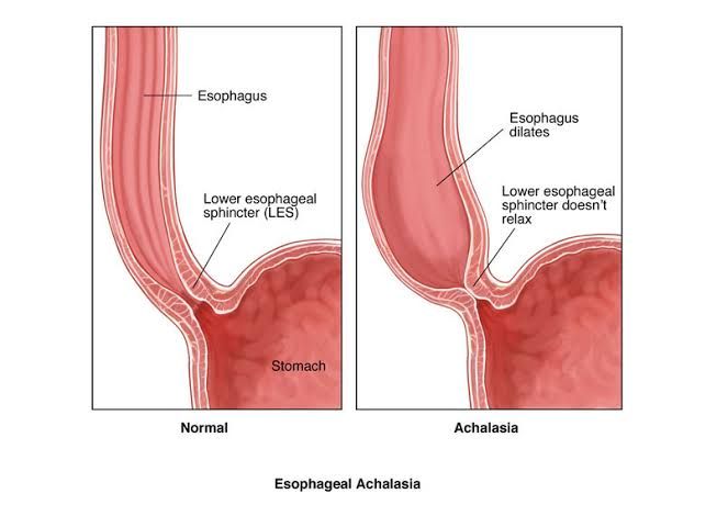
Cardia Achalasia, commonly known as achalasia, is a rare and chronic primary esophageal motility disorder characterized by two key physiological failures: the inability of the lower esophageal sphincter (LES) to relax upon swallowing and the absence of effective, coordinated muscle contractions (peristalsis) in the body of the esophagus. This dysfunction leads to impaired passage of food and liquid from the esophagus into the stomach, causing progressive symptoms that can significantly impact a patient’s quality of life.
Etiology: The Origins of a Motility Failure
The precise cause of primary achalasia remains largely unknown, classifying it as an idiopathic disorder. However, extensive research points toward a multifactorial process involving autoimmune, infectious, and genetic factors that culminate in a specific type of nerve damage.
- The Core Pathophysiology: The fundamental problem in achalasia is the selective destruction of inhibitory ganglion cells within the myenteric (Auerbach’s) plexus of the esophageal wall. These specialized neurons are responsible for producing nitric oxide (NO) and Vasoactive Intestinal Polypeptide (VIP), neurotransmitters that signal the smooth muscle of the LES to relax during swallowing. Without these inhibitory signals, the excitatory (cholinergic) signals dominate, resulting in a hypertensive, non-relaxing sphincter. The concurrent loss of these ganglion cells along the esophageal body leads to the failure of peristalsis.
- The Autoimmune Hypothesis: The most widely accepted theory suggests that achalasia is an autoimmune disease. It is proposed that in genetically susceptible individuals, an environmental trigger—most likely a viral infection such as Herpes Simplex Virus type 1 (HSV-1)—initiates an inflammatory cascade. The body’s immune system, in its attempt to fight the virus, mistakenly cross-reacts with proteins on the surface of the myenteric neurons, leading to a chronic, T-cell mediated inflammatory response that gradually destroys these vital nerve cells. This theory is supported by the presence of inflammatory infiltrates in the myenteric plexus of achalasia patients and associations with certain human leukocyte antigen (HLA) class II alleles.
- Infectious Causes of Secondary Achalasia: While primary achalasia is idiopathic, a similar condition known as secondary achalasia (or pseudoachalasia) can be caused by other diseases. The most famous example is Chagas disease, endemic to South America, which is caused by the parasite Trypanosoma cruzi. This parasite directly infects and destroys autonomic ganglion cells throughout the body, including the esophagus, leading to clinical features identical to primary achalasia. Other causes of pseudoachalasia include malignancies, particularly adenocarcinoma of the gastric cardia or distal esophagus, which can invade or compress the myenteric plexus, mimicking the functional obstruction of achalasia.
Clinical Features: Recognizing the Signs and Symptoms
The onset of achalasia is typically insidious, with symptoms developing gradually over months or even years. The clinical presentation is dominated by the consequences of esophageal obstruction.
- Dysphagia (Difficulty Swallowing): This is the hallmark symptom, experienced by nearly all patients. A key diagnostic clue is that dysphagia in achalasia occurs with both solids and liquids from the outset. Patients often describe a sensation of food or drink “sticking” or “getting held up” in the middle of their chest. Over time, individuals may unconsciously adopt maneuvers to aid esophageal emptying, such as eating slowly, drinking large amounts of water, or arching their back and neck.
- Regurgitation: As food and saliva accumulate in the dilated, aperistaltic esophagus, regurgitation of undigested food is common. This often occurs hours after a meal and is particularly problematic when lying down at night, which can lead to coughing, choking, and a high risk of aspiration pneumonia.
- Chest Pain: A significant number of patients report substernal chest pain, often described as a squeezing or pressure-like sensation that can radiate to the back, neck, or jaw. This pain can be severe enough to mimic a cardiac event (angina) and is thought to be caused by esophageal spasm or distension from retained contents.
- Weight Loss: Due to dysphagia, fear of eating (sitophobia), and regurgitation, unintentional and significant weight loss is a common and concerning feature.
- Other Symptoms: Patients may also experience heartburn, which is paradoxical as it is caused by the fermentation of retained food into lactic acid rather than gastric acid reflux. Halitosis (bad breath) and recurrent respiratory infections can also occur due to food stasis and aspiration.
Investigations: The Diagnostic Pathway
Diagnosing achalasia requires a combination of tests to confirm the motility disorder and rule out other conditions.
- Step 1: Barium Esophagram (Barium Swallow): This is often the initial diagnostic test. The patient swallows a barium-containing liquid while a series of X-rays are taken. In classic achalasia, the esophagram reveals several characteristic findings:
- Dilation of the esophageal body (a “megaesophagus” in advanced cases).
- Poor emptying of barium from the esophagus into the stomach.
- The absence of peristaltic waves.
- A smooth, tapered narrowing at the gastroesophageal junction, classically described as a “bird’s beak” or “rat’s tail” appearance.
- Step 2: Upper Endoscopy (Esophagogastroduodenoscopy – EGD): Endoscopy is crucial for two reasons. First, it helps rule out pseudoachalasia caused by a malignancy at the gastroesophageal junction. Second, it allows for direct visualization of the esophagus, which may appear dilated and contain retained food and saliva. The LES will feel tight to the passage of the endoscope but will typically open with gentle pressure (a “pop”), unlike a fixed cancerous stricture. Biopsies may be taken to rule out eosinophilic esophagitis or cancer.
- Step 3: Esophageal Manometry: This test is the gold standard for confirming the diagnosis of achalasia. A thin, pressure-sensitive catheter is passed through the nose into the esophagus to measure muscle contractions and LES pressure. The definitive manometric criteria for achalasia, according to the modern Chicago Classification, are:
- Incomplete LES Relaxation: Measured as an elevated Integrated Relaxation Pressure (IRP).
- 100% Failed Peristalsis (Aperistalsis): No coordinated swallowing waves are observed.
High-resolution manometry further allows for the sub-classification of achalasia into three types (Type I, II, and III), which have prognostic value in predicting treatment response.
Management: Therapeutic Strategies
There is no cure for achalasia, as the lost nerve cells cannot be regenerated. Therefore, treatment is palliative and aims to relieve symptoms by reducing the pressure and functional obstruction at the LES.
- Pharmacological Therapy: Medications like nitrates and calcium channel blockers can help relax the smooth muscle of the LES. However, their efficacy is modest and often short-lived, and they are associated with side effects like headaches and hypotension. They are typically reserved for patients who are poor candidates for more definitive therapies.
- Endoscopic Therapies:
- Pneumatic Dilation (PD): This involves passing a balloon through an endoscope and forcefully inflating it across the LES to stretch and disrupt the muscle fibers. It has a good success rate (around 70-90%) but may require multiple sessions over a patient’s lifetime. The main risk is esophageal perforation (2-3%).
- Botulinum Toxin (Botox) Injection: Botox is injected directly into the LES during endoscopy, where it works by blocking the release of acetylcholine, a neurotransmitter that causes muscle contraction. This is a very safe procedure but its effects are temporary, typically lasting only 6-12 months, making it most suitable for frail, elderly patients.
- Surgical and Advanced Endoscopic Myotomy:
- Laparoscopic Heller Myotomy (LHM): Considered a gold standard treatment, this is a minimally invasive surgical procedure where the outer muscle layers of the LES and distal esophagus are cut (a myotomy), permanently relieving the obstruction. To prevent severe post-operative gastroesophageal reflux disease (GERD), the myotomy is almost always combined with a partial anti-reflux surgery (fundoplication). LHM offers excellent long-term symptom relief in over 90% of patients.
- Per-Oral Endoscopic Myotomy (POEM): This is a newer, incisionless endoscopic technique that achieves the same goal as a Heller myotomy. An endoscopist creates a tunnel within the esophageal wall, advances the endoscope through it, and performs the myotomy from the inside. POEM has shown efficacy comparable to LHM but may be associated with a higher rate of post-procedure GERD, as an anti-reflux procedure is not typically performed concurrently.
In conclusion, Cardia Achalasia is a debilitating esophageal motility disorder stemming from the irreversible loss of myenteric neurons. Its diagnosis hinges on a classic symptom profile confirmed by barium swallow and, definitively, by esophageal manometry. While incurable, a range of highly effective treatments, from pneumatic dilation to Heller myotomy and POEM, are available to disrupt the non-relaxing lower esophageal sphincter, providing patients with significant and lasting relief and restoring their ability to eat and drink comfortably.
References:
- Vaezi, M. F., Pandolfino, J. E., & Vela, M. F. (2013). ACG clinical guideline: diagnosis and management of achalasia. The American journal of gastroenterology, 108(8), 1238–1249.
- Pandolfino, J. E., & Gawron, A. J. (2015). Achalasia: a systematic review. JAMA, 313(18), 1841–1852.
- Boeckxstaens, G. E., Zaninotto, G., & Richter, J. E. (2014). Achalasia. The Lancet, 383(9911), 83–93.
- Richter, J. E. (2010). Achalasia – an update. Journal of neurogastroenterology and motility, 16(3), 232–242.
- Kahrilas, P. J., Bredenoord, A. J., Fox, M., et al. (2015). The Chicago Classification of esophageal motility disorders, v3.0. Neurogastroenterology & Motility, 27(2), 160-174.


