Tetralogy of Fallot (TOF) is the most common cyanotic congenital heart defect, representing approximately 10% of all congenital heart anomalies. Named after the French physician Étienne-Louis Arthur Fallot, who first described it in 1888, this condition is characterized by a combination of four specific structural heart defects present at birth. Advances in cardiac diagnostics and surgical techniques have transformed the prognosis for individuals with TOF from a life-threatening condition in infancy to one where most patients can lead full and productive adult lives.
Understanding the Etiology and Pathophysiology
The development of Tetralogy of Fallot is a complex process rooted in early fetal development. While the precise cause is often unknown (idiopathic), it is understood to be multifactorial, involving a combination of genetic predispositions and environmental influences.
1. Genetic Factors: A significant number of TOF cases are associated with specific genetic syndromes and chromosomal abnormalities. The most prominent of these is the 22q11.2 deletion syndrome (also known as DiGeorge syndrome or velocardiofacial syndrome), which is found in up to 15% of patients with TOF. Other associated genetic conditions include:
- Trisomy 21 (Down syndrome)
- Trisomy 18 (Edwards syndrome)
- Alagille syndrome
A family history of congenital heart defects can also increase the risk, suggesting a hereditary component even in the absence of a known syndrome.
2. Environmental and Maternal Factors: Certain maternal conditions and environmental exposures during pregnancy are considered risk factors that may interfere with normal heart development. These include:
- Maternal viral infections, such as rubella, during the first trimester.
- Poor maternal nutrition or deficiencies in folic acid.
- Maternal alcohol consumption (fetal alcohol syndrome).
- Poorly controlled maternal diabetes mellitus.
- Maternal age over 40.
- Use of certain medications, such as retinoic acids for acne.
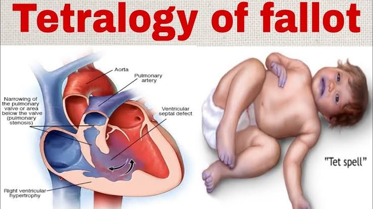
3. The Four Anatomical Defects (The “Tetralogy”): The underlying developmental error in TOF is the anterior and cephalad (upward) deviation of the conal septum, which divides the ventricles’ outflow tracts. This single embryological anomaly leads to the four characteristic defects:
- Ventricular Septal Defect (VSD): A large hole in the septum (the wall) that separates the two lower chambers of the heart (the right and left ventricles).
- Pulmonary Stenosis (PS): A narrowing of the pulmonary valve and the outflow tract from the right ventricle to the pulmonary artery. This obstruction restricts blood flow to the lungs.
- Overriding Aorta: The aorta, the main artery carrying oxygenated blood to the body, is displaced. Instead of arising exclusively from the left ventricle, it sits over the VSD, receiving blood from both the right and left ventricles.
- Right Ventricular Hypertrophy (RVH): The muscular wall of the right ventricle becomes thicker than normal. This is a secondary, adaptive change, as the right ventricle must pump harder to push blood past the narrowed pulmonary valve.
The severity of the clinical symptoms is almost entirely determined by the degree of pulmonary stenosis. This narrowing dictates how much blood can get to the lungs and, consequently, how much deoxygenated (“blue”) blood is shunted through the VSD into the overriding aorta and out to the body.
Recognizing the Clinical Presentation
The signs and symptoms of Tetralogy of Fallot can vary widely depending on the severity of the pulmonary stenosis.
1. Cyanosis: This is the hallmark sign of TOF. Cyanosis is a bluish discoloration of the skin, lips, and nail beds caused by an insufficient concentration of oxygen in the arterial blood.
- “Pink Tets”: Infants with mild pulmonary stenosis may have minimal shunting of blue blood and appear pink and healthy at birth. Their cyanosis may only become apparent weeks or months later as the pulmonary stenosis worsens.
- “Blue Babies”: Infants with severe pulmonary stenosis will present with noticeable cyanosis shortly after birth, which is often exacerbated by crying or feeding.
2. Hypercyanotic Spells (“Tet Spells”): These are acute, life-threatening episodes of profound cyanosis and hypoxia, most common in infants between 2 and 4 months of age. Triggers often include activities that decrease systemic vascular resistance or increase oxygen demand, such as crying, feeding, or defecation. During a Tet spell, a spasm of the muscle beneath the pulmonary valve dramatically worsens the outflow obstruction, forcing nearly all deoxygenated blood from the right ventricle into the aorta. This presents as:
- Sudden onset of deep cyanosis.
- Rapid and deep breathing (hyperpnea).
- Irritability and uncontrollable crying.
- In severe cases, limpness, loss of consciousness, or seizures. Older children instinctively learn to alleviate these spells by squatting. This posture increases systemic vascular resistance, which in turn reduces the right-to-left shunting of blood across the VSD and forces more blood through the narrowed pulmonary artery to the lungs.
3. Other Common Signs:
- Heart Murmur: A loud, harsh, systolic ejection murmur is typically heard at the upper left sternal border, caused by blood flow across the narrowed pulmonary valve.
- Poor Weight Gain and Failure to Thrive: Due to the increased metabolic demands of breathing and the poor oxygenation, infants may feed poorly and fail to grow at a normal rate.
- Clubbing: In older, uncorrected children, a painless rounding and thickening of the fingertips and toes may develop as a chronic sign of low blood oxygen levels.
The Diagnostic Workup
A diagnosis of TOF is typically suspected based on clinical presentation and confirmed with a series of diagnostic tests.
- Physical Examination and Pulse Oximetry: Initial suspicion arises from observing cyanosis and hearing the characteristic heart murmur. Pulse oximetry, a non-invasive test measuring blood oxygen saturation, will show low levels (often 75-90%).
- Chest X-ray (CXR): The chest radiograph often reveals a classic “boot-shaped heart” (cœur en sabot). This shape is created by the upturned cardiac apex due to right ventricular hypertrophy and a concave main pulmonary artery segment due to its underdevelopment.
- Electrocardiogram (ECG): An ECG measures the heart’s electrical activity. In TOF, it will typically show evidence of right axis deviation and right ventricular hypertrophy.
- Echocardiogram (Cardiac Ultrasound): This is the definitive diagnostic tool. An echocardiogram uses sound waves to create detailed, real-time images of the heart’s structure and function. It can clearly visualize all four anatomical defects, assess the severity of the pulmonary stenosis, identify the VSD’s size and location, and map the coronary artery anatomy, which is critical for surgical planning.
- Cardiac Catheterization and MRI: In complex cases or when the anatomy is not fully clear on echocardiogram, a cardiac MRI or cardiac catheterization may be performed. These provide more detailed information about the pulmonary arteries and coronary artery anatomy before surgical intervention.
Management and Treatment
The management of Tetralogy of Fallot is a two-pronged approach involving medical stabilization and definitive surgical correction.
1. Medical Management of Tet Spells: The primary goal is to interrupt the hypercyanotic spell. Interventions include:
- Knee-to-Chest Position: Placing the infant in this position mimics squatting, increasing systemic vascular resistance.
- Calming the Infant: Reducing anxiety decreases oxygen demand.
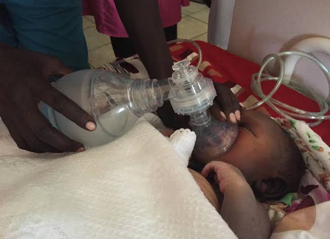
- Supplemental Oxygen: Helps to improve oxygenation, although its primary effect is pulmonary vasodilation.
- Medications: If the spell persists, intravenous medications like morphine (to sedate and reduce respiratory drive), propranolol (a beta-blocker to relax the sub-pulmonary muscle), or phenylephrine (to increase systemic vascular resistance) may be administered.
2. Surgical Management: Surgery is the only definitive treatment for TOF. The timing and type of surgery depend on the infant’s size, symptoms, and specific anatomy.
- Palliative Shunt (e.g., Blalock-Taussig-Thomas Shunt): In the past, this was a common first step. Today, it is reserved for specific cases, such as low-birth-weight infants or those with complex coronary anatomy. A synthetic tube (shunt) is placed between a major body artery (like the subclavian artery) and the pulmonary artery. This creates a reliable, secondary pathway for blood to reach the lungs for oxygenation, alleviating cyanosis until the infant is large enough for full repair.
- Complete Intracardiac Repair: This is the standard of care and is typically performed between 3 and 6 months of age. The open-heart procedure involves two main steps:
- Closure of the VSD: The hole between the ventricles is closed with a synthetic patch, stopping the mixing of oxygenated and deoxygenated blood.
- Relief of the Right Ventricular Outflow Tract Obstruction (RVOTO): The surgeon widens the narrowed pulmonary valve and outflow passage, often by removing excess muscle tissue and sometimes placing a patch across the area to enlarge it.
3. Long-Term Follow-Up: Surgical repair of TOF is corrective, not a cure. All patients require lifelong follow-up with a cardiologist, preferably one specializing in adult congenital heart disease (ACHD). Long-term concerns include:
- Pulmonary Regurgitation: The repaired or replaced pulmonary valve may become leaky over time, leading to right ventricle enlargement and dysfunction. This is the most common reason for re-operation decades later.
- Arrhythmias: Scar tissue from surgery can lead to irregular heart rhythms.
- Residual VSD or Stenosis: Small leaks or areas of narrowing may persist.
Despite these potential issues, the vast majority of individuals who undergo successful surgical repair for Tetralogy of Fallot have an excellent long-term prognosis and can enjoy active, high-quality lives.
References:
- Bailliard, F., & Anderson, R. H. (2009). Tetralogy of Fallot. Orphanet Journal of Rare Diseases, 4(1), 2.
- Apitz, C., Webb, G. D., & Redington, A. N. (2009). Tetralogy of Fallot. The Lancet, 374(9699), 1462-1471.
- Villafañe, J., Feinstein, J. A., Jenkins, K. J., Vincent, R. N., Walsh, E. P., Dubin, A. M., … & Cohen, M. S. (2013). Hot topics in tetralogy of Fallot. Journal of the American College of Cardiology, 62(23), 2155-2166.
- Centers for Disease Control and Prevention (CDC). (2022). Facts about Tetralogy of Fallot. Retrieved from https://www.cdc.gov/ncbddd/heartdefects/tetralogyoffallot.html
- Mayo Clinic. (2022). Tetralogy of Fallot. Retrieved from https://www.mayoclinic.org/diseases-conditions/tetralogy-of-fallot/symptoms-causes/syc-20353477


