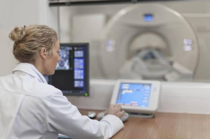
In the field of medical imaging, precision is paramount. Every step, from the moment a patient is referred for an examination to the final archiving of their images and records, is part of a critical pathway designed to ensure patient safety, diagnostic accuracy, and regulatory compliance. This systematic, standardized procedure is often referred to as the radiographic work drill. Understanding this workflow is fundamental for every radiologic technologist, administrator, and healthcare professional involved in the imaging process.
Definition of the Radiographic Work Drill
The radiographic work drill is a standardized sequence of operations and verification checks performed by a radiologic technologist before, during, and after a diagnostic imaging procedure. It is not merely a routine but a deliberate, safety-critical protocol designed to minimize errors, ensure the correct procedure is performed on the correct patient, and produce high-quality diagnostic images.
Think of it as the equivalent of a pilot’s pre-flight checklist. Each step is methodical and serves a specific purpose, collectively ensuring a safe and effective outcome. This drill encompasses patient identification, request form verification, patient preparation, equipment setup, image acquisition, and post-procedure documentation. Adherence to this work drill is a hallmark of a professional and safe imaging department.
The Significance, Purpose, and Process of Patient Identification
Patient identification is arguably the most critical step in the entire radiographic workflow. A failure at this stage can lead to catastrophic consequences, including misdiagnosis, unnecessary radiation exposure to the wrong individual, and delayed treatment for the correct patient.
Significance and Purpose: The primary purpose of rigorous patient identification is to unequivocally confirm that the individual undergoing the examination is the same person for whom the procedure was ordered. This prevents “wrong-patient” events, which are considered sentinel events by regulatory bodies like The Joint Commission. Proper identification ensures that the resulting medical images are correctly assigned to the patient’s electronic health record (EHR), forming an accurate diagnostic history.
The Process (A Step-by-Step Approach): A robust patient identification process requires at least two unique patient identifiers. Reliance on a single identifier, such as the patient’s room number, is insufficient and unsafe.
- Request Verification: The technologist begins by reviewing the radiographic request form.
- Greeting the Patient: The technologist greets the patient by name.
- Active Identification: The technologist must use active, open-ended questions. Instead of asking, “Are you John Smith?” (which can elicit a false positive from a confused or hard-of-hearing patient), the technologist should ask, “Please state your full name and date of birth.”
- Cross-Verification: The patient’s verbal response is then cross-referenced against the information on the radiographic request form and the patient’s identification wristband (for inpatients).
- Final Confirmation: For outpatients, a photo ID can be used as an additional verifier. This two-identifier method (e.g., name and date of birth) is the industry standard for ensuring patient safety.
Usage and Significance of the Radiographic Request Form
The radiographic request form (or electronic order) is the primary legal and medical document that authorizes a radiographic examination. It serves as the formal communication channel between the referring physician and the radiology department.
Usage: The technologist meticulously reviews the request form to understand:
- Patient Demographics: Name, date of birth, hospital number, etc., for identification.
- Ordering Physician: To know who will receive the final report.
- Clinical History/Indication: This is crucial. It answers the “why” of the exam. A history of “cough and fever” for a chest X-ray is far more informative than just “rule out pneumonia,” as it guides the radiologist’s interpretation.
- Specific Examination Ordered: E.g., “Left Wrist, 3 Views” or “Chest X-ray, PA and Lateral.”
- Special Instructions: Notes on patient isolation, allergies (especially for contrast studies), or mobility limitations.
Significance:
- Legal Authorization: An examination performed without a valid request can be considered battery.
- Diagnostic Guidance: The clinical history provides context for the radiologist, leading to a more accurate and relevant diagnostic report. For example, knowing a patient has a history of trauma to the ankle directs the radiologist to look for subtle fractures.
- Justification for Radiation Exposure: The request form documents the medical necessity of the procedure, justifying the use of ionizing radiation under the ALARA (As Low As Reasonably Achievable) principle.
Common Medical Terms and Abbreviations in Radiographic Request Forms
Radiographic requests are filled with medical shorthand. Technologists must be fluent in this language to perform the correct examination.
| Abbreviation | Full Term | Meaning/Context |
|---|---|---|
| CXR | Chest X-ray | Standard imaging of the chest. |
| AP | Anteroposterior | X-ray beam enters the anterior (front) and exits the posterior (back). |
| PA | Posteroanterior | X-ray beam enters the posterior (back) and exits the anterior (front). |
| Lat | Lateral | A side view of the body part. |
| KUB | Kidneys, Ureters, Bladder | An abdominal X-ray, typically to assess for stones or bowel gas patterns. |
| Fx | Fracture | A suspected or confirmed break in a bone. |
| SOB | Shortness of Breath | A common clinical indication for a chest X-ray. |
| R/O | Rule Out | To perform an exam to confirm or deny the presence of a specific condition. |
| Hx | History | Refers to the patient’s clinical history (e.g., “Hx of falls”). |
| NPO | Nil Per Os (Nothing by Mouth) | An instruction for patients, often for exams requiring contrast or sedation. |
| STAT | Statim (Immediately) | Indicates the exam is urgent and must be performed without delay. |
Patient Registration and Record Keeping Process
Patient Registration: This is the administrative process of entering a patient into the healthcare facility’s system. It typically occurs at a central registration desk or within the radiology department for outpatients.
- Process: The registrar collects demographic information (name, address, date of birth), insurance details, and contact information. A unique Hospital Number (also known as a Medical Record Number or MRN) is assigned to the patient. This number becomes the primary key linking all of the patient’s records, appointments, and billing information within that healthcare system.
- Importance: Accurate registration is the foundation of the patient’s electronic health record. Errors at this stage can lead to billing issues, fragmented medical records, and potential patient identification errors downstream.
Record Keeping Process: Modern record keeping is predominantly digital, managed by two key systems:
- Radiology Information System (RIS): This system manages all radiology-specific data, including patient scheduling, exam orders, report dictation and distribution, and billing information.
- Picture Archiving and Communication System (PACS): This is the system that stores, retrieves, and displays the medical images themselves.
The process ensures that every image is permanently linked to the correct patient’s file, accessible for future reference, comparison studies, and legal purposes.
Patient Identification and Verification with Specific Identifiers
This section expands on Step 2 by defining the specific identifiers used in the verification process.
- Patient’s Name: The full legal name. It is a primary but non-unique identifier (many people can have the same name).
- Hospital Number (MRN): A unique number assigned by the healthcare facility during registration. It is a highly reliable identifier specific to that institution.
- X-ray Identification Number (Accession Number): This is a unique number assigned by the RIS specifically for a single imaging examination or series. While the Hospital Number follows the patient for life, a new Accession Number is generated for each new exam order. It links the images, the request, and the final report for a particular visit.
- Cross-Reference Bill with Patient’s Name: In some outpatient settings, the patient’s bill or receipt for the procedure may contain their name and the service ordered. While not a primary identifier, it can serve as an additional layer of verification, confirming that the patient who paid for a “Left Hand X-ray” is indeed the person present for that exam.
The gold standard verification involves matching the patient’s verbal confirmation of their Name and Date of Birth with the data on the request form, which also contains their Hospital Number and the exam’s unique Accession Number.
The Usage and Importance of the Radiographic Examination Logbook
Despite the move to digital systems like RIS and PACS, a physical or digital radiographic examination logbook remains a vital tool in many departments.
Usage: The logbook is a chronological record of every examination performed in a specific X-ray room. For each entry, the technologist typically records:
- Date and time of the exam
- Patient’s full name and hospital/X-ray number
- The specific examination performed (e.g., “CXR PA/Lat”)
- The name or initials of the performing technologist
- Exposure factors used (kVp, mAs)
- Number of images taken, including any repeats
- Notes on any incidents or difficulties during the procedure
Importance:
- Legal Document: The logbook serves as a legal record of departmental activity. In case of litigation, it can provide evidence of what procedure was performed, by whom, and when.
- Quality Assurance (QA): By tracking repeat images and reasons for them (e.g., patient motion, technique error), supervisors can identify trends, troubleshoot equipment problems, and provide targeted training for technologists.
- Workload and Staffing Analysis: The log provides a raw data set for managers to analyze room usage, procedure volume, and peak activity times, which helps in making informed decisions about staffing and resource allocation.
- Radiation Dose Tracking: Recording exposure factors contributes to dose monitoring programs, helping to ensure that departmental protocols adhere to the ALARA principle.
In conclusion, the radiographic work drill is a comprehensive system where each step is interlinked. From the accuracy of the initial registration to the final entry in a logbook, these processes work in concert to protect the patient, support the clinician, and produce a diagnostic record of the highest quality and integrity.
References
- The Joint Commission. (2023). National Patient Safety Goals. Retrieved from https://www.jointcommission.org/standards/national-patient-safety-goals/
- Adler, A. M., & Carlton, R. R. (2018). Introduction to Radiologic and Imaging Sciences and Patient Care (7th ed.). Elsevier.
- American Society of Radiologic Technologists (ASRT). (2021). Practice Standards for Medical Imaging and Radiation Therapy. ASRT.
- Ballinger, P. W., Frank, E. D., & Merrill, V. (2016). Merrill’s Atlas of Radiographic Positioning & Procedures (13th ed.). Mosby Elsevier.
- World Health Organization (WHO). (2016). Patient Identification: WHO Guidelines on Hand Hygiene in Health Care. Geneva: World Health Organization.


