Urolithiasis, commonly known as kidney stone disease, is a prevalent and often recurrent condition characterized by the formation of solid crystalline structures, or calculi, within the urinary tract. These stones can develop anywhere from the kidneys to the bladder, causing significant pain, obstruction, infection, and, if left untreated, potential long-term kidney damage. Understanding the multifaceted nature of urolithiasis, including its causes, types, and metabolic associations, is crucial for effective prevention and management.
Understanding Urolithiasis
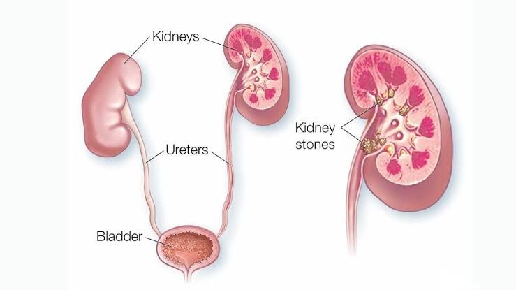
Urolithiasis refers to the pathological condition involving the formation of “stones” (calculi) within the urinary system. These stones are concretions of mineral and organic matrix, typically forming when certain substances in the urine become highly concentrated and crystallize. While they can occur at any point along the urinary tract, from the renal calyces to the urethra, the most common site of origin is the kidney, hence the frequent use of the term “renal stones” or “kidney stones.”
The incidence of urolithiasis is increasing globally, influenced by dietary changes, lifestyle factors, and environmental conditions. It affects individuals across all age groups, though it is most common in adults aged 30-50 years, with a higher prevalence in males. The clinical presentation often involves excruciating pain (renal colic) due to stone movement and obstruction, hematuria (blood in urine), and sometimes urinary tract infections.
Types of Renal Stones
Renal stones are not homogenous; they are composed of various chemical substances, each with distinct characteristics and predisposing factors. The primary types of renal stones include:
- Calcium Stones: These are the most common type, accounting for approximately 70-80% of all kidney stones. They are further subdivided into:
- Calcium Oxalate Stones: The predominant form, occurring as monohydrate (whewellite) or dihydrate (weddellite).
- Calcium Phosphate Stones: Less common than oxalate stones, often associated with alkaline urine.
- Struvite Stones: Also known as magnesium ammonium phosphate stones or “infection stones,” comprising about 10-15% of cases.
- Uric Acid Stones: Representing 5-10% of stones, often linked to metabolic conditions.
- Cystine Stones: A rare type, accounting for 1-2% of stones, resulting from a genetic metabolic disorder.
- Other Rare Stones: Including xanthine stones, drug-induced stones, and silicate stones.
Etiology and Pathogenesis of Renal Stones
The formation of renal stones is a complex process involving a confluence of genetic, environmental, dietary, and metabolic factors. The fundamental principle revolves around the supersaturation of urine with stone-forming constituents, which allows for crystal nucleation, growth, aggregation, and ultimate retention within the renal parenchyma or collecting system.
Key Pathogenic Mechanisms:
- Supersaturation: Urine becomes supersaturated when the concentration of stone-forming ions (e.g., calcium, oxalate, uric acid, phosphate, magnesium, ammonium) exceeds their solubility limits. This can occur due to low urine volume (dehydration) or excessive excretion of these substances.
- Nucleation: Once supersaturation is reached, crystals begin to form. This can be:
- Homogeneous Nucleation: Spontaneous formation of crystals without a pre-existing template, requiring very high levels of supersaturation.
- Heterogeneous Nucleation: Crystal formation on a pre-existing solid surface or “nidus.” This is more common and occurs at lower levels of supersaturation. A common nidus is Randall’s plaque, an interstitial apatite deposit in the renal papilla.
- Crystal Growth and Aggregation: Once formed, small crystals can grow by further deposition of ions or aggregate together to form larger particles. Inhibitors of crystal growth and aggregation (e.g., citrate, pyrophosphate, Tamm-Horsfall protein, osteopontin) normally present in urine help prevent this process. A deficiency or absence of these inhibitors increases stone risk.
- Retention: For a stone to become clinically significant, the formed crystals must be retained within the urinary tract. This often involves adherence of crystals to the renal tubular epithelium, particularly at sites of injury or inflammation, or attachment to Randall’s plaques.
- Urine pH: The pH of urine significantly influences the solubility of various stone-forming compounds. For example, uric acid is less soluble in acidic urine, while calcium phosphate and struvite stones form more readily in alkaline urine.
- Dietary Factors: High intake of sodium, animal protein, and oxalate-rich foods can increase stone risk. Conversely, a diet rich in fruits, vegetables, and adequate fluid intake can reduce the risk.
- Fluid Intake: Chronic dehydration leads to decreased urine volume, increasing solute concentration and promoting supersaturation.
- Urinary Tract Infections (UTIs): Certain bacteria, particularly urea-splitting organisms (e.g., Proteus mirabilis, Klebsiella spp.), produce urease, an enzyme that hydrolyzes urea into ammonia and carbon dioxide. This process raises urine pH and increases ammonium and phosphate concentrations, directly leading to the formation of struvite stones.
- Anatomical Abnormalities: Conditions causing urinary stasis or obstruction (e.g., ureteropelvic junction obstruction, medullary sponge kidney, horseshoe kidney, strictures) increase the risk of stone formation by allowing more time for crystal aggregation and growth.
- Genetic Predisposition: A family history of kidney stones significantly increases an individual’s risk, suggesting a genetic component. Specific genetic disorders, such as cystinuria and primary hyperoxaluria, directly cause stone formation.
Co-relating the Occurrence of Renal Stones with Different Metabolic Diseases
Renal stone formation is frequently a manifestation of underlying systemic metabolic disorders, highlighting the importance of a comprehensive metabolic evaluation in recurrent stone formers.
- Hypercalciuria: This is the most common metabolic abnormality in calcium stone formers, defined as excessive calcium excretion in the urine.
- Absorptive Hypercalciuria: Increased intestinal absorption of calcium, leading to elevated serum calcium and subsequent renal excretion.
- Resorptive Hypercalciuria: Primarily due to hyperparathyroidism, where excessive parathyroid hormone (PTH) causes increased bone resorption and renal calcium reabsorption, leading to hypercalcemia and hypercalciuria.
- Renal Hypercalciuria: Impaired renal tubular reabsorption of calcium, resulting in excessive calcium excretion even with normal calcium intake. This leads to secondary hyperparathyroidism.
- Association: Primarily with calcium oxalate and calcium phosphate stones.
- Hyperoxaluria: Elevated urinary oxalate excretion.
- Primary Hyperoxaluria: A rare genetic disorder involving enzyme defects (e.g., alanine-glyoxylate aminotransferase deficiency in Type I PH) that lead to overproduction of oxalate by the liver.
- Enteric Hyperoxaluria: Occurs in individuals with malabsorption syndromes (e.g., Crohn’s disease, ulcerative colitis, celiac disease, bariatric surgery). Unabsorbed fatty acids bind calcium in the gut, leaving free oxalate to be absorbed and excreted in urine.
- Dietary Hyperoxaluria: High intake of oxalate-rich foods (e.g., spinach, rhubarb, nuts, chocolate).
- Association: Almost exclusively with calcium oxalate stones.
- Hyperuricosuria: High levels of uric acid in the urine.
- Causes: High purine intake (animal protein), gout, certain myeloproliferative disorders, rapid tissue breakdown.
- Association: Directly causes uric acid stones, especially in persistently acidic urine. Uric acid crystals can also act as heterogeneous nucleators for calcium oxalate stones.
- Hypocitraturia: Low levels of citrate in the urine. Citrate is a crucial inhibitor of calcium stone formation as it binds to calcium, reducing its availability for crystal formation, and also inhibits crystal aggregation.
- Causes: Metabolic acidosis (e.g., renal tubular acidosis), chronic diarrhea, high animal protein diet.
- Association: Predisposes to calcium oxalate and calcium phosphate stones.
- Renal Tubular Acidosis (RTA) Type 1 (Distal RTA): A defect in the renal tubules’ ability to excrete acid, leading to systemic metabolic acidosis and persistently alkaline urine.
- Association: Strongly linked to calcium phosphate stones due to alkaline urine and associated hypocitraturia.
- Cystinuria: An inherited autosomal recessive disorder characterized by defective transport of the amino acids cystine, ornithine, lysine, and arginine (COLA) in the renal tubules and intestinal tract. Cystine is poorly soluble in urine.
- Association: Exclusively causes cystine stones, which are often recurrent and can form large staghorn calculi.
- Gout: A metabolic disorder characterized by hyperuricemia (high uric acid in blood) and deposition of uric acid crystals in joints.
- Association: Individuals with gout frequently develop uric acid stones due to chronically high uric acid levels and often, persistently acidic urine.
- Obesity and Metabolic Syndrome: These conditions are associated with systemic insulin resistance and may lead to a lower urine pH, increased uric acid excretion, and altered ammonium handling.
- Association: Increased risk of uric acid stones and, to a lesser extent, calcium oxalate stones.
Differentiating Between the Different Renal Stones
Differentiating between stone types is crucial for guiding targeted therapeutic and preventive strategies. This table summarizes key characteristics:
| Stone Type | Frequency | Predisposing Factors | Urine pH | Morphology (Gross & Microscopic) | Radiopacity |
|---|---|---|---|---|---|
| Calcium Oxalate | 70-80% | Hypercalciuria, hyperoxaluria, hypocitraturia, low urine volume, high sodium/protein diet. | Normal to slightly acidic (5.5-6.8) | Varied; rough, spiky, dark brown/black (monohydrate, Whewellite); yellow/tan, blocky, “envelope” shape (dihydrate, Weddellite). | Radiopaque (dense) |
| Calcium Phosphate | 5-15% | Renal Tubular Acidosis (Type 1), hyperparathyroidism, persistently alkaline urine. | Persistently alkaline (>7.0) | White, amorphous, smooth, often crystalline or granular. Can be lamellar (apatite) or needle-like (brushite). | Radiopaque (dense) |
| Struvite | 10-15% | Chronic UTIs with urea-splitting bacteria (Proteus, Klebsiella, Pseudomonas, Staphylococcus). | Highly alkaline (>7.5-9.0) | White/grey, soft, friable, often large “staghorn” calculi filling renal pelvis/calyces. | Fairly radiopaque |
| Uric Acid | 5-10% | Hyperuricosuria, persistently acidic urine (<5.5), gout, dehydration, metabolic syndrome, high purine diet. | Consistently acidic (<5.5) | Yellow to reddish-brown, smooth or finely granular. Rhomboid, barrel, or rosette-shaped crystals. | Radiolucent (poorly visible on X-ray, visible on CT) |
| Cystine | 1-2% | Genetic disorder (Cystinuria) affecting renal amino acid transport. | Variable (5.0-7.0), solubility increases at alkaline pH. | Yellowish, waxy, “grapefruit” appearance, often recurrent. Hexagonal crystals. | Moderately radiopaque (ground glass appearance) |
Conclusion
Urolithiasis is a complex and often debilitating condition with significant healthcare implications. Its pathogenesis is multifactorial, stemming from an interplay of genetic predispositions, dietary habits, environmental factors, and underlying metabolic derangements. A thorough understanding of the different types of renal stones, their specific etiologies, and their correlation with various metabolic diseases is paramount. Accurate diagnosis and stone analysis are critical, as they inform tailored preventive strategies and treatment plans, ultimately aiming to reduce stone recurrence and preserve renal function. Managing urolithiasis extends beyond acute symptom relief, emphasizing comprehensive metabolic evaluation and patient education for long-term stone prevention.


