Peripheral nerves are the intricate network responsible for transmitting signals between the brain and spinal cord and the rest of the body, including muscles, organs, and skin. They facilitate movement, sensation, and autonomic functions critical for daily life. Injuries to these vital pathways can significantly impair function and quality of life. Understanding the types, causes, and management of peripheral nerve injuries, including common conditions like entrapment neuropathies and specifically Carpal Tunnel Syndrome, is crucial for effective diagnosis and treatment.
Types and Management of Peripheral Nerve Injury
A peripheral nerve injury refers to damage to any part of the peripheral nervous system, which includes all nerves outside the brain and spinal cord. These injuries can range from mild to severe, impacting the nerve’s ability to transmit signals.
Classification of Peripheral Nerve Injuries
Peripheral nerve injuries are typically classified based on the degree of damage to the nerve’s structure. Two widely recognized classification systems are Seddon’s and Sunderland’s, which provide a framework for understanding prognosis and guiding treatment.
(a) Seddon’s Classification (1943)
Seddon’s classification categorizes nerve injuries into three grades:
- Neuropraxia:
- Description: This is the mildest form of nerve injury, involving a temporary conduction block without anatomical disruption of the axon. The myelin sheath may be locally compressed or demyelinated.
- Mechanism: Typically caused by transient compression, stretch, or ischemia.
- Clinical Features: Patients experience temporary motor weakness or paralysis, sensory deficits (numbness, tingling), but autonomic function is usually preserved.
- Prognosis: Full recovery is expected within hours to weeks, as the axon and its supporting structures remain intact. Wallerian degeneration (degeneration of the axon distal to the injury) does not occur.
- Axonotmesis:
- Description: This injury involves disruption of the axon and its myelin sheath, but the surrounding connective tissue sheaths (endoneurium, perineurium, epineurium) remain intact.
- Mechanism: Caused by more severe crushing, prolonged compression, or stretching injuries.
- Clinical Features: Complete motor, sensory, and autonomic loss distal to the injury site occurs. Wallerian degeneration takes place, meaning the axon distal to the injury site degenerates.
- Prognosis: Recovery is possible as the intact connective tissue sheaths provide a pathway for axonal regeneration. Regeneration occurs at a rate of approximately 1 mm per day or 1 inch per month, making recovery slow and potentially incomplete depending on the distance regeneration needs to cover and the quality of regeneration. Surgical exploration is usually not required unless the extent of the injury is unclear.
- Neurotmesis:
- Description: This is the most severe type of nerve injury, involving complete anatomical transection or severe disruption of the entire nerve trunk, including the axon, myelin, and all connective tissue sheaths.
- Mechanism: Often results from lacerations, avulsions, or severe crush injuries.
- Clinical Features: Complete and permanent loss of motor, sensory, and autonomic function distal to the injury. Wallerian degeneration occurs.
- Prognosis: Spontaneous recovery is highly unlikely or very poor due to the complete disruption of the nerve’s architecture. Surgical intervention (nerve repair or grafting) is almost always required to facilitate any meaningful recovery.
(b) Sunderland’s Classification (1951)
Sunderland expanded on Seddon’s classification, dividing nerve injuries into five (and sometimes six, including mixed injuries) degrees, offering a more precise understanding of the extent of damage and aiding in prognosis prediction:
- First Degree: Equivalent to Seddon’s Neuropraxia. Isolated conduction block, no Wallerian degeneration.
- Second Degree: Equivalent to Seddon’s Axonotmesis. Axon disrupted, but the endoneurium (innermost connective tissue sheath) is intact. Wallerian degeneration occurs, good prognosis for recovery.
- Third Degree: Axon and endoneurium are disrupted, but the perineurium (sheath around nerve fascicles) remains intact. Wallerian degeneration occurs. Recovery is less predictable than second degree due to potential scarring within the endoneurial tubes, which can hinder regenerating axons.
- Fourth Degree: Axon, endoneurium, and perineurium are disrupted, but the epineurium (outermost connective tissue sheath) remains intact. Wallerian degeneration occurs. Spontaneous recovery is minimal as scar tissue within the nerve prevents axonal regrowth across the injury site. Surgical repair is often necessary.
- Fifth Degree: Equivalent to Seddon’s Neurotmesis. Complete transection of the entire nerve trunk. No spontaneous recovery without surgical intervention.
- Sixth Degree (Mixed Injury): A combination of the above injury types within a single nerve trunk.
Clinical Presentation and Diagnosis of Peripheral Nerve Injury
Patients with peripheral nerve injuries typically present with a combination of motor, sensory, and autonomic deficits corresponding to the distribution of the affected nerve. Symptoms may include weakness or paralysis, numbness, tingling (paresthesias), burning pain, and sometimes changes in skin temperature, sweating, or texture.
Diagnosis involves:
- Detailed History and Physical Examination: Assessing the mechanism of injury, onset, severity, and distribution of symptoms. A thorough neurological examination mapping sensory and motor deficits is crucial.
- Electrodiagnostic Studies (Nerve Conduction Studies & Electromyography – NCS/EMG): These tests measure the electrical activity of nerves and muscles, helping to localize the lesion, determine the type and severity of nerve damage, and assess prognosis. They can differentiate between demyelination and axonal loss.
- Imaging Studies: Magnetic Resonance Imaging (MRI) or High-Resolution Ultrasound can visualize nerve continuity, swelling, compression, or the presence of neuromas (nerve tumors).
Management of Peripheral Nerve Injury
Management strategies depend heavily on the type and severity of the nerve injury, as well as the time elapsed since injury.
- Non-Surgical Management:
- Observation: For neuropraxia and mild axonotmesis, observation is often the primary approach, as spontaneous recovery is expected.
- Conservative Care: This includes rest, immobilization (splinting or bracing) to protect the nerve and prevent further damage, pain management (NSAIDs, neuropathic pain medications), and early physical and occupational therapy.
- Physical and Occupational Therapy: Essential for maintaining joint range of motion, preventing muscle atrophy (through electrical stimulation if needed), and re-educating muscles and sensory pathways as regeneration occurs.
- Surgical Management:
- Indications: Typically reserved for neurotmesis, severe axonotmesis (Sunderland third, fourth, or fifth degree), or when there’s no sign of recovery after a reasonable observation period.
- Nerve Repair (Neurorrhaphy): If the nerve ends are clean and close, they can be directly sutured together (epineurial repair or fascicular repair).
- Nerve Grafting: If there’s a gap between the severed nerve ends that cannot be bridged directly, a segment of nerve from another part of the body (autograft, e.g., sural nerve) or a cadaveric nerve (allograft) is used to bridge the gap.
- Nerve Transfer: Involves transferring a healthy, less critical nerve or nerve branch to reinnervate a denervated muscle or sensory territory, especially when direct repair or grafting is not feasible.
- Decompression: For nerve compression injuries (like entrapment neuropathies), surgical release of the compressing structure can alleviate pressure and allow the nerve to recover.
- Rehabilitation:
- Post-surgical or prolonged conservative management, rehabilitation is paramount. It aims to maximize functional recovery, prevent complications (e.g., contractures, chronic pain), and facilitate return to daily activities. This often involves intense physical and occupational therapy, sensory re-education, and adaptive equipment.
Entrapment Neuropathies
Entrapment neuropathies are a common subset of peripheral nerve injuries where a specific nerve is compressed, stretched, or pinched as it passes through a narrow anatomical space. This chronic compression can lead to nerve dysfunction, causing pain, sensory disturbances, and weakness in the area supplied by the nerve.
General Principles of Entrapment Neuropathies
- Mechanism: The common underlying mechanism is mechanical compression, often exacerbated by repetitive movements, sustained postures, or swelling in the confined space. This compression compromises blood supply to the nerve (ischemia) and can directly damage the myelin sheath (demyelination) or, in severe cases, the axons themselves (axonal degeneration).
- Common Sites: Entrapment neuropathies can occur at various anatomical sites where nerves pass through narrow tunnels or tight fibrous bands. Examples include:
- Median Nerve: Carpal Tunnel Syndrome (at the wrist).
- Ulnar Nerve: Cubital Tunnel Syndrome (at the elbow).
- Radial Nerve: Radial Tunnel Syndrome or Saturday Night Palsy (at the forearm/arm).
- Peroneal Nerve: At the fibular head (common in leg crossing).
- Tibial Nerve: Tarsal Tunnel Syndrome (at the ankle).
- Lateral Femoral Cutaneous Nerve: Meralgia Paresthetica (at the groin).
- Clinical Presentation: Symptoms typically follow the distribution of the affected nerve and include:
- Sensory: Numbness, tingling, burning pain, or altered sensation.
- Motor: Weakness, muscle atrophy, or difficulty with fine motor control (in later stages).
- Symptoms often worsen with specific activities or positions and may be exacerbated at night.
- Management Principles:
- Conservative: Rest, activity modification, splinting/bracing, NSAIDs, physical therapy, and corticosteroid injections (to reduce inflammation).
- Surgical: Decompression of the nerve by releasing the compressing structure is often curative when conservative measures fail or in cases of severe nerve compression with progressive deficits.
Carpal Tunnel Syndrome (CTS)
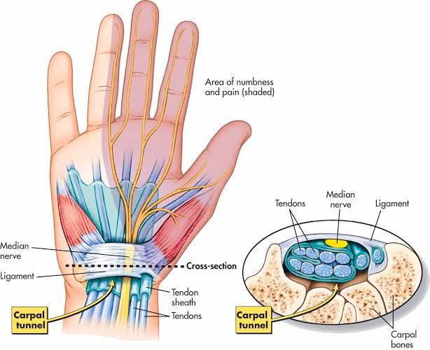
Carpal Tunnel Syndrome (CTS) is the most prevalent entrapment neuropathy, affecting millions worldwide. It results from the compression of the median nerve as it passes through the carpal tunnel at the wrist.
Anatomy of the Carpal Tunnel
The carpal tunnel is a narrow, rigid passageway located on the palm side of the wrist. Its floor and sides are formed by the carpal (wrist) bones, creating a bony arch. The roof is formed by the strong, fibrous transverse carpal ligament (also known as the flexor retinaculum). Passing through this confined space are nine flexor tendons (responsible for finger and thumb movement) and the median nerve. Any condition that reduces the space within this tunnel or increases the volume of its contents can compress the median nerve.
Risk Factors for Carpal Tunnel Syndrome
CTS is often multifactorial, with several factors contributing to its development:
- Repetitive and Forceful Hand/Wrist Use: Occupations involving repetitive wrist flexion/extension, forceful gripping, or prolonged vibration (e.g., assembly line work, computer use, carpentry, hairdressing) are frequently associated with CTS.
- Systemic Medical Conditions:
- Diabetes Mellitus: Causes changes in nerve structure and microvasculature, making nerves more susceptible to compression.
- Hypothyroidism: Can lead to fluid retention and tissue swelling.
- Rheumatoid Arthritis and other Inflammatory Arthropathies: Inflammation of the flexor tendon sheaths (tenosynovitis) can increase pressure within the tunnel.
- Obesity: Associated with increased systemic inflammation and pressure.
- Pregnancy: Hormonal changes and fluid retention can lead to transient CTS, which often resolves after childbirth.
- Kidney Failure (especially with dialysis): Amyloid deposition can occur.
- Acromegaly: Enlargement of tissues.
- Anatomical Factors:
- Smaller Carpal Tunnel Size: Some individuals may have naturally narrower carpal tunnels.
- Wrist Fractures or Dislocations: Can alter the anatomy of the carpal tunnel, leading to narrowing or direct nerve impingement.
- Ganglion Cysts or Tumors: Rarely, space-occupying lesions within the tunnel can cause compression.
- Genetics: A family history of CTS may indicate a genetic predisposition.
- Age and Sex: More common in middle-aged and older adults, and significantly more prevalent in women, possibly due to hormonal factors, smaller wrist size, and higher prevalence of related systemic conditions.
Clinical Features of Carpal Tunnel Syndrome
The symptoms of Carpal Tunnel Syndrome typically follow the distribution of the median nerve in the hand:
- Sensory Symptoms:
- Numbness and Tingling (Paresthesias): Most common initial symptoms, usually affecting the thumb, index finger, middle finger, and the radial (thumb) half of the ring finger. The little finger is generally spared.
- Pain: Can occur in the affected fingers, palm, wrist, and sometimes radiate up the forearm, arm, and even shoulder.
- Nocturnal Exacerbation: Symptoms are often worse at night, waking the patient from sleep. This is thought to be due to fluid shifts and wrist positioning during sleep.
- “Flick Sign”: Patients often report shaking or “flicking” their hands to relieve symptoms.
- Motor Symptoms (usually later stages):
- Weakness: Difficulty with fine motor tasks, gripping objects, or dropping items due to weakness in the muscles supplied by the median nerve (especially the abductor pollicis brevis and opponens pollicis, part of the thenar eminence).
- Muscle Atrophy: Visible wasting of the thenar muscles (the fleshy mound at the base of the thumb) is a late and severe sign, indicating chronic nerve compression and axonal loss.
- Provocative Tests (used during physical examination):
- Phalen’s Maneuver: Holding the wrists in maximal flexion for 30-60 seconds. A positive test reproduces numbness or tingling in the median nerve distribution.
- Tinel’s Sign: Tapping directly over the median nerve at the carpal tunnel. A positive test elicits tingling or “pins and needles” sensations in the median nerve distribution.
- Durkan’s Compression Test: Direct pressure over the median nerve in the carpal tunnel for up to 30 seconds. Reproduction of symptoms indicates a positive test and is highly sensitive.
Diagnosis of Carpal Tunnel Syndrome
Diagnosis is primarily clinical, based on the patient’s history and a thorough physical examination.
- Electrodiagnostic Studies (NCS/EMG): While not strictly necessary for mild cases, NCS/EMG are often performed to confirm the diagnosis, determine the severity of nerve compression, rule out other neuropathies (e.g., cervical radiculopathy), and provide a baseline before treatment.
- Ultrasound: High-resolution ultrasound can visualize nerve swelling and compression, and may be used in some centers.
Management of Carpal Tunnel Syndrome
Management of CTS follows a step-wise approach, starting with conservative measures and progressing to surgery if conservative treatments fail or if the condition is severe.
- Non-Surgical (Conservative) Management:
- Wrist Splinting: Wearing a rigid wrist splint, especially at night, to keep the wrist in a neutral position. This reduces pressure on the median nerve during sleep and often provides significant symptom relief.
- Activity Modification and Ergonomics: Identifying and avoiding activities that aggravate symptoms. Adjusting workstation ergonomics to promote neutral wrist postures.
- Non-Steroidal Anti-Inflammatory Drugs (NSAIDs): May provide temporary pain relief but are generally not effective in resolving the underlying compression.
- Corticosteroid Injections: Injection of corticosteroids directly into the carpal tunnel can reduce inflammation and swelling, providing temporary relief (often weeks to months). They can also be diagnostic, confirming the carpal tunnel as the source of symptoms. However, frequent injections are not recommended due to potential side effects and diminishing returns.
- Physical Therapy: Nerve gliding exercises and manual therapy techniques may be beneficial for some patients.
- Surgical Management (Carpal Tunnel Release):
- Indications: Surgery is considered when conservative treatments fail to provide lasting relief, symptoms are severe and progressive, or there is objective evidence of median nerve damage (e.g., persistent numbness, muscle weakness, or significant findings on NCS/EMG).
- Procedure: Carpal Tunnel Release involves surgically dividing (transecting) the transverse carpal ligament, which forms the roof of the carpal tunnel. This releases pressure on the median nerve, decompressing it.
- Open Carpal Tunnel Release: Involves a small incision in the palm.
- Endoscopic Carpal Tunnel Release: Involves smaller incisions and uses an endoscope (a small camera) to visualize and cut the ligament. Both techniques have high success rates.
- Prognosis: The vast majority of patients experience significant symptom relief and improvement in nerve function after surgery. Recovery of sensation and strength can take weeks to months, especially if nerve damage was severe. Post-operative hand therapy is often crucial for optimal recovery, strengthening, and preventing stiffness.
Conclusion
Peripheral nerve injuries, including entrapment neuropathies like Carpal Tunnel Syndrome, represent a broad spectrum of conditions that can severely impact an individual’s motor and sensory function. A thorough understanding of their classification, risk factors, clinical presentation, and management strategies is paramount for effective patient care. Early and accurate diagnosis, coupled with appropriate conservative or surgical interventions and timely rehabilitation, offers the best chance for alleviating symptoms, restoring nerve function, and improving the overall quality of life for affected individuals.


