Choroidal melanoma, the most common primary intraocular malignancy in adults, is a serious condition originating from the melanocytes of the choroid, the vascular layer of the eye. While relatively rare, its potential for metastasis, particularly to the liver, significantly impacts patient mortality. Understanding this complex disease requires a comprehensive overview of its underlying causes, clinical presentation, diagnostic modalities, therapeutic strategies, and profound implications for survival.
Etiology of Choroidal Melanoma
The exact etiology of choroidal melanoma remains largely unknown, but a combination of genetic predisposition and environmental factors is believed to contribute to its development.
- Genetic Factors:
- BAP1 Gene Mutation: Mutations in the BAP1 (BRCA1-associated protein 1) tumor suppressor gene are the most significant genetic risk factor for aggressive choroidal melanoma with a high propensity for metastasis. Germline BAP1 mutations are associated with a familial cancer syndrome (BAP1 tumor predisposition syndrome), increasing the risk of various cancers, including uveal melanoma, mesothelioma, renal cell carcinoma, and cutaneous melanoma.
- GNAQ/GNA11 Mutations: Somatic mutations in the GNAQ and GNA11 genes, encoding G-protein alpha subunits, are found in approximately 80-90% of choroidal melanomas. While these mutations are typically driver mutations for tumor development, they are generally associated with less aggressive tumors compared to BAP1 mutations.
- Chromosomal Abnormalities: Specific chromosomal aberrations, particularly monosomy of chromosome 3 (loss of one copy of chromosome 3) and gain of chromosome 8q, are strong prognostic markers for metastasis and poor survival. These aberrations often correlate with BAP1 mutations.
- Environmental Factors:
- Ultraviolet (UV) Radiation: Unlike cutaneous melanoma, the role of UV radiation in choroidal melanoma is less definitively established. While some studies suggest a weak association, the choroid’s shielded location within the eye makes direct UV exposure less impactful. However, indirect effects or cumulative lifetime exposure might play a subtle role.
- Occupational Exposures: Some research has explored links to certain occupational exposures, but no conclusive evidence points to a specific carcinogen.
- Risk Factors:
- Fair Skin and Light Eye Color: Individuals of Caucasian descent with fair skin, light hair, and light eye color (blue, green) have a higher incidence of choroidal melanoma, similar to cutaneous melanoma.
- Ocular Melanocytosis (Nevus of Ota): This congenital condition, characterized by increased melanocyte numbers in the uvea and periocular skin, significantly increases the risk of developing uveal melanoma within the affected eye.
- Atypical Mole Syndrome/Dysplastic Nevus Syndrome: While primarily associated with cutaneous melanoma, individuals with these conditions may have a slightly increased risk of choroidal melanoma.
- Precursor Lesions (Choroidal Nevi): Approximately 10% of the population has choroidal nevi (benign freckles of the choroid). While most nevi remain benign, a small percentage can transform into melanoma. Features indicating a higher risk of transformation include thickness >2 mm, presence of subretinal fluid, symptoms, orange pigment (lipofuscin), and halo absence (mnemonic: “To Find Small Ocular Melanomas, Use Helpful Hints Daily” – Thickness, Fluid, Symptoms, Orange pigment, Margin near optic disc, Ultrasonographic hollowness, Halo absence, Drusen absence).
Clinical Features
The clinical presentation of choroidal melanoma can be highly variable, ranging from asymptomatic discovery to significant visual impairment. Early detection is often challenging due to the tumor’s location and slow growth.
- Asymptomatic Presentation: Many small to medium-sized choroidal melanomas are discovered incidentally during routine ophthalmic examinations, especially in their early stages.
- Visual Symptoms: As the tumor grows or causes complications, patients may experience:
- Blurred Vision: The most common symptom, occurring when the tumor involves or affects the fovea (central retina).
- Scotoma (Blind Spot): A localized area of visual field loss.
- Flashes of Light (Photopsia) and Floaters: Caused by retinal irritation or detachment.
- Reduced Visual Acuity: Progressive loss of sight as the tumor enlarges or causes secondary retinal detachment.
- Metamorphopsia: Distortion of straight lines, often due to subretinal fluid affecting the macula.
- Pain: Ocular pain is uncommon and typically indicates complications such as secondary glaucoma (due to tumor-induced angle closure or neovascularization) or extensive inflammation.
- Ophthalmoscopic Signs (Visible on Fundus Examination):
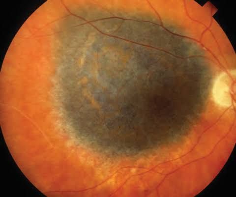
- Pigmented or Amelanotic Mass: The tumor typically appears as a solitary, elevated, dome-shaped, or mushroom-shaped lesion. While usually pigmented (brown to black), amelanotic (non-pigmented, yellowish) melanomas can occur, making diagnosis more challenging.
- Subretinal Fluid: Accumulation of fluid under the retina, often leading to serous retinal detachment, a common complication.
- Orange Pigment (Lipofuscin): Clumps of orange-yellow pigment on the tumor surface, representing degenerated retinal cells or macrophages, are a strong indicator of malignancy.
- Drusen: Absences of drusen (small yellowish deposits) on the tumor surface, despite presence on adjacent choroid, can be a subtle sign distinguishing melanoma from a benign nevus.
- Prominent Feeding Vessels: Dilated episcleral or sentinel vessels may be observed, indicating increased blood supply to the tumor.
- “Collar-button” or “Mushroom” Shape: Occurs when the tumor breaks through Bruch’s membrane, creating a narrow neck and a broader domed cap.
- Extraocular Extension: Rare but serious, indicating advanced disease with tumor growth outside the globe.
- Secondary Glaucoma or Cataract: Late-stage complications.
Investigations
A multi-modal approach combining clinical examination with advanced imaging is crucial for accurate diagnosis, staging, and management planning of choroidal melanoma.
- Ophthalmic Examination:
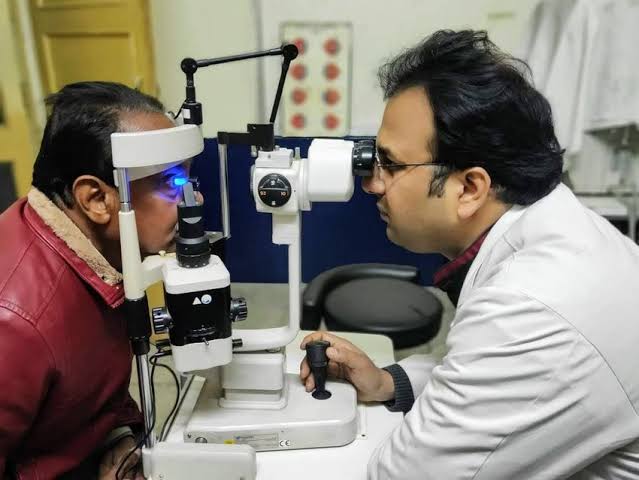
- Slit-lamp Biomicroscopy: To assess the anterior segment for any signs of tumor extension or secondary complications.
- Dilated Fundus Examination: Performed using indirect ophthalmoscopy with scleral indentation and slit-lamp biomicroscopy with a contact or non-contact lens to visualize the tumor’s size, shape, pigmentation, and associated features.
- Fundus Photography: Baseline and serial photographs are essential for documenting the tumor’s appearance and monitoring growth or changes over time.
- Ocular Imaging Modalities:
- Ocular Ultrasonography (B-scan): The cornerstone of diagnosis. Characteristic features include a solid, dome-shaped or mushroom-shaped mass with low to medium internal reflectivity, often with choroidal excavation (acoustic hollowing) and orbital shadowing. A-scan ultrasonography measures tumor dimensions (thickness, base) crucial for staging and treatment planning.
- Optical Coherence Tomography (OCT): Provides high-resolution cross-sectional images of the tumor-retina interface, revealing subretinal fluid, photoreceptor atrophy, intraretinal edema, and the presence of overlying orange pigment.
- Fluorescein Angiography (FFA): Demonstrates early choroidal filling followed by “double circulation” (arterial phase filling of choroidal and retinal circulations) and late leakage in the tumor, often with pinpoint hyperfluorescence.
- Indocyanine Green Angiography (ICG-A): Superior to FFA for visualizing choroidal circulation and can show intrinsic tumor vasculature and filling defects.
- Fundus Autofluorescence (FAF): Identifies areas of lipofuscin accumulation (orange pigment) overlying the tumor, appearing as hyperautofluorescent spots, indicating metabolic activity.
- Systemic Evaluation for Metastasis: Choroidal melanoma commonly metastasizes hematogenously, predominantly to the liver. Therefore, systemic workup is crucial at diagnosis and for ongoing surveillance.
- Liver Function Tests (LFTs): To screen for liver involvement.
- Abdominal Ultrasound/MRI of Liver: The primary imaging modalities for detecting liver metastases. MRI is more sensitive than ultrasound.
- Chest X-ray or CT Chest: To rule out lung metastases, though less common than liver involvement.
- Positron Emission Tomography (PET) Scan: May be used in cases of suspected metastasis or for equivocal findings on other imaging, but not routinely for initial staging of primary tumor.
- Biopsy and Genetic Profiling:
- Fine Needle Aspiration Biopsy (FNAB): While not routinely performed for primary diagnosis due to potential risks (e.g., tumor seeding, hemorrhage) and high diagnostic accuracy of imaging, FNAB can be used in atypical cases or for prognostic genetic profiling.
- Genetic Profiling: Analysis of tumor tissue (often obtained via FNAB or during enucleation) for BAP1 status, GNAQ/GNA11 mutations, and chromosomal aberrations (e.g., monosomy 3, 8q gain) provides crucial prognostic information regarding the risk of metastasis.
Management
The management of choroidal melanoma is complex, aiming to eradicate the primary tumor, preserve useful vision if possible, and prevent metastasis. Treatment decisions depend on tumor size, location, patient’s general health, and visual potential of the affected eye. A multidisciplinary team approach is often employed.
- Observation: For very small, suspicious lesions (often categorized as “small choroidal nevus with suspicious features”), careful observation with serial photography, ultrasonography, and OCT is an option. Treatment is initiated upon documented growth or development of malignant features.
- Local Treatment (Eye-Sparing Modalities): The goal is to destroy the tumor while preserving the eye and, ideally, vision.
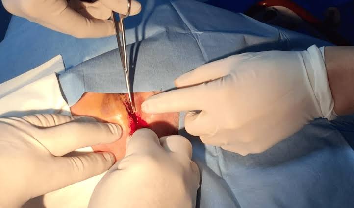
- Brachytherapy (Plaque Radiotherapy): The most common eye-sparing treatment for small to medium-sized tumors. A radioactive plaque (e.g., Iodine-125, Ruthenium-106) is surgically sutured to the sclera over the tumor and left in place for several days, delivering a high dose of radiation directly to the tumor while minimizing exposure to surrounding healthy tissues.
- External Beam Radiotherapy (EBRT):
- Proton Beam Radiotherapy (PBRT): Utilizes accelerated proton beams, which deposit most of their energy at a specific depth (Bragg peak), allowing highly conformal dose delivery to the tumor with minimal damage to adjacent structures. Effective for medium to large tumors.
- Stereotactic Radiosurgery (SRS): A form of highly focused photon radiation, delivering a high dose in a single or few fractions, typically for smaller or peripapillary tumors.
- Transpupillary Thermotherapy (TTT): Uses a diode laser applied through the pupil to heat and destroy the tumor. Primarily effective for small tumors, often used as an adjunct to brachytherapy or for very small lesions.
- Local Resection (Endoresection/Exoresection): Surgical removal of the tumor while preserving the eye. Technically challenging and carries risks of complications (hemorrhage, retinal detachment, seeding), typically reserved for selected cases in specialized centers.
- Enucleation: Surgical removal of the entire eye. This is indicated for:
- Large tumors (typically >10 mm thickness or >16 mm basal diameter) where eye-sparing therapies are unlikely to be effective or carry unacceptable risks.
- Tumors with extensive retinal detachment or secondary neovascular glaucoma, leading to a painful, blind eye.
- Recurrence after primary eye-sparing treatment.
- Tumors with documented extraocular extension.
- Systemic Treatment (for Metastatic Disease): Once choroidal melanoma metastasizes, particularly to the liver, prognosis is poor, and treatment options are limited and often palliative.
- Immunotherapy: Checkpoint inhibitors (e.g., nivolumab, pembrolizumab, ipilimumab) have shown limited response rates in metastatic uveal melanoma compared to cutaneous melanoma, though research is ongoing.
- Targeted Therapies: Studies targeting specific pathways (e.g., MEK inhibitors, protein kinase C inhibitors) have shown some promise in specific patient subsets but are not widely curative.
- Chemotherapy: Generally ineffective in controlling metastatic choroidal melanoma.
- Hepatic-directed Therapies: Given the liver’s role as the primary site of metastasis, therapies directly targeting liver metastases include surgical resection, chemoembolization (TACE), radioembolization (TARE), and isolated hepatic perfusion. These aim to control local liver disease and improve quality of life, but rarely cure.
- Clinical Trials: Patients with metastatic disease are often encouraged to participate in clinical trials exploring novel therapies.
- Follow-up: Regular ophthalmic examinations are essential to monitor for recurrence or complications. Systemic surveillance (e.g., liver imaging, LFTs) is crucial to detect metastases early, as this is the primary determinant of survival. The frequency and type of surveillance depend on the initial tumor and its genetic risk profile.
Importance of Choroidal Melanoma on Mortality
Choroidal melanoma’s most significant impact on mortality stems directly from its high propensity for metastatic spread, which transforms a potentially curable localized disease into a life-threatening systemic illness.
- High Metastatic Potential:
- Unlike many solid tumors, choroidal melanoma typically metastasizes hematogenously (via the bloodstream) rather than via the lymphatic system.
- The liver is the most common site of metastasis, accounting for approximately 90% of all metastatic cases. Less commonly, metastases can occur in the lungs, bone, skin, and brain.
- Metastasis can occur many years after successful treatment of the primary ocular tumor, making long-term surveillance mandatory.
- Poor Prognosis for Metastatic Disease:
- Once choroidal melanoma metastasizes, the prognosis is grim. The median survival time after documented metastasis is typically less than one year.
- The lack of highly effective systemic therapies for metastatic uveal melanoma, unlike the advances seen in cutaneous melanoma, is a critical factor contributing to high mortality. Uveal melanoma is often resistant to conventional chemotherapy and shows limited response to many immunotherapies.
- Prognostic Factors for Metastasis:
- Tumor Size: Larger primary tumors are associated with a higher risk of metastasis. Tumors >10 mm in thickness or >16 mm in basal diameter carry a significantly increased metastatic risk.
- Histopathological Features: The presence of epithelioid cells (rather than spindle cells) in the tumor, high mitotic activity, and microvascular density are associated with a higher metastatic risk.
- Genetic Markers: This is arguably the most powerful predictor.
- Monosomy 3: Loss of one copy of chromosome 3 is the strongest independent predictor of metastasis, present in approximately 50% of tumors, and associated with a very poor prognosis.
- BAP1 Mutation: Loss of BAP1 function, often correlated with monosomy 3, is directly linked to increased metastatic potential and significantly reduced survival.
- Gain of 8q: An additional copy of the long arm of chromosome 8 is also associated with an increased risk of metastasis.
- Class 2 Tumors: Based on gene expression profiling (GEP), “Class 2” tumors (distinguished by their gene expression profile) have a high risk of metastasis, correlating strongly with monosomy 3 and BAP1 mutations.
- Challenges in Early Detection of Metastasis:
- Micrometastases can be present at the time of primary diagnosis but are undetectable by current imaging modalities. These silent seeds can grow and become clinically apparent years later.
- The predominantly hepatic tropism means that even small, asymptomatic liver lesions can progress rapidly and fatally.
- Survival Rates:
- For localized choroidal melanoma, the 5-year survival rate is generally good (ranging from 80-90% depending on tumor size and type).
- However, the 10-year survival rate drops significantly due to late metastases, highlighting the persistent threat of the disease.
- Once distant metastasis is confirmed, the 5-year survival rate plummets to less than 15%.
In conclusion, while significant advancements have been made in the diagnosis and local treatment of choroidal melanoma, its profound impact on patient mortality remains a critical challenge. The aggressive metastatic potential, particularly to the liver, coupled with the current limitations in effective systemic therapies for advanced disease, underscores the urgent need for continued research into novel targeted therapies and improved surveillance strategies to mitigate this life-threatening consequence. Multidisciplinary care, including ophthalmic oncologists, radiation oncologists, medical oncologists, and genetic counselors, is paramount for optimizing patient outcomes and managing the complex implications of this disease on survival.


