The production and maturation of red blood cells (RBCs), a process known as erythropoiesis, is a meticulously regulated physiological mechanism essential for maintaining oxygen homeostasis within the body. This intricate process involves a complex interplay of hormonal signals, nutrient availability, and environmental cues, all coordinated to ensure an optimal supply of oxygen-carrying erythrocytes. Understanding the multifaceted factors governing erythropoiesis and RBC maturation is fundamental to comprehending both normal physiological function and various hematological disorders.
Overview of Erythropoiesis: The Developmental Journey of a Red Blood Cell
Erythropoiesis is the specific lineage within hematopoiesis that leads to the formation of mature red blood cells. This process occurs primarily in the red bone marrow of adults and involves a series of differentiation and maturation steps from a pluripotent hematopoietic stem cell (HSC) to a fully functional erythrocyte. The key stages include:
- Hematopoietic Stem Cell (HSC): The multipotent precursor residing in the bone marrow, capable of self-renewal and differentiation into various blood cell lineages.
- Common Myeloid Progenitor (CMP): HSCs differentiate into CMPs, which are committed precursors for myeloid cells, including the erythroid lineage.
- Erythroid Burst-Forming Unit (BFU-E): The earliest recognizable erythroid progenitor, highly sensitive to erythropoietin (EPO), and capable of forming large colonies in culture.
- Erythroid Colony-Forming Unit (CFU-E): A more mature progenitor than BFU-E, with fewer proliferative capabilities but a higher sensitivity to EPO, leading to smaller colonies.
- Proerythroblast (Pronormoblast): The first morphologically identifiable erythroid precursor, characterized by a large nucleus and basophilic cytoplasm.
- Basophilic Erythroblast: Further differentiation leads to increased hemoglobin synthesis and accumulation, with the cytoplasm becoming less basophilic.
- Polychromatophilic Erythroblast: Hemoglobin synthesis continues, causing the cytoplasm to exhibit a mixed basophilic and eosinophilic (polychromatic) appearance. Nuclear chromatin condensation accelerates.
- Orthochromatophilic Erythroblast (Normoblast): The nucleus becomes pyknotic (condensed and inactive), and the cytoplasm is predominantly eosinophilic due to maximal hemoglobin content. The nucleus is eventually extruded from the cell.
- Reticulocyte: An anucleated immature RBC that still contains residual ribosomal RNA (reticulum), visible with supravital stains. Reticulocytes are released from the bone marrow into the peripheral blood and mature into erythrocytes within 1-2 days.
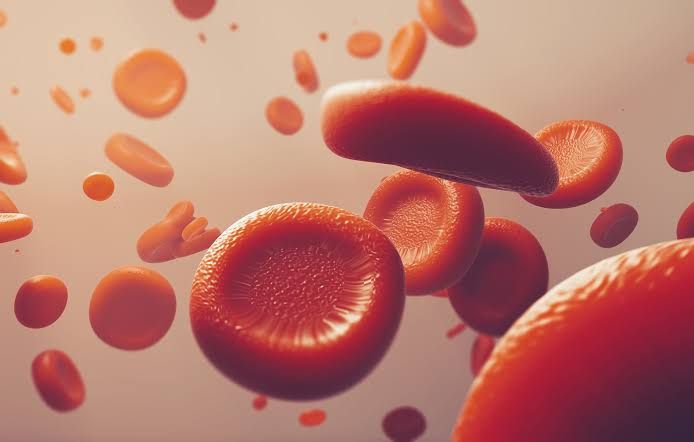
- Mature Erythrocyte: A biconcave disc, anucleated cell, rich in hemoglobin, designed for efficient oxygen transport.
This orderly progression is tightly controlled by a range of factors that influence proliferation, differentiation, and survival at each stage.
Factors Regulating Erythropoiesis
The regulation of erythropoiesis is a dynamic process ensuring that the rate of RBC production matches the body’s oxygen demands. Several critical factors orchestrate this balance:
- a. Oxygen Availability (Hypoxia) – The Primary Stimulus: Hypoxia, defined as a deficiency in the amount of oxygen reaching the tissues, is the single most potent physiological stimulus for erythropoiesis. The body’s sensor for oxygen levels is primarily located in the renal cortex, although other tissues like the liver also play a role. When oxygen delivery to the kidneys is reduced (e.g., due to anemia, high altitude, respiratory disease, or cardiac failure), specialized peritubular interstitial cells in the renal cortex detect this decrease.
- Mechanism of Hypoxia-Induced Erythropoiesis: The cellular mechanism involves a complex pathway centered around Hypoxia-Inducible Factors (HIFs). Under normoxic conditions, HIF-α subunits are hydroxylated by prolyl hydroxylases (PHDs), marking them for degradation by the ubiquitin-proteasome pathway. However, under hypoxic conditions, PHDs are inhibited, leading to the stabilization and accumulation of HIF-α. HIF-α then translocates to the nucleus, heterodimerizes with HIF-β, and binds to hypoxia-responsive elements (HREs) in the promoter region of target genes, most notably the erythropoietin (EPO) gene. This transcriptional activation leads to a significant increase in EPO synthesis and secretion.
- Relative Importance of Hypoxia: Hypoxia stands out as the paramount physiological inducer of erythropoiesis due to its direct link to the primary function of RBCs – oxygen transport. The entire regulatory system is geared towards maintaining tissue oxygenation. Any condition that compromises oxygen delivery, whether a decrease in atmospheric oxygen (high altitude), impaired lung function (respiratory disease), reduced cardiac output (heart failure), or a reduced number of RBCs (anemia), immediately triggers the hypoxic response, leading to increased EPO production and subsequent erythropoiesis. This robust feedback loop ensures a rapid and effective compensatory mechanism to restore oxygen homeostasis. Its ability to directly modulate EPO production, the master regulator of erythropoiesis, underscores its unmatched significance.
- b. Hormonal Regulation: Erythropoietin (EPO) Erythropoietin (EPO) is the principal hormone regulating erythropoiesis. It is a glycoprotein hormone primarily produced by specialized peritubular interstitial cells in the kidneys (90%) and, to a lesser extent, by perisinusoidal cells in the liver (10%).
- Role in Regulating RBC Production: EPO acts as the “master switch” for RBC production. Its secretion is inversely proportional to the tissue oxygen levels. When oxygen levels decrease (hypoxia), EPO synthesis and release increase dramatically.
- Mechanism of Action: EPO circulates in the blood and binds to specific EPO receptors (EPORs) expressed on the surface of erythroid progenitor cells (BFU-E and CFU-E) in the bone marrow. This binding triggers a signaling cascade, primarily through the JAK2-STAT5 pathway, which promotes:
- Proliferation: Stimulates the rapid division of erythroid progenitor cells.
- Differentiation: Directs these cells towards the erythroid lineage.
- Survival: Inhibits apoptosis (programmed cell death) of erythroid precursors, allowing more cells to mature.
- Hemoglobin Synthesis: Enhances the rate of hemoglobin production within the developing cells.
- Premature Release: Under severe hypoxic stress, it can also promote the early release of reticulocytes from the bone marrow. The combined effects of EPO lead to an accelerated production of mature RBCs, thereby increasing the oxygen-carrying capacity of the blood and alleviating the initial hypoxic stimulus.
- c. Nutritional Factors: Adequate nutrition is critical for the robust production and maturation of RBCs. Deficiencies in these nutrients can lead to various forms of anemia.
- Iron: Absolutely essential for hemoglobin synthesis. Each hemoglobin molecule contains four heme groups, and each heme group contains an iron atom. Iron is incorporated into protoporphyrin to form heme. Iron deficiency is the most common cause of anemia worldwide (iron-deficiency anemia), resulting in microcytic, hypochromic RBCs.
- Proteins/Amino Acids: Required for the synthesis of globin chains (the protein component of hemoglobin) and the structural components of the cell membrane and enzymes.
- Vitamins:
- Pyridoxine (Vitamin B6): A coenzyme required for heme synthesis.
- Riboflavin (Vitamin B2): Involved in various enzymatic reactions, including those related to iron metabolism.
- Niacin (Vitamin B3): Involved in metabolic pathways.
- Ascorbic Acid (Vitamin C): Enhances iron absorption from the gut and is involved in maintaining the ferric state of iron in tissues.
- Vitamin E: An antioxidant that protects RBC membranes from oxidative damage.
- d. Bone Marrow Environment and Other Growth Factors: The bone marrow provides a crucial microenvironment (stromal cells, extracellular matrix) that supports erythropoiesis. Various cytokines and growth factors, besides EPO, play roles, particularly in the earlier stages of progenitor cell development:
- Interleukin-3 (IL-3): Supports the proliferation of early hematopoietic stem cells and progenitor cells, including BFU-E.
- Granulocyte-Macrophage Colony-Stimulating Factor (GM-CSF): Also supports the proliferation of early myeloid progenitors.
- Stem Cell Factor (SCF): Synergizes with EPO and other cytokines to promote the proliferation and survival of early erythroid precursors.
Role of Erythropoietin in Regulating RBC Production
As highlighted earlier, EPO is the principal regulator. To elaborate:
- Primary Sensor and Effector: The kidneys act as the primary oxygen sensors and the main producers of EPO. This unique arrangement ensures a direct and rapid response to changes in blood oxygen levels.
- Feedback Loop: The EPO regulatory system operates as a classic negative feedback loop. When tissue oxygen delivery is adequate, EPO production is basal. When tissue oxygen levels drop, the kidneys release more EPO. This increased EPO stimulates erythropoiesis, leading to an increase in RBC mass and thus increased oxygen-carrying capacity. As oxygen delivery improves, the renal oxygen sensors detect this correction, and EPO production returns to basal levels. This feedback mechanism ensures that RBC mass is maintained within a narrow physiological range, precisely matching the body’s oxygen demands.
- Clinical Significance: The understanding of EPO’s role has revolutionized the management of anemia, particularly in chronic kidney disease (CKD). Patients with CKD often develop severe anemia due to impaired renal EPO production. Recombinant human erythropoietin (rHuEPO), administered therapeutically, effectively stimulates erythropoiesis in these patients, reducing the need for blood transfusions and improving their quality of life. rHuEPO is also used in certain cancer-related anemias and for patients undergoing specific surgical procedures.
Role of Vitamin B12 (Cobalamin) and Folic Acid in Maturation of RBC
While iron and EPO are critical for RBC production quantity, Vitamin B12 and Folic Acid are indispensable for the quality and proper maturation of RBCs, specifically for nuclear maturation and cell division. Deficiencies in either lead to megaloblastic anemia, characterized by abnormally large (macrocytic) red blood cells with immature nuclei and ineffective erythropoiesis.
- a. Their Crucial Role in DNA Synthesis: Both Vitamin B12 and Folic Acid (in its active form, tetrahydrofolate or THF) are vital coenzymes in the synthesis of purines and pyrimidines, the building blocks of DNA. They are specifically involved in the one-carbon metabolism pathways essential for rapid cell proliferation.
- Impaired DNA Synthesis: In the absence of adequate B12 or folate, DNA replication is impaired. Cells attempt to divide but cannot complete nuclear maturation and division efficiently. This leads to a delay in nuclear development (maturation arrest) relative to cytoplasmic maturation. The cells continue to grow in size due to ongoing protein synthesis (including hemoglobin), resulting in large, immature cells known as megaloblasts in the bone marrow and macrocytes in the peripheral blood.
- b. Vitamin B12 (Cobalamin):
- Absorption and Metabolism: Vitamin B12 is unique as its absorption requires intrinsic factor, a glycoprotein secreted by parietal cells in the stomach. The B12-intrinsic factor complex is absorbed in the terminal ileum.
- Functions in Erythropoiesis: Vitamin B12 acts as a coenzyme in two critical metabolic reactions:
- Conversion of Methylmalonyl-CoA to Succinyl-CoA: This reaction is important for fatty acid metabolism and myelin sheath maintenance. Its disruption contributes to the neurological symptoms seen in B12 deficiency.
- Conversion of Homocysteine to Methionine: This reaction is vital for regenerating tetrahydrofolate (THF) from methyl-THF. THF is essential for DNA synthesis. Without B12, methyl-THF accumulates (“folate trap”), making THF unavailable for purine and pyrimidine synthesis.
- Deficiency: Vitamin B12 deficiency (e.g., due to pernicious anemia, malabsorption, or strict vegan diet) leads to megaloblastic anemia and can also cause demyelination of nerves, leading to neurological manifestations.
- c. Folic Acid (Folate):
- Absorption and Metabolism: Folic acid is absorbed primarily in the duodenum and jejunum. It’s converted to its active form, tetrahydrofolate (THF), within cells.
- Functions in Erythropoiesis: THF is a crucial coenzyme in one-carbon unit transfer reactions, which are indispensable for:
- De Novo Purine Synthesis: Essential for DNA and RNA.
- Thymidylate Synthesis: Conversion of deoxyuridylate (dUMP) to deoxythymidylate (dTMP), a critical step for DNA synthesis. This reaction requires 5,10-methylenetetrahydrofolate.
- Deficiency: Folic acid deficiency (e.g., due to inadequate dietary intake, malabsorption, increased demand like pregnancy, or certain medications) also results in megaloblastic anemia. Unlike B12 deficiency, it typically does not cause neurological symptoms.
- d. Interdependence of B12 and Folic Acid: The metabolic pathways of Vitamin B12 and Folic Acid are intricately linked. The conversion of homocysteine to methionine, a reaction critical for maintaining methionine levels (and subsequent S-adenosylmethionine for methylation reactions), requires both methyl-B12 and methyl-THF. When B12 is deficient, methyl-THF cannot donate its methyl group, leading to the “folate trap” phenomenon. This traps folate in its unusable methyl-THF form, effectively causing a functional folate deficiency, even if total folate levels are normal. Therefore, adequate levels of both vitamins are essential for proper DNA synthesis and, consequently, for the normal maturation and division of erythroid precursors.
In conclusion, erythropoiesis and RBC maturation are highly regulated processes governed by a sophisticated network of factors. Hypoxia serves as the paramount physiological stimulus, prompting the kidney to secrete erythropoietin, the primary hormonal regulator. EPO then acts on bone marrow progenitor cells to enhance their proliferation, differentiation, and survival, ultimately increasing oxygen-carrying capacity. Concurrently, essential nutritional elements, particularly iron for hemoglobin synthesis and Vitamins B12 and Folic Acid for proper DNA replication and nuclear maturation, are indispensable for the production of healthy, functional red blood cells. A disruption in any of these critical factors can lead to various forms of anemia, highlighting the delicate balance required for maintaining oxygen homeostasis.


