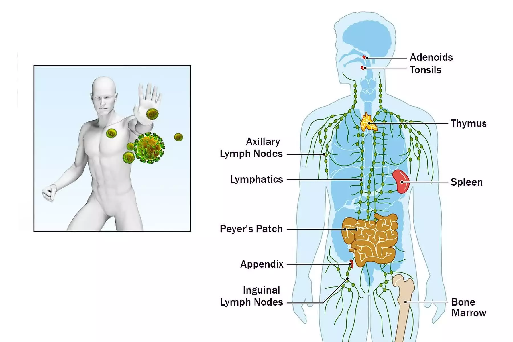
The human immune system is a complex network of cells and molecules designed to protect the body from pathogens and harmful substances. It is broadly divided into two main branches: the innate (non-specific) immune system and the adaptive (specific) immune system. The innate immune system serves as the body’s first line of defense, providing rapid, non-specific responses to potential threats. Unlike adaptive immunity, which develops memory and targets specific antigens, innate immunity recognizes general patterns associated with pathogens or tissue damage.
Cells Involved in Non-Specific Immunity
The innate immune system relies on a diverse array of cell types, primarily belonging to the myeloid lineage of hematopoietic stem cells. These cells patrol the bloodstream and reside in tissues, ready to detect and respond to danger signals. The principal cells involved in non-specific immunity include:
- Phagocytic Cells:
- Neutrophils
- Macrophages (differentiated from Monocytes)
- Granulocytes (besides Neutrophils):
- Eosinophils
- Basophils
- Other Cells:
- Natural Killer (NK) cells (possess some adaptive-like features, but generally considered part of innate immunity due to non-MHC restricted killing)
- Mast cells
- Dendritic cells (act as a bridge between innate and adaptive immunity)
This guide will focus primarily on the most prominent innate immune cells: neutrophils, eosinophils, basophils, monocytes, and macrophages.
Function of Neutrophils, Eosinophils, and Basophils
These three cell types are collectively known as granulocytes due to the prominent granules in their cytoplasm, which contain various cytotoxic molecules and mediators.
- Neutrophils:
- Neutrophils are the most abundant type of white blood cell in circulation and are typically the first responders recruited to sites of infection or inflammation.
- They are highly effective phagocytes, meaning they can engulf and internalize pathogens, cellular debris, and foreign particles. Once internalized within a phagosome, the neutrophil fuses the phagosome with lysosomes containing antimicrobial substances, leading to the destruction of the engulfed material.
- Beyond phagocytosis, neutrophils employ other killing mechanisms. They can release the contents of their granules (a process called degranulation) into the extracellular environment to directly attack larger extracellular pathogens. These granules contain enzymes like myeloperoxidase, lysozyme, and defensins.
- Another significant mechanism is the formation of Neutrophil Extracellular Traps (NETs). Upon activation, neutrophils can release their nuclear contents (DNA, histones) along with granule proteins to form a web-like structure that traps and immobilizes pathogens, particularly bacteria, allowing for their subsequent killing and degradation. Neutrophils have a relatively short lifespan at the site of infection and often undergo apoptosis or die after deploying NETs, which are then cleared by macrophages.
- Eosinophils:
- Eosinophils are less numerous than neutrophils but play a crucial role in defense against parasitic infections, such as helminths (worms).
- They are not typically effective phagocytes of large parasites. Instead, they combat these pathogens primarily through degranulation, releasing cytotoxic proteins from their prominent eosinophilic granules onto the parasite’s surface. Key granule proteins include Major Basic Protein (MBP), Eosinophil Cationic Protein (ECP), Eosinophil Peroxidase (EPO), and Eosinophil-Derived Neurotoxin (EDN). These proteins can damage the parasite’s tegument (outer layer) and disrupt its function.
- Eosinophils are also involved in allergic reactions and asthma, where their excessive activation and release of mediators can contribute to tissue damage and inflammation.
- Basophils:
- Basophils are the least common type of granulocyte in circulation.
- They are best known for their role in allergic responses. Similar to mast cells (which reside in tissues), basophils express receptors for IgE antibodies. When an allergen binds to IgE on the basophil surface, it triggers degranulation and the release of potent inflammatory mediators stored in their basophilic granules.
- Key mediators released by basophils include histamine, heparin, and various cytokines (like IL-4 and IL-13). Histamine increases vascular permeability and causes smooth muscle contraction, contributing to symptoms like swelling, redness, and bronchoconstriction in allergic reactions. Heparin is an anticoagulant.
- While primarily associated with allergy, basophils also contribute to innate immunity by releasing mediators and cytokines that can influence other immune cells and help initiate immune responses, particularly against certain parasites.
Function of Monocytes and Macrophages and Differentiating One from the Other
Monocytes and macrophages are closely related phagocytic cells that play central roles in innate immunity, inflammation, and initiating adaptive responses.
- Monocytes:
- Monocytes are circulating leukocytes that originate in the bone marrow.
- They are relatively large cells with a characteristic kidney-shaped or indented nucleus and abundant cytoplasm containing fine granules.
- They circulate in the bloodstream for a short period (typically 1-3 days).
- Their primary function is to migrate from the blood into various tissues throughout the body in response to inflammatory signals.
- Macrophages:
- Once monocytes enter tissues, they differentiate and mature into macrophages.
- Macrophages are larger than monocytes, have a more rounded nucleus, and possess more extensive cytoplasm with numerous lysosomes.
- Unlike the transient monocytes, macrophages are long-lived residents of tissues. They are found in almost every tissue, often adopting specific names based on their location (e.g., Kupffer cells in the liver, alveolar macrophages in the lungs, microglia in the central nervous system, osteoclasts in bone, histiocytes in connective tissue).
- Macrophages are highly versatile phagocytes capable of engulfing larger particles than neutrophils, including apoptotic cells, cellular debris, and large pathogens. They are crucial for clearing tissue damage and maintaining homeostasis.
- Macrophages serve as professional antigen-presenting cells (APCs). Although this function primarily links innate and adaptive immunity, their ability to process engulfed material and present components (antigens) on their surface (via MHC molecules) is initiated by their innate recognition and phagocytosis.
- Macrophages are potent producers of various cytokines, chemokines, enzymes, and other mediators that regulate inflammation, recruit other immune cells, promote tissue repair, and influence the differentiation of adaptive immune cells (see Step 5).
- Differentiation:
- The key difference lies in their location and state of maturity/differentiation. Monocytes are the circulating precursors found in the blood. Macrophages are the mature, differentiated cells that reside in tissues.
- Upon receiving specific signals (often related to inflammation or tissue microenvironment), monocytes exit the bloodstream (extravasate) and undergo significant morphological and functional changes to become macrophages tailored to the needs of the specific tissue.
- Functionally, macrophages are generally considered more robust and versatile phagocytes than monocytes and are the primary architects of the tissue-level inflammatory response and subsequent repair processes.
Mechanisms by Which Neutrophils and Macrophages Distinguish Pathogens and Normal Host Tissue
A fundamental challenge for the innate immune system is distinguishing between potentially harmful entities (pathogens, damaged cells) and healthy self-tissue to launch an appropriate attack while avoiding autoimmunity. Neutrophils and macrophages achieve this discrimination primarily through specialized receptors and context recognition.
- Pattern Recognition Receptors (PRRs): The primary mechanism involves Pattern Recognition Receptors (PRRs) expressed on the surface and within the endosomes/cytoplasm of neutrophils and macrophages. These receptors do not recognize specific molecular structures unique to a single pathogen strain (like adaptive immunity’s antigen receptors) but instead recognize conserved molecular patterns shared by broad groups of microbes or associated with cellular damage.
- Pathogen-Associated Molecular Patterns (PAMPs): These are molecular structures essential for microbial survival or pathogenicity that are not found in host cells. Examples include:
- Lipopolysaccharide (LPS) found on the outer membrane of Gram-negative bacteria.
- Peptidoglycan, a major component of bacterial cell walls.
- Flagellin, the protein subunit of bacterial flagella.
- Unmethylated CpG DNA sequences (common in bacterial DNA, rare in mammalian DNA).
- Double-stranded RNA (dsRNA), often produced during viral replication.
- Zymosan, a component of fungal cell walls.
- PRRs like Toll-like Receptors (TLRs, located on the cell surface and in endosomes) and NOD-like Receptors (NLRs, located in the cytoplasm) recognize specific PAMPs. Ligation of these receptors triggers signaling cascades within the neutrophil or macrophage, leading to activation, phagocytosis, cytokine production, and antimicrobial activities.
- Damage-Associated Molecular Patterns (DAMPs): These are molecules released or exposed by host cells undergoing stress, injury, or necrotic death, rather than controlled apoptosis. They signal “danger” or tissue damage. Examples include:
- Intracellular proteins released into the extracellular space (e.g., Heat Shock Proteins).
- Nuclear components released from damaged cells (e.g., DNA, HMGB1).
- Metabolic products released inappropriately (e.g., ATP released extracellularly, uric acid crystals).
- Certain PRRs also recognize DAMPs, initiating inflammatory responses to clear damaged tissue and promote repair.
- Pathogen-Associated Molecular Patterns (PAMPs): These are molecular structures essential for microbial survival or pathogenicity that are not found in host cells. Examples include:
- Why Normal Host Tissue Isn’t Attacked: Healthy, living host cells do not express PAMPs. While they contain the molecules that can become DAMPs, these molecules are typically contained within the cell or in specific intracellular compartments. Only upon injury or stress are DAMPs released or exposed to a level that triggers PRRs. Furthermore, host tissues express various regulatory molecules and “don’t eat me” signals (like CD47) that inhibit phagocytosis by macrophages. The presence of opsonins (see below) also strongly biases phagocytosis towards particles decorated with these tags.
- Opsonization: While not a direct recognition of the pathogen by the cell’s surface receptor (like a PRR-PAMP interaction), opsonization significantly enhances and directs phagocytosis. Pathogens can be coated with host proteins called opsonins, such as complement proteins (like C3b) or antibodies (from adaptive immunity, though complement is a key innate opsonin). Neutrophils and macrophages express receptors for these opsonins (e.g., complement receptors, Fc receptors). Binding of the opsonized pathogen to these receptors facilitates efficient engulfment, effectively marking the pathogen for destruction and helping to distinguish it from non-opsonized self-tissue.
In essence, neutrophils and macrophages discriminate by recognizing molecular patterns indicative of “non-self” (PAMPs) or “danger” (DAMPs) and are guided by opsonins that tag targets for removal, while healthy self-tissue lacks these specific “danger” or “eat me” signals and may express “don’t eat me” signals.
Pro-inflammatory Molecules Secreted by Macrophages and Their Role in Tissue Damage
Activated macrophages are potent secretors of a wide range of signaling molecules that orchestrate the inflammatory response. While inflammation is a vital host defense mechanism, excessive or chronic inflammation driven by these molecules can cause significant tissue damage.
Key pro-inflammatory molecules secreted by macrophages include:
- Tumor Necrosis Factor-alpha (TNF-alpha):
- Role in Inflammation: A central mediator of inflammation. It promotes fever, increases vascular permeability (allowing immune cells access to tissues), induces the expression of adhesion molecules on endothelial cells (facilitating leukocyte extravasation), stimulates the production of other cytokines, and can directly induce apoptosis in some cells.
- Role in Tissue Damage: Chronic or systemic release of TNF-alpha contributes significantly to cachexia (wasting), tissue remodeling, granuloma formation (which can impair organ function), and plays a major role in the pathogenesis of inflammatory diseases like rheumatoid arthritis and inflammatory bowel disease, promoting joint destruction and gut damage, respectively.
- Interleukin-1beta (IL-1beta):
- Role in Inflammation: Similar to TNF-alpha, IL-1beta is a major pyrogen (induces fever), increases vascular permeability, stimulates the production of chemokines and adhesion molecules, and promotes the activation and differentiation of various immune cells.
- Role in Tissue Damage: Persistent or excessive IL-1beta signaling contributes to chronic inflammation, cartilage degradation, bone resorption (e.g., in arthritis), and systemic inflammation that can lead to organ dysfunction.
- Interleukin-6 (IL-6):
- Role in Inflammation: A key mediator of the acute-phase response by the liver (production of proteins like C-reactive protein). It stimulates proliferation and differentiation of lymphocytes and contributes to fever.
- Role in Tissue Damage: Chronic elevation of IL-6 is associated with several inflammatory and autoimmune diseases. It contributes to systemic inflammation, anemia of chronic disease, and can promote the survival and proliferation of certain cell types involved in pathology like plasma cells in multiple myeloma or fibroblasts in fibrosis.
- Chemokines (e.g., IL-8/CXCL8, CCL2/MCP-1):
- Role in Inflammation: These are chemoattractant cytokines that guide the migration of specific immune cells from the bloodstream into the inflamed tissue. IL-8 primarily attracts neutrophils, while CCL2 attracts monocytes and T cells.
- Role in Tissue Damage: While essential for recruiting protective cells, sustained chemokine production leads to excessive accumulation of leukocytes, which can release cytotoxic molecules and contribute to damage. For instance, prolonged neutrophil accumulation can lead to bystander tissue injury from released enzymes and reactive oxygen species.
- Reactive Oxygen Species (ROS) and Reactive Nitrogen Species (RNS):
- Role in Inflammation/Killing: Produced by macrophages (and neutrophils) via the “respiratory burst,” these highly reactive molecules (e.g., superoxide anion, hydrogen peroxide, nitric oxide) are potent antimicrobial agents used to kill engulfed pathogens.
- Role in Tissue Damage: If released extracellularly in large quantities, ROS and RNS can directly damage host cells, lipids, proteins, and DNA, contributing to oxidative stress and tissue injury in inflammatory conditions.
- Proteolytic Enzymes (e.g., Matrix Metalloproteinases – MMPs):
- Role in Inflammation: Macrophages secrete enzymes that degrade the extracellular matrix (ECM), which is necessary for immune cell migration through tissues and for tissue remodeling after injury.
- Role in Tissue Damage: Uncontrolled or excessive release of these enzymes can lead to widespread degradation of connective tissues, contributing to pathology in diseases like periodontitis, atherosclerosis, and chronic wounds.
In summary, while these molecules are critical for mounting an effective immune response and clearing threats, their dysregulated or prolonged production by activated macrophages is a major driver of tissue pathology in chronic inflammatory and autoimmune diseases.
The Roles of the Classical and Alternative Pathways of Complement Activation in Host Defenses
The complement system is a crucial component of innate immunity, comprising a cascade of plasma proteins that act together to identify and eliminate pathogens. It can also be activated by the adaptive immune system. The system has three main pathways of activation: the classical, alternative, and lectin pathways. Here, we focus on the classical and alternative pathways and their roles. All pathways converge on the activation of the protein C3, leading to similar effector functions.
- Overview of Complement Functions: Regardless of the activation pathway, the main outcomes of complement activation include:
- Opsonization: Coating pathogens with complement proteins (especially C3b) to enhance phagocytosis by cells like macrophages and neutrophils, which have receptors for C3b.
- Lysis: Formation of the Membrane Attack Complex (MAC – C5b, C6, C7, C8, multiple C9 molecules) which inserts into the membrane of target cells (like bacteria), creating pores that disrupt osmotic balance and lead to cell lysis.
- Inflammation: Release of small complement fragments (anaphylatoxins like C3a and C5a) that act as chemoattractants for leukocytes (especially neutrophils and monocytes) and induce mast cell and basophil degranulation, increasing vascular permeability and promoting the inflammatory response.
- Alternative Pathway:
- Role in Host Defenses: This pathway is entirely innate and provides a rapid, antibody-independent mechanism for detecting and attacking pathogens. It is a first-line defense that is constantly operating at a low level.
- Mechanism: The alternative pathway is initiated by the spontaneous hydrolysis of C3 in plasma (“C3 tickover”) into C3a and C3b. This generated C3b is rapidly inactivated in the fluid phase. However, if this C3b encounters the surface of a pathogen (e.g., bacterial cell wall, fungal cell wall), it can bind to it. Pathogen surfaces generally lack the complement-regulatory proteins found on host cells. Bound C3b then binds Factor B, which is cleaved by Factor D, creating the C3 convertase of the alternative pathway (C3bBb). This convertase is stabilized by properdin and is highly efficient at cleaving more C3 into C3a and C3b, leading to an amplification loop on the pathogen surface. This cascade generates large amounts of C3b for opsonization and leads to the formation of the C5 convertase (C3bBbC3b), which initiates the formation of the MAC for lysis and releases C5a for inflammation.
- Significance: Its spontaneous initiation and amplification on microbial surfaces allow for immediate detection and attack of invaders without prior exposure or antibody production.
- Classical Pathway:
- Role in Host Defenses: While often associated with adaptive immunity (antibody-dependent), the classical pathway can also be activated by certain innate molecules, providing another layer of innate defense and a crucial link between innate and adaptive responses.
- Mechanism: The classical pathway is typically activated when the C1 complex binds to its activators. The most common activators are antibody-antigen complexes (specifically IgM or certain IgG subclasses). However, C1q (a component of the C1 complex) can also bind directly to certain microbial surface structures or to innate immune proteins like C-reactive protein (CRP) bound to pathogens. Binding of C1q activates associated proteases (C1r and C1s), which cleave C4 and C2. Fragments C4b and C2a combine to form the C3 convertase of the classical pathway (C4b2a). Like the alternative pathway convertase, this enzyme cleaves C3, leading to opsonization (C3b), formation of the C5 convertase (C4b2aC3b), and subsequent MAC formation (lysis) and release of anaphylatoxins (C3a, C5a).
- Significance: The classical pathway provides a mechanism for complement activation triggered by antibodies specific to a pathogen (adaptive immunity), but its ability to be activated by innate pattern recognition molecules like CRP means it also functions as an innate surveillance tool, particularly against pathogens recognized by these acute-phase proteins.
Both the classical and alternative pathways are essential for effective innate immunity, providing rapid mechanisms for identifying, tagging, and eliminating pathogens through opsonization, lysis, and the induction of inflammation, thereby clearing infections and mobilizing further immune responses.
By understanding the roles of these diverse cellular and protein components, we gain appreciation for the complexity and effectiveness of the innate immune system as the vital first line of defense against a constant barrage of potential threats.


