Hypotonic Duodenography (HD) is a specialized radiological imaging technique used for the detailed examination of the duodenum. It is a valuable tool in gastroenterology and radiology, providing enhanced visualization of the duodenal lumen and mucosal surface compared to standard barium meal studies. The key principle behind Hypotonic Duodenography is the temporary pharmacological paralysis of the duodenal smooth muscle, rendering the bowel hypotonic (relaxed and without peristalsis). This allows for sustained distension of the duodenum with contrast material and air (in double-contrast technique), enabling meticulous assessment of subtle anatomical and pathological changes.
This procedure is typically employed when standard upper gastrointestinal (UGI) barium studies (barium meal) are insufficient for diagnosing suspected duodenal pathology, or when greater detail of the duodenal anatomy is required. Conditions often investigated using HD include inflammatory diseases like Crohn’s disease, strictures, ulcers, polyps, diverticula, and suspected neoplastic lesions.
Procedure of Hypotonic Duodenography with Filming
The Hypotonic Duodenography procedure is a multi-step process requiring careful patient preparation, precise timing of contrast and pharmacological agents, and skilled radiographic technique including targeted filming. The goal is to distend the duodenum and eliminate peristalsis to obtain high-quality, static images.
1. Patient Preparation:
- Fasting: The patient is typically required to fast for a specified period (usually 6-8 hours) prior to the examination. This ensures the stomach and duodenum are empty of food and fluids, which could otherwise obscure visualization of the mucosal lining and lumen.
- Medication Review: The patient’s medication list is reviewed, particularly for drugs that might interfere with the procedure or interact with the hypotonic agent (e.g., anticholinergics, medications for glaucoma, heart conditions, or prostatic hypertrophy). Relevant medical history, including allergies and pre-existing conditions, is also assessed to identify potential contraindications.
- Explanation: The procedure is explained to the patient, including the purpose, steps involved, the sensation of receiving the injection for the hypotonic agent, and potential temporary side effects (like blurred vision or dry mouth).
2. Initial Positioning and Fluoroscopy:
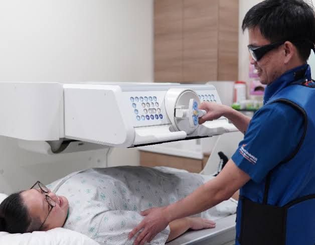
- The patient is typically positioned on the fluoroscopy table, often starting in an upright or semi-recumbent position.
- Initial fluoroscopic views of the upper abdomen may be taken to confirm that the stomach and duodenum are empty.
3. Contrast Administration:
- A high-density barium sulfate suspension is typically used as the positive contrast agent. The patient is instructed to swallow the barium.
- The radiologist uses fluoroscopy to observe the passage of barium from the esophagus into the stomach and then into the duodenum.
- Enough barium is administered to coat the duodenal mucosa and partially fill the lumen. Depending on the technique (single or double contrast), the volume of barium may vary.
4. Administration of Hypotonic Agent:
- Once sufficient barium has entered the duodenum (usually reaching at least the distal part of the C-loop), the hypotonic agent is administered.
- Commonly used agents include:
- Hyoscine-N-butylbromide (Buscopan): Administered intravenously (IV) or intramuscularly (IM). It is an anticholinergic agent that inhibits smooth muscle contraction. Its effect is usually rapid (within a few minutes) and lasts for 15-20 minutes.
- Glucagon: Administered IV or IM. It is a polypeptide hormone that inhibits smooth muscle motility. Onset is typically within 5-10 minutes, with effects lasting 10-20 minutes. Glucagon is often preferred in patients with contraindications to anticholinergics.
- The timing of the injection is critical to ensure the duodenum is adequately filled with barium before the muscle relaxation takes effect.
5. Patient Manipulation and Positioning:
- After the hypotonic agent has taken effect (confirmed by observing cessation of peristalsis fluoroscopically), the radiologist guides the patient through various positions.
- Positions typically include supine, prone, oblique (right and left), and left lateral.
- The purpose of changing positions is multifactorial:
- To optimally distribute the barium throughout the duodenal loop, ensuring complete coating of the mucosa.
- To distend different segments of the duodenum by gravity and positioning.
- To demonstrate the flexibility of the duodenal wall and distinguish fixed lesions (e.g., strictures, tumors) from transient spasms.
- If a double-contrast technique is being performed, air is introduced (either by having the patient swallow air or effervescent granules that produce gas in the stomach/duodenum). Repositioning helps distribute the air to create a thin layer of barium coating the mucosa, outlined by air in the lumen, for maximal mucosal detail.
6. Filming:
- This is where detailed imaging is captured using X-ray. Filming is done while the duodenum is hypotonic and optimally distended/coated.
- Fluoroscopy: Live imaging is continuously used by the radiologist to guide the procedure, observe barium flow, assess hypotonia, identify areas of interest, and select optimal projections for filming.
- Spot Films: These are high-resolution, targeted X-ray images of specific segments of the duodenum, particularly areas showing suspected pathology or requiring detailed assessment of mucosal patterns. Multiple spot films are taken in different projections (angles) to visualize the area of interest without superimposition.
- Overhead Films: Larger, overhead X-ray films may also be taken to provide a broader overview of the duodenal sweep and its relationship to surrounding structures. Both single-contrast (lumen filled with barium) and double-contrast (barium-coated mucosa outlined by air) films are acquired depending on the technique used and the area being evaluated.
7. Post-Procedure:
- Once sufficient high-quality images have been obtained, the procedure is complete.
- Patients are advised to drink plenty of fluids to help the barium pass through the digestive tract and prevent constipation.
- Temporary side effects from the hypotonic agent (e.g., blurred vision, dry mouth) are explained and usually resolve within a few hours.
Advantages of Hypotonic Duodenography over Standard Barium Meal
Hypotonic Duodenography offers several distinct advantages when a detailed assessment of the duodenum is required, surpassing the capabilities of a standard UGI barium meal series:
- Elimination of Peristalsis: This is the primary advantage. Standard barium meal studies are often hampered by rapid duodenal peristalsis, which can distort the anatomy, make it difficult to sustain distension, and obscure subtle lesions or mucosal detail. By arresting peristalsis, HD allows for prolonged distension and static imaging, enabling thorough and unhurried examination.
- Enhanced Mucosal Detail: With peristalsis suppressed, a thin, uniform coating of barium can be achieved on the mucosal surface, particularly with the double-contrast technique (using barium and air). This highlights mucosal folds, villi, and any subtle irregularities like erosions, aphthous ulcers, or granular changes that might indicate early inflammatory disease (e.g., Crohn’s) or superficial lesions.
- Improved Visualization of the Duodenal C-Loop: The duodenal C-loop and distal duodenum can be challenging to visualize adequately on standard UGI studies due to their position, overlying structures, and rapid peristalsis. Hypotonic agents allow for better control over the distension and coating of these segments, leading to more complete and clearer visualization.
- Differentiation of Fixed vs. Transient Abnormalities: By eliminating muscular contraction, HD can definitively distinguish between fixed structural abnormalities (such as a stricture caused by fibrosis or tumor) and transient findings due to spasm or peristalsis. A fixed narrowing will persist despite the hypotonic state, whereas a spasm will resolve.
- Better Characterization of Lesions: Lesions, such as ulcers, polyps, or diverticula, can be more clearly defined, measured, and characterized in their morphology (shape, size, depth, relationship to mucosal folds) when the duodenum is distended and static. This improves diagnostic accuracy.
- Increased Detection of Subtle Pathology: The enhanced detail and controlled distension significantly increase the sensitivity of detecting small or superficial lesions that might be missed on a standard, dynamic barium meal study. This includes early inflammatory changes, minute polyps, or subtle stenoses.
- Evaluation of Extrinsic Compression: While primarily focused on the lumen, HD can sometimes help assess the degree and nature of extrinsic compression on the duodenum from surrounding structures (e.g., pancreas, lymph nodes) by showing smooth indentation of the hypotonic, distended wall.
Complications of Hypotonic Duodenography
While generally safe when performed correctly and with appropriate patient selection, Hypotonic Duodenography carries potential complications, primarily associated with the pharmacological hypotonic agent used.
- Complications Related to Hypotonic Agents:
- Hyoscine-N-butylbromide (Buscopan):
- Anticholinergic Side Effects: These are the most common side effects and include transient dry mouth, blurred vision (due to cycloplegia/mydriasis), tachycardia (increased heart rate), and sometimes dizziness or lightheadedness.
- Urinary Retention: Anticholinergics can exacerbate symptoms in men with prostatic hypertrophy; thus, it is a relative contraindication.
- Precipitation of Acute Angle-Closure Glaucoma: Anticholinergics can cause pupil dilation, which can precipitate an acute attack in individuals with narrow anterior chamber angles. Acute angle-closure glaucoma is a strong contraindication to the use of anticholinergic agents like Buscopan.
- Contraindications: Besides angle-closure glaucoma and significant prostatic hypertrophy, other contraindications include myasthenia gravis, paralytic ileus, and toxic megacolon. Caution is advised in patients with significant heart disease prone to tachycardia.
- Glucagon:
- Nausea and Vomiting: These are the most frequent side effects, although usually mild and transient.
- Hyperglycemia: Glucagon increases blood glucose levels. While generally not problematic in fasting patients, caution is needed in diabetics, although significant hyperglycemia from a single dose is rare. Conversely, although rare, there have been reports of delayed hypoglycemia in fasting patients receiving glucagon.
- Allergic Reactions: Theoretically possible but very rare.
- Contraindication: Glucagon can stimulate the release of catecholamines from pheochromocytoma, potentially causing a hypertensive crisis. Pheochromocytoma is a contraindication.
- Hyoscine-N-butylbromide (Buscopan):
- Complications Related to Barium Sulfate:
- Constipation or Impaction: Barium can absorb water in the colon, leading to hardening and potential impaction, especially in dehydrated patients or those with pre-existing bowel motility issues. Adequate post-procedure hydration is crucial.
- Aspiration: In patients with swallowing difficulties or impaired gag reflex, there is a small risk of aspirating the barium into the lungs, which can cause pneumonitis.
- Leakage in Case of Perforation: Barium is not absorbed by the body and is highly irritating if it enters the mediastinum or peritoneal cavity through a perforation. Suspected gastrointestinal perforation is an absolute contraindication to any barium study, including HD.
- Allergic Reaction: True allergic reactions to barium sulfate itself are extremely rare.
- General Procedure-Related Risks:
- Radiation Exposure: As an X-ray-based procedure, HD involves exposure to ionizing radiation. The dose is controlled by the radiologist to be as low as reasonably achievable (ALARA principle). The risks associated with this level of exposure are generally considered small relative to the diagnostic benefit.
- Discomfort: Some patients may experience mild discomfort from the barium distension or lying in various positions on the hard table.
In summary, Hypotonic Duodenography is a powerful, specialized technique offering superior diagnostic detail of the duodenum compared to standard methods, particularly valuable for evaluating subtle mucosal or structural abnormalities. However, its use requires careful patient selection, thorough understanding of the procedure, and awareness of the potential complications, primarily related to the pharmacological agent used to induce hypotonia.


