Definition of a Barium Meal
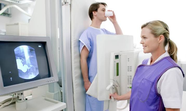
A Barium Meal, also known as an Upper GI series or Esophagogastroduodenal (EGD) study using barium, is a radiological examination that uses X-rays and a contrast medium called barium sulfate to visualize the inner lining and function of the esophagus, stomach, and the first part of the small intestine (duodenum).
The procedure involves the patient swallowing a liquid containing barium sulfate, which is an inert, white, chalky substance that does not absorb X-rays. As the barium coats the lining of the upper GI tract, it outlines the shape, size, and contour of these organs, making them visible on X-ray images and fluoroscopy (a type of X-ray that shows continuous motion). This allows healthcare professionals to identify abnormalities such as ulcers, inflammation, polyps, strictures (narrowing), and masses.
Indications for a Barium Meal
A Barium Meal is typically ordered to investigate persistent or unexplained symptoms related to the upper digestive system. Common indications include:
- Dysphagia: Difficulty or pain when swallowing.
- Odynophagia: Painful swallowing.
- Persistent Heartburn or Reflux (GERD): To identify potential complications like esophagitis or strictures.
- Unexplained Abdominal Pain in the upper abdomen.
- Chronic Nausea and Vomiting.
- Unexplained Weight Loss suspected to be related to a GI issue.
- Suspected Ulcers: In the esophagus, stomach, or duodenum.
- Investigation of Hiatal Hernia.
- Detection of Strictures or Narrowing: Caused by inflammation, scarring, or tumors.
- Identification of Masses or Tumors: Although often followed by endoscopy/biopsy for confirmation.
- Evaluation of Motility Disorders: Although often less precise than manometry.
- Follow-up after certain types of gastric or esophageal surgery.
- Suspected Diverticula: Small pouches that bulge outward from the wall of the GI tract.
Contraindications for a Barium Meal
While generally safe, a Barium Meal is not suitable for all patients. Contraindications include:
- Suspected Bowel Perforation (Rupture): Barium sulfate is not water-soluble and can cause severe, life-threatening peritonitis if it leaks into the abdominal cavity through a hole in the GI tract. In such cases, a water-soluble contrast agent (like Gastrografin) is typically used instead.
- Suspected Complete Bowel Obstruction: Barium can solidify behind an obstruction, potentially worsening it or causing impaction. Water-soluble contrast is usually preferred if contrast is needed.
- Severe Dehydration: Barium can absorb water from the bowel, potentially worsening dehydration and increasing the risk of impaction.
- Patients Unable to Cooperate: The procedure requires the patient to follow instructions (drink on command, hold positions) and tolerate lying/standing on a tilting table.
- Pregnancy: As the procedure involves X-ray radiation, it is generally contraindicated in pregnant women unless absolutely necessary, and then only with clear justification and precautions.
- Recent Biopsy of the Esophagus or Stomach: There is a theoretical increased risk of perforation shortly after a biopsy.
- Severe Swallowing Difficulties with High Risk of Aspiration: While care is taken, a high risk of aspirating (inhaling) the barium into the lungs can be a contraindication.
Equipment Used
The Barium Meal procedure requires specialized radiological equipment and materials:
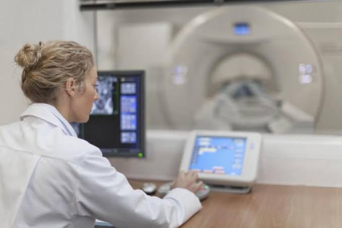
- Fluoroscopy Unit: A sophisticated X-ray machine capable of producing real-time, dynamic X-ray images. It typically includes:
- Radiographic Table: Often tiltable from horizontal to vertical positions.
- X-ray Tube: Generates the X-rays.
- Image Intensifier or Flat-Panel Detector: Converts the X-rays into a visible image.
- Monitoring Screen: Displays the fluoroscopic images in real-time.
- Image Recording System: To capture static (spot) images and/or dynamic (cine) loops.
- Barium Sulfate Suspension: The contrast medium (discussed below).
- Effervescent Agent (optional but common for double-contrast studies): Granules or tablets that produce carbon dioxide gas when swallowed, distending the stomach.
- Cups and Straws: For the patient to drink the barium.
- Positioning Aids: Pillows, sponges, or wedges for patient comfort and positioning.
- Protective Lead Aprons and Shields: For the radiologist, technologist, and potentially certain patient areas to minimize radiation exposure.
- Emergency Equipment: Although uncommon, equipment for managing potential complications (e.g., aspiration) should be readily available.
Contrast Media: Barium Sulfate
The primary contrast medium used for a Barium Meal is Barium Sulfate (BaSO4).
- Properties: It is a heavy metal salt that is radiopaque, meaning it effectively blocks X-rays. In its pure form, it’s insoluble in water and biologically inert, which is why it can be safely ingested (as long as it stays within the GI tract).
- Formulations: Barium sulfate is prepared as a suspension – fine particles mixed with water and often flavoring agents, thickeners, and stabilizers to make it palatable and ensure it coats the mucosal lining effectively. Different viscosities are available:
- Thin Barium: A less concentrated, more liquid suspension used to evaluate the rapid passage through the esophagus and visualize the general shape of the stomach and duodenum.
- Thick Barium: A more concentrated, paste-like suspension used to coat the mucosal lining for detailed visualization of subtle abnormalities (like ulcers or polyps) and to demonstrate strictures.
- Mechanism: When swallowed, the barium suspension coats the inner surface (mucosa) of the esophagus, stomach, and duodenum. On an X-ray, the barium-coated lumen appears white, outlining the structure.
- Water-Soluble Contrast Agents: In cases where perforation is suspected or the patient cannot tolerate barium, water-soluble contrast agents (e.g., iodine-based solutions like Gastrografin) may be used. These are absorbed if they enter the bloodstream or peritoneal cavity, making them safer outside the lumen but providing less mucosal detail compared to barium.
Procedure/Technique for Barium Meal with Filming
The Barium Meal procedure is performed in the radiology department by a radiologist (a medical doctor specializing in interpreting medical images) and a radiologic technologist. The procedure typically takes 30-60 minutes.
Step-by-Step Guide:
- Patient Preparation:
- The patient is usually instructed to fast for 8-12 hours prior to the examination (no food or drink). This ensures the stomach is empty, allowing for better visualization of the lining and preventing dilution of the barium.
- Patients should inform the healthcare team of any allergies, medical conditions (especially diabetes requiring medication timing adjustments), and medications they are taking.
- The patient will likely be asked to remove clothing, jewelry, and any metallic objects that could interfere with the X-rays and change into a hospital gown.
- Arrival and Explanation:
- Upon arrival in the radiology suite, the radiologic technologist and/or radiologist will explain the procedure to the patient, answer questions, and ensure they understand what is expected.
- Initial Positioning and Scout Images:
- The patient is typically positioned on the fluoroscopy table, which is usually in an upright (standing) or semi-upright position initially.
- A “scout” X-ray image (without contrast) may be taken to check patient positioning, view the abdominal area, and ensure the stomach is empty.
- Swallowing Thin Barium (Esophageal Phase):
- The patient is given a cup of the thin barium suspension.
- They are instructed to take sips and swallow the barium on command.
- The radiologist observes the passage of the barium through the esophagus using fluoroscopy, looking for smooth movement, any hold-ups, narrowing, or abnormalities in the esophageal wall.
- Spot images (still X-ray images taken during fluoroscopy) are captured as the barium passes through the esophagus in different views (e.g., straight anterior-posterior (AP), oblique views) to document its appearance and any findings. The patient may be asked to hold their breath briefly during image acquisition.
- Evaluating the Stomach and Duodenum (Single-Contrast and Double-Contrast):
- The patient continues to drink the thin barium as it enters the stomach. The radiologist observes how the stomach fills and empties.
- The table is often tilted to different positions (horizontal, head down/Trendelenburg, head up) and the patient is asked to change positions (lie on their back, side, or stomach, turn over) to ensure the barium coats all surfaces of the stomach and duodenum. This helps visualize the contours and folds.
- Single-Contrast Technique: Involves filling the stomach and duodenum with barium to visualize their overall size, shape, and any large filling defects (masses) or outlines of ulcers. Spot images are taken in various positions.
- Double-Contrast Technique: This is often performed to enhance visualization of the mucosal lining and detect subtle lesions like small ulcers, polyps, or erosions. After drinking some thin barium, the patient is given an effervescent agent (often granules with water). This produces carbon dioxide gas, which distends the stomach. More thin barium (or sometimes thick barium) is then given. The gas provides a “negative” contrast (appears dark), while the barium coating the mucosa provides “positive” contrast (appears white), creating a double-contrast effect that outlines the fine details of the stomach wall. Spot images are taken rapidly in various orientations as the stomach wall is distended.
- Following Barium into the Duodenum and Beyond:
- The radiologist follows the barium as it empties from the stomach into the duodenum. The shape and pattern of the duodenal bulb and C-loop are examined.
- Spot images of the duodenum are crucial, as this area is a common site for ulcers.
- Depending on the clinical question, the study may track the barium further into the small intestine (this extended examination is sometimes called a Barium Follow-Through, though often performed as a separate or combined study).
- Completion:
- Once sufficient images have been obtained in various positions and techniques (usually 10-20 or more spot films), the procedure is concluded.
Complications
While generally safe, potential complications, though uncommon, can occur:
- Constipation: The most common complication. Barium is not absorbed and can solidify in the intestines, leading to constipation or, rarely, impaction.
- Aspiration: Accidental inhalation of barium into the lungs, which can cause pneumonitis (inflammation of the lungs). This risk is higher in patients with swallowing difficulties or impaired consciousness.
- Barium Leakage (Peritonitis): A life-threatening complication if there is an unsuspected perforation in the GI tract and barium leaks into the abdominal cavity. This is why water-soluble contrast is used if perforation is suspected.
- Allergic Reaction: True allergy to barium sulfate is exceedingly rare. Reactions are more likely due to additives (flavorings, stabilizers) or related to the effervescent agent.
Aftercare
Proper aftercare is important to help the patient recover and minimize the risk of constipation:
- Hydration: The patient is strongly advised to drink plenty of fluids (water, juice, etc.) immediately after the procedure and for the next 24-48 hours. This helps move the barium through the digestive tract and reduces the risk of constipation.
- Bowel Movements: The patient should be informed that their stool will likely be white or light-colored for a day or two due to the passage of barium.
- Laxatives: A mild laxative may be recommended or prescribed by the healthcare provider, especially if the patient is prone to constipation or did not drink sufficient fluids. This helps expel the barium.
- Monitoring Symptoms: Patients should be advised to contact their healthcare provider if they experience severe constipation, abdominal pain, inability to pass gas or stool, or any other concerning symptoms after the procedure.
- Return to Normal Activities: Patients can typically resume their normal diet and activities immediately after the procedure, unless otherwise instructed by their doctor.
In conclusion, the Barium Meal is a valuable diagnostic tool providing detailed anatomical and functional information about the upper GI tract. By understanding the procedure, its purpose, and the necessary steps before, during, and after, patients can approach the examination with greater confidence.


