Kidney transplantation is the preferred treatment for many patients with end-stage renal disease. While the vast majority of transplants function well immediately, a significant complication known as Delayed Graft Function (DGF) can occur. DGF is generally defined as the need for dialysis within the first week post-transplantation or a failure of serum creatinine to decline significantly over this period. It can impact up to 50% of deceased donor kidney recipients, and less frequently, living donor recipients.
Identifying and managing DGF is crucial because it is associated with increased rates of acute rejection, longer hospital stays, higher costs, and potentially worse long-term graft survival compared to immediate graft function. DGF is not a diagnosis in itself, but rather a clinical syndrome indicative of underlying issues that require investigation and management.
Understanding DGF
The primary underlying cause of DGF is often ischemia-reperfusion injury sustained during the procurement, preservation, and transplantation process, leading to Acute Tubular Necrosis (ATN). However, other factors can contribute or be primary causes, including:
- Donor factors (age, comorbidities, cause of death, kidney quality)
- Procurement and preservation factors (cold ischemia time, warm ischemia time, machine perfusion)
- Recipient factors (age, comorbidities, pre-sensitization, severity of ESRD)
- Immunological factors (acute rejection)
- Technical factors (vascular thrombosis, urine leak, ureteral obstruction)
- Recurrent disease
Effective management requires a systematic approach to differentiate between these potential causes while supporting the patient and the recovering graft.
Recognize and Confirm the Presence of DGF
The first step is clinical suspicion and confirmation. This typically occurs in the immediate post-operative period.
- Monitor Graft Function: Closely monitor serum creatinine and urine output trends.
- Establish Baseline: Understand the expected trajectory of creatinine decline post-transplant with immediate function (often halving every 1-2 days initially).
- Identify Deviation: A persistent or rising creatinine, inadequate urine output (though output can be preserved in high-output ATN), or the need for dialysis within the first 7 days post-transplant strongly suggests DGF. Use a working definition standard within your transplant center.
Once DGF is suspected or confirmed based on these criteria, the management pathway diverges from that of immediate function. Supportive care and proactive monitoring become paramount, alongside preparation for potential interventions and diagnostics.
Implement Initial Supportive Management
While investigating the cause, immediate supportive care is essential.
- Fluid and Electrolyte Balance: Meticulously manage fluid status. Avoid both volume overload (can exacerbate hypertension, cardiac strain) and hypovolemia (can worsen ischemia). Manage hyperkalemia, metabolic acidosis, hyperphosphatemia, and hypocalcemia resulting from poor kidney function.
- Blood Pressure Management: Aim for appropriate blood pressure. While avoiding severe hypertension is important, permissive hypertension might be tolerated or even beneficial in some cases to ensure adequate graft perfusion, balancing this against cardiac risk.
- Immunosuppression: Administer standard induction immunosuppression as per protocol. Maintenance immunosuppression (e.g., calcineurin inhibitors – CNIs, antiproliferatives) needs careful management. CNI levels may be slow to rise or unpredictable with DGF due to poor clearance, and potential nephrotoxicity is a concern. Adjust dosing based on therapeutic drug monitoring and overall clinical picture. Consider delaying full-dose CNIs or using alternative agents initially in select high-risk cases per center protocol.
- Monitor for Complications: Vigilantly monitor for surgical complications (bleeding, hematoma, vascular issues, urine leak), infection, and cardiovascular events.
Determine the Need for Post-Operative Dialysis
This is a critical and often the first decision point in DGF management. Dialysis in the setting of DGF is a supportive measure, primarily aimed at managing life-threatening complications of kidney failure while providing time for the graft to potentially recover function. It does not directly treat the underlying cause of DGF, nor does it typically improve graft function itself, but it is vital for patient survival and stability.
The indications for initiating post-operative dialysis in a patient with DGF are the standard, absolute or relative, indications for dialysis in acute kidney injury:
- Severe or Refractory Hyperkalemia: Typically serum potassium >6.0-6.5 mEq/L, especially with ECG changes, unresponsive to medical therapies (insulin/glucose, calcium gluconate, potassium binders, bicarbonate).
- Severe Volume Overload: Causing respiratory distress (pulmonary edema) or significant cardiovascular compromise, refractory to diuretic therapy (diuretics are often ineffective with DGF).
- Severe or Refractory Metabolic Acidosis: Arterial pH <7.1-7.2 or serum bicarbonate <15 mEq/L, particularly if rapidly worsening, unresponsive to bicarbonate administration.
- Uremic Symptoms: Signs of significant uremia such as pericarditis, encephalopathy (confusion, asterixis), severe intractable nausea or vomiting, uremic coagulopathy with bleeding.
- Severe Electrolyte Imbalances: Other severe, refractory electrolyte disturbances that cannot be corrected medically due to lack of kidney function (e.g., severe hyperphosphatemia).
Timing of Dialysis:
Dialysis should be initiated promptly when any of these indications develop. The frequency and duration of dialysis sessions are tailored to the individual patient’s needs, aiming to maintain biochemical stability and euvolemia while minimizing hemodynamic instability that could compromise graft perfusion. Modalities like intermittent hemodialysis (HD) or continuous renal replacement therapy (CRRT), particularly in critically ill or hemodynamically unstable patients, may be used. Close collaboration with nephrology services is essential.
Investigate the Cause of DGF
Simultaneously with supportive care, efforts must be made to determine the underlying reason for DGF, as this dictates specific treatment strategies.
- Review Patient and Donor Factors: Re-evaluate recipient comorbidities, pre-transplant dialysis vintage, donor factors, and specifics of the procurement and implant surgery.
- Rule out Technical Issues: This is critical, especially in cases of anuria or sudden decline after initial output. Perform a Doppler ultrasound of the renal allograft vessels promptly to assess blood flow and rule out arterial or venous thrombosis. Assess for hydronephrosis potentially indicating obstruction. Monitor for signs of urine leak (fluid collection around graft). Surgical exploration may be necessary if there is high suspicion despite imaging, or if imaging is inconclusive.
- Consider Immunological Issues (Acute Rejection): While ATN is the most common cause, acute rejection can occur early, especially in highly sensitized recipients or with certain immunosuppression regimens. Clinical signs are often indistinguishable from ATN in the setting of DGF, making biopsy important (see Step 5).
- Assess Drug Levels: Check immunosuppressant levels (particularly CNIs) if therapy has started, although levels may be low due to poor absorption or high despite poor function. Ensure appropriate dosing adjustments.
Determine When a Biopsy Should Be Performed
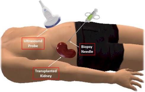
Performing a kidney allograft biopsy is often necessary to definitively diagnose the cause of DGF, particularly once technical issues have been reasonably excluded and supportive care is underway. The timing of the biopsy is a clinical decision based on several factors:
- Purpose: The primary purpose of the biopsy is to distinguish between ATN, acute rejection (cellular or antibody-mediated), calcineurin inhibitor toxicity, recurrent disease, or other less common causes. Identification of rejection is crucial as it requires specific anti-rejection therapies distinct from supportive care for ATN.
- Typical Timing: There is no single mandatory time point for biopsy in DGF. Common approaches include:
- Early Biopsy (within first 3-7 days): May be considered in specific situations:
- High suspicion for early acute rejection (e.g., highly sensitized recipient, specific donor-recipient HLA mismatches).
- Unusual clinical course (e.g., increasing lactate dehydrogenase suggesting microangiopathy, sudden change in output not explained by technical issues).
- To obtain a baseline histological assessment, although waiting for better tissue quality is often preferred.
- If contemplating specific early interventions whose utility depends on the underlying histology.
- Delayed Biopsy (often around 7-14 days or later): This is a more common approach if DGF persists without significant signs of recovery and technical issues have been ruled out.
- Allows time for a proportion of ATN cases to show signs of spontaneous recovery.
- Tissue quality for histological assessment is often better than in the immediate post-op period.
- Helps confirm persistent ATN or identify rejection (which could be primary or secondary to the initial injury).
- Guides further management if creatinine is plateauing or worsening and dialysis dependency continues.
- Triggered Biopsy: Performed at any time if there is a change in the clinical picture (e.g., unexplained fever, change in output, suspicion of specific complications) or if initial investigative steps are inconclusive.
- Early Biopsy (within first 3-7 days): May be considered in specific situations:
- Considerations Before Biopsy:
- Ensure vascular patency via Doppler ultrasound immediately prior.
- Assess patient’s coagulation status and platelet count to minimize bleeding risk.
- Patient cooperation is required.
- Interpretation: The biopsy findings, integrated with clinical context and imaging results, provide the diagnosis guiding subsequent, cause-specific therapy (e.g., anti-rejection treatment if cellular or antibody-mediated rejection is found, continued supportive care if significant ATN is the predominant finding).
Tailor Management Based on Diagnosis
Once the cause of DGF is determined (or if strongly suspected based on clinical context while awaiting biopsy results), management is adjusted.
- Acute Tubular Necrosis (ATN): If ATN is the primary diagnosis (often after excluding other causes, or confirmed by biopsy), the cornerstone of management remains supportive care: continued dialysis if needed, fluid/electrolyte balance, optimizing immunosuppression, and allowing time for tubular recovery. Recovery from ATN can take days to several weeks.
- Acute Rejection: If biopsy demonstrates acute cellular or antibody-mediated rejection, specific anti-rejection therapy (e.g., high-dose corticosteroids, antibody therapies like antithymocyte globulin or rituximab, plasma exchange for antibody-mediated rejection) must be initiated promptly according to established protocols.
- Technical Issues: Vascular thrombosis requires urgent surgical intervention (thrombectomy or revision), though graft salvage rates are low. Ureteral obstruction or leak requires urological intervention (e.g., stent placement, surgical repair).
- Other Causes: Management is directed at the specific identified pathology (e.g., stopping nephrotoxic agents, treating infection).
Monitor Recovery and Long-Term Outcomes
- Continued Monitoring: Regardless of the initial diagnosis, vigilant monitoring of creatinine, urine output, electrolytes, immunosuppressant levels, and blood pressure continues. Look for the first signs of graft recovery (a sustained fall in creatinine, increased urine output).
- Weaning from Dialysis: As kidney function improves and creatinine begins to fall consistently, the need for dialysis will decrease. Sessions can be spaced out and eventually discontinued when the graft can adequately manage fluid, electrolytes, and uremia.
- Long-term Follow-up: Patients who experience DGF require careful long-term follow-up. They are at higher risk for subsequent acute rejection episodes and may have a slightly reduced long-term graft survival compared to those with immediate function. Optimization of immunosuppression and aggressive management of cardiovascular risk factors are critical.
Conclusion
Delayed Graft Function is a common and significant complication following kidney transplantation that requires a structured and professional approach. Identifying DGF promptly, providing meticulous supportive care, and systematically investigating the underlying cause through imaging and timely biopsy are crucial steps. Post-operative dialysis serves as a vital supportive measure for managing the consequences of kidney failure while investigations and potential graft recovery are underway. Ultimately, tailoring management based on the specific diagnosis offers the best chance for graft recovery and improved long-term outcomes for the recipient. A multidisciplinary team approach involving transplant surgeons, nephrologists, nurses, and radiologists is essential for optimal care of patients experiencing DGF.


