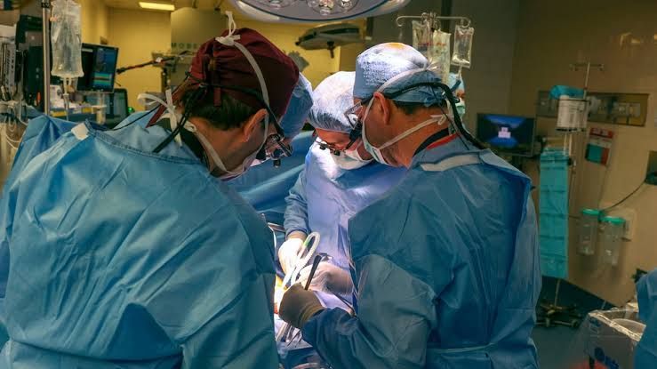
Kidney transplantation stands as the preferred therapeutic option for many individuals suffering from End-Stage Renal Disease (ESRD). It offers the potential for improved quality of life and increased longevity compared to long-term dialysis.
Overview of Kidney Transplantation
Kidney transplantation involves surgically placing a healthy kidney from a deceased or living donor into the recipient’s body to take over the function of their failed native kidneys. The recipient’s native kidneys are typically not removed unless they are causing specific problems (e.g., infection, uncontrolled hypertension, large size due to polycystic kidney disease).
The process begins with a comprehensive evaluation of both the potential recipient and the donor (if living). This evaluation assesses their overall health, suitability for surgery, tissue compatibility (HLA matching), and the likelihood of successful outcomes.
There are two primary types of kidney transplants:
- Deceased Donor Transplant: A kidney is recovered from a person who has been declared brain dead or, less commonly, circulatory death, and whose family has consented to organ donation. These organs are allocated based on strict criteria managed by organizations like the United Network for Organ Sharing (UNOS) in the United States.
- Living Donor Transplant: A kidney is donated by a living person, typically a family member, friend, or even a stranger (altruistic donation). Living donation offers several advantages, including the ability to schedule the surgery, shorter cold ischemia time (the time the kidney is without blood supply), and often better long-term outcomes.
Regardless of the donor type, the recipient implantation procedure follows a largely standardized process, with specific differences related to organ procurement and timing.
The Standard Recipient Kidney Transplant Procedure
The standard kidney transplant surgery focuses on placing the new kidney, connecting its blood vessels to the recipient’s circulation, and connecting its ureter to the recipient’s bladder.
- Step 1: Recipient Preparation & Anesthesia
- The recipient undergoes final pre-operative checks, including confirming blood type and crossmatch compatibility, reviewing imaging, and receiving pre-operative medications, including immunosuppressants to prevent immediate rejection.
- General anesthesia is administered.
- The surgical area, typically the lower abdomen, is prepped and draped in a sterile manner. A Foley catheter is inserted into the bladder.
- Step 2: Surgical Incision and Exposure
- A curvilinear incision is made in the lower abdomen, usually in the right or left iliac fossa (groin area). The choice of side may depend on previous surgeries or surgeon preference; the right side is common.
- The abdominal muscles are carefully divided or retracted to expose the retroperitoneal space, where the iliac blood vessels (external iliac artery and vein) are located.
- The peritoneum (lining of the abdominal cavity) is identified and pushed aside to access the iliac vessels.
- Step 3: Preparing the Recipient Vessels
- The external iliac artery and vein are identified and carefully dissected free from surrounding tissues.
- Small branches emanating from these vessels in the surgical field are ligated (tied off) and divided to create clear segments for connection.
- Vascular control is achieved using soft clamps to temporarily stop blood flow in the selected segments of the external iliac artery and vein.
- Step 4: Preparing the Donor Kidney
- While the recipient is being prepared, the donor kidney is brought to the operating room.
- The donor kidney, which has been preserved in a cold solution, is carefully inspected on a back table. The renal artery(ies), renal vein, and ureter are identified.
- Any extra tissues or redundant donor vessels are trimmed. If there are multiple renal arteries, they may need to be reconstructed on the back table into a single vessel opening or connected separately to the recipient vessels.
- Step 5: Vascular Anastomosis – Venous Connection
- The donor renal vein is typically connected first.
- A precise opening (anastomosis) is created in the side of the recipient’s external iliac vein.
- The donor renal vein is then sewn end-to-side onto the recipient external iliac vein using fine, non-absorbable sutures. This connection allows blood returning from the new kidney to flow into the recipient’s systemic circulation.
- Step 6: Vascular Anastomosis – Arterial Connection
- Next, the donor renal artery (or reconstructed artery) is connected.
- An opening is created in the side of the recipient’s external iliac artery (end-to-side anastomosis) or occasionally end-to-end to the internal iliac artery (hypogastric artery).
- The donor renal artery is meticulously sewn onto the recipient iliac artery using fine sutures. This connection supplies arterial blood flow to the new kidney.
- Once both vascular anastomoses are complete, the vascular clamps are carefully released, first from the vein, then the artery. Blood flow is re-established to the transplanted kidney. The kidney should pink up and become firm, indicating good perfusion. Urine production may begin almost immediately.
- Step 7: Ureteral Anastomosis (Ureteroneocystostomy)
- The donor ureter is connected to the recipient’s bladder to allow urine to drain.
- The bladder is partially filled with sterile saline via the Foley catheter.
- An incision is made in the bladder wall. The donor ureter is passed through a tunnel created in the bladder wall muscle (an anti-reflux technique like Politano-Leadbetter or Lich-Gregoir is often used) before being sewn to the inner lining of the bladder (mucosa). This technique helps prevent urine from flowing backward up to the kidney from the bladder, which could cause infection or damage. Alternatively, a direct anastomosis without a formal tunnel may be used.
- A small stent may be placed temporarily in the ureter crossing the anastomosis into the bladder to ensure proper drainage and help with healing; this stent is typically removed a few weeks after surgery.
- Step 8: Closure
- The surgical area is irrigated, and meticulous hemostasis (control of bleeding) is ensured.
- A surgical drain may be placed near the kidney to collect any fluid accumulation in the immediate post-operative period.
- The layers of the abdominal wall are closed with sutures. The skin incision is closed with sutures or staples.
- Step 9: Immediate Post-operative Care
- The recipient is transferred to a recovery area or intensive care unit for close monitoring of kidney function, fluid balance, blood pressure, and signs of bleeding or infection.
- Immunosuppressant medications are continued and adjusted based on patient status and kidney function.
3. Specifics of Donor Procedures
- Living Donor Nephrectomy:
- This can be performed via either laparoscopic (keyhole) or open surgery. Laparoscopic nephrectomy is now the standard approach due to faster recovery and less pain for the donor.
- For laparoscopic nephrectomy, several small incisions are made to insert instruments and a camera. The kidney is carefully dissected free, and the renal artery and vein are clipped or stapled and divided. A larger incision (typically 5-10 cm) is made, usually in the lower abdomen or groin, to extract the kidney.
- Open nephrectomy involves a larger incision (flank or abdominal) to directly access and remove the kidney.
- The chosen kidney (usually the left due to a longer renal vein) is flushed with a cold preservation solution and prepared for immediate transfer to the recipient operating room.
- Deceased Donor Procurement:
- This procedure occurs in an operating room after the donor has been declared dead.
- A large incision is made to access the abdominal organs.
- Major blood vessels are identified and prepared.
- Cold preservation solution is rapidly infused into the renal artery (and other arteries for multi-organ recovery) to cool the organs and flush out blood.
- The kidney, along with segments of the donor aorta and vena cava, and the attached ureter and a patch of bladder, is carefully removed.
- The kidney is packaged in sterile bags and kept cold in an ice chest for transport to the recipient center. Cold ischemia time begins the moment the cold solution enters the artery and is a critical factor affecting kidney function.
Strategies for Highly Complex Recipients
Transplanting a kidney into recipients with specific anatomical, surgical, or immunological challenges requires specialized planning and surgical techniques.
- A. Re-transplantation:
- Challenges: Previous surgery creates scar tissue (adhesions), altering normal anatomy and increasing dissection difficulty and bleeding risk. The previous transplant site may be unusable or suboptimal. Recipients may be highly sensitized to human leukocyte antigens (HLAs) from previous transplants, blood transfusions, or pregnancies, increasing the risk of hyperacute or acute rejection.
- Strategies:
- Detailed Pre-operative Imaging: CT scans and MRIs are crucial to assess previous surgical fields, identify scar tissue, map vascular anatomy, and locate potential recipient vessel sites free from previous dissection.
- Alternative Transplant Site: If the standard iliac fossa site used for the primary transplant is heavily scarred or contains the failed allograft (transplanted kidney) that isn’t being removed, the opposite iliac fossa is typically used. In rare, highly complex cases, alternative sites or techniques for vascular connection may be considered.
- Meticulous Dissection: The surgeon must be extremely careful during dissection to avoid injury to surrounding structures, especially the iliac vessels embedded in scar tissue. This often takes significantly longer than a primary transplant.
- Immunological Management: Pre-transplant immunological assessment is critical. Strategies may include desensitization protocols (e.g., plasmapheresis, IVIG) to reduce existing antibodies in highly sensitized patients before or immediately after transplant. Immunosuppression regimens are often more intensive in the post-transplant period.
- B. Pediatric Recipients:
- Challenges: Size mismatch is common, especially when transplanting an adult kidney into a small child or infant. Pediatric vessels are smaller and more fragile, requiring microsurgical techniques. Growth potential needs to be protected (e.g., avoiding damaging growth plates during dissection or placement). Underlying causes of ESRD may be different and require specific consideration.
- Strategies:
- Intraperitoneal Placement: For a large adult kidney in a small child, the kidney may be placed entirely within the abdominal cavity (intraperitoneally) rather than in the retroperitoneal iliac fossa because there is more space.
- Vascular Anastomosis Technique: Meticulous surgical technique with fine sutures and often magnification is required for connecting the small donor or recipient vessels. The aorta and vena cava (central vessels) may be used for anastomosis in infants and small children rather than the iliac vessels to accommodate larger adult donor vessels.
- Ureteral Management: Special care is taken with the ureteroneocystostomy due to smaller bladder size or potential underlying bladder issues related to congenital conditions causing ESRD.
- Growth Considerations: Surgical planning considers the child’s future growth. The kidney is placed so that it does not interfere with musculoskeletal development.
- Specialized Team: Pediatric kidney transplant requires a multidisciplinary team with expertise in pediatric nephrology, surgery, anesthesia, critical care, nursing, and psychosocial support.
- C. Complex Urinary Reconstruction:
- Challenges: Recipients may have pre-existing conditions affecting the bladder or urethra, such as neurogenic bladder, prior cystectomy (bladder removal), bladder augmentation, urethral strictures, or persistent high-grade vesicoureteral reflux not amenable to standard ureteral implantation techniques. A bladder may be non-functional or non-existent.
- Strategies:
- Pre-operative Urological Assessment: Urodynamic studies are essential to evaluate bladder function, capacity, and pressure.
- Managing the Native Bladder: If the native bladder is small, high-pressure, or non-compliant, augmentation cystoplasty (increasing bladder volume with a segment of intestine) may be necessary before or at the time of transplant.
- Ileal Conduit: If the native bladder is absent, irreparable, or clearly unsuitable, the donor ureter is connected to a segment of isolated small intestine (ileum), which is brought out to the abdominal wall as a stoma (ileal conduit or urostomy). This provides a pathway for urine drainage into an external bag.
- Careful Ureteroneocystostomy or Uretero-ureterostomy: If the bladder is usable but requires specific techniques, the ureteroneocystostomy must be adapted. In some situations, the donor ureter can be connected directly to one of the recipient’s native ureters (uretero-ureterostomy) if the native ureter is healthy and patent down to the bladder.
- Multidisciplinary Approach: Close collaboration with urologists specializing in reconstructive procedures is vital in planning and executing the surgery.
Conclusion
Kidney transplantation is a complex and life-changing surgical procedure requiring detailed planning, precise execution, and expert post-operative management. While the standard procedure involves meticulous vascular and ureteral anastomoses, successfully extending this therapy to complex recipients like those requiring re-transplantation, pediatric patients, or those with challenging urinary anatomy necessitates advanced strategies, multidisciplinary collaboration, and significant surgical expertise. These specialized approaches aim to overcome unique challenges and ensure the best possible outcome for every patient benefiting from the gift of a transplanted kidney.
Disclaimer: This guide is intended for educational purposes only and provides a simplified overview of complex medical procedures. It is not a substitute for professional medical training, advice, or treatment. Surgical techniques and strategies may vary based on patient factors, surgeon experience, and institutional protocols.


