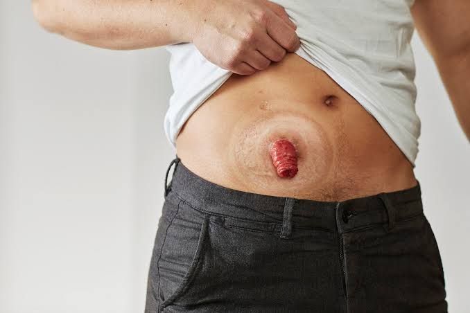
A stoma, derived from the Greek word for “mouth” or “opening,” is a surgically created opening in the body that connects an internal organ to the surface of the skin. This procedure, known as an ostomy, is performed to divert the normal flow of bodily waste (feces or urine) when the natural pathway is diseased, damaged, or needs time to heal. Stomas are critical interventions in various medical conditions affecting the gastrointestinal or urinary systems, significantly impacting patients’ lives and requiring specialized care and understanding from healthcare professionals.
1. Defining Stomas
A stoma is a surgically constructed opening that serves as an alternative exit route for bodily contents. It is formed by bringing a segment of the internal organ (usually intestine or urinary tract) through the abdominal wall and suturing it to the skin. This creates a new opening through which waste material can exit the body and be collected in an external pouching system.
Key characteristics of a healthy stoma include:
- Appearance: Typically red or pink, similar to the moist tissue lining inside the mouth (mucosa).
- Sensation: A healthy stoma has no nerve endings for pain, so touching it should not be painful.
- Moisture: The stoma surface should appear moist.
- Shape and Size: Varies depending on the type and location, but usually round or oval and slightly protuberant (budded) or flush with the skin.
Understanding the basic definition and appearance of a healthy stoma is fundamental to recognizing deviations and potential problems.
2. Different Types of Stomas
Stomas are primarily categorized based on the organ involved. The most common types involve the bowel (intestine) and the urinary tract.
2.1 Intestinal Stomas
These divert fecal matter. They are named based on the section of the bowel brought to the surface:
- Colostomy: A stoma formed from a section of the large intestine (colon). The location of the colostomy along the colon affects the consistency of the output:
- Ascending Colostomy: Rare. Located on the right side of the abdomen. Output is typically liquid to semi-liquid.
- Transverse Colostomy: Often temporary, located in the upper abdomen (midline or slightly to the right/left). Output is typically semi-liquid to semi-formed.
- Descending Colostomy: Located on the left side of the abdomen. Output is typically semi-formed to formed.
- Sigmoid Colostomy: Most common type of permanent colostomy, located in the lower left abdomen. Output is typically formed stool.
- Types based on construction:
- End Colostomy: The proximal end of the bowel is brought out as a single opening. The distal end is either removed or sewn closed (Hartmann’s pouch).
- Loop Colostomy: Often temporary. A loop of bowel is brought through the abdominal wall, supported by a rod or bridge temporarily, and opened in two places, creating two openings (proximal for stool, distal for mucus) or one large opening with two lumens.
- Double-Barreled Colostomy: Two separate stomas created side-by-side from the divided ends of the bowel (proximal functioning, distal non-functioning). Less common than loop colostomy.
- Ileostomy: A stoma formed from a section of the small intestine (ileum). Ileostomies are typically located on the right side of the abdomen.
- End Ileostomy: The most common type, usually permanent. The entire colon and rectum are removed, and the end of the ileum is brought out as a stoma. Output is continuous, watery to paste-like, and contains digestive enzymes which are very irritating to the skin.
- Loop Ileostomy: Often temporary. A loop of ileum is brought through the abdominal wall and usually creates two lumens (proximal for stool, distal for mucus), similar to a loop colostomy. Often created to protect a distal anastomosis (surgical join).
2.2 Urinary Stomas (Urostomy)
These divert urine from the bladder.
- Ileal Conduit (or Bricker Conduit): The most common type of urostomy. A small segment of the ileum (about 6-8 inches) is removed from the intestinal tract (blood supply kept intact). The ureters are disconnected from the bladder and reconnected to this isolated ileal segment. One end of the ileal segment is closed, and the other end is brought through the abdominal wall to form a stoma. The ileal segment does not absorb urine; it acts as a passive conduit for urine flow from the kidneys to the stoma. The bladder is usually removed. Output is continuous urine flow.
- Other less common types: Include cutaneous ureterostomy (ureters brought directly to the skin) or nephrostomy (tube inserted directly into the kidney pelvis).
3. Physical Examination of Stomas
A systematic physical examination of the stoma and surrounding skin is crucial for monitoring function, identifying complications, and ensuring the effectiveness of the pouching system. This should be performed regularly during pouch changes.
Step-by-Step Guide to Stoma Examination:
- Preparation:
- Gather necessary supplies: new pouching system (pouch and/or wafer), stoma measuring guide, adhesive remover spray/wipes, barrier wipes/spray, gentle cleaning solution (usually just water), gauze pads or soft wipes, gloves, adequate lighting.
- Ensure patient privacy. Explain the procedure and obtain consent.
- Wash hands and don gloves.
- Remove the Old Pouching System:
- Gently peel the old pouch and skin barrier away from the skin, working from top to bottom while supporting the skin. Adhesive remover can be used cautiously if needed.
- Note the amount and type of output in the old pouch.
- Dispose of the old system appropriately.
- Clean the Stoma and Peristomal Skin:
- Gently cleanse the stoma and the 2-3 inches of skin surrounding it using warm water and soft wipes or non-soapy gauze. Pat dry gently. Avoid vigorous rubbing.
- Observe the output from the stoma during cleaning (amount, consistency, color).
- Visual Inspection (The Core Stoma Assessment):
- Peristomal Skin: Examine the skin immediately surrounding the stoma (peristomal skin). Assess color (should be intact, without redness, blue/purple discoloration, or breakdown), integrity (no rashes, open areas, pustules, or irritation), and presence of any discharge, leakage residue, or fungal growth. Note any scarring or existing issues.
- Stoma Color: Crucially assess the color. A healthy stoma is pink or red. Report immediately if the stoma is dark red, purple, black (ischemia/necrosis), or pale/white (anemia, poor perfusion).
- Stoma Size and Shape: Note if the stoma appears round, oval, or irregular. Is it the expected size? New stomas shrink over the first 6-8 weeks post-operatively. Use a measuring guide to measure the widest point of the stoma.
- Stoma Protrusion: Is the stoma budded (protruding outwards), flush with the skin, or retracted (pulled inwards below skin level)? A slightly budded stoma (1-2 cm) is often ideal for pouching.
- Stoma Opening: Observe the lumen (opening). Is it patent? Can you see inside?
- Bleeding: Note if there is any bleeding. Minor bleeding with touch is normal due to the rich blood supply. Significant or spontaneous bleeding is abnormal.
- Sutures: If the stoma is recent (within weeks), note the presence and condition of any dissolving sutures at the base of the stoma.
- Palpation (Optional but helpful):
- Gently palpate the abdomen around the peristomal area. Note any tenderness, swelling, firmness, or palpable masses (e.g., potential hernia edge). Never probe inside the stoma unless specifically instructed and trained for a particular assessment (e.g., assessing stricture).
- Auscultation (For Intestinal Stomas):
- Listen for bowel sounds in the abdomen around the stoma site using a stethoscope. This helps assess bowel motility.
- Measurement:
- Use a stoma measuring guide to determine the exact size and shape of the stoma opening. This is essential to cut the opening in the skin barrier accurately, ensuring a snug fit (no skin exposed to output) without impinging on the stoma itself. Measure at the base where the stoma meets the skin.
- Apply New Pouching System:
- Apply any necessary skin prep (barrier wipes/spray) to protect the clean, dry peristomal skin outside the stoma opening.
- Ensure the opening in the skin barrier is the correct size based on your measurement.
- Apply the new pouching system securely, ensuring a good seal around the stoma.
- Documentation:
- Record all findings: date and time, appearance of peristomal skin (clear, irritated, location/description of irritation), stoma color, size, shape, protrusion, presence/absence of bleeding, characteristics of output, any complications noted, and the type and size of the pouching system applied.
4. Permanent and Temporary Indications for Stomas
Stomas are created for various reasons, which dictate whether the stoma is intended to be temporary or permanent.
4.1 Permanent Indications:
A permanent stoma is created when the underlying condition requires irreversible removal or bypass of a section of the bowel or bladder, making restoration of normal function impossible or inadvisable.
- Irreversible Damage/Disease:
- Low Rectal Cancer: Requiring abdominoperineal resection where the anus and rectum are removed. Requires a permanent sigmoid colostomy.
- Severe Inflammatory Bowel Disease (IBD) (Crohn’s, Ulcerative Colitis): Extensive disease, multiple complications (fistulas, strictures), or failure of medical treatment may necessitate the removal of the entire colon and rectum, resulting in a permanent end ileostomy.
- Severe Trauma: Irreparable damage to the rectum or anal sphincter.
- Severe Neurological Conditions: Conditions causing irreversible loss of bowel or bladder control (e.g., spinal cord injury, multiple sclerosis) where other management strategies are insufficient.
- Congenital Abnormalities: Conditions present at birth that cannot be surgically corrected to allow normal function.
- Bladder Cancer: Requiring removal of the bladder (cystectomy) necessitates a permanent urinary diversion, most commonly an ileal conduit urostomy.
- Severe Bladder Dysfunction: Due to neurological issues or chronic retention unresponsive to other treatments.
4.2 Temporary Indications:
A temporary stoma is created to divert stool or urine flow for a limited time, allowing a distal section of the bowel or urinary tract to heal or recover from surgery, inflammation, or radiation. The stoma is later closed or reversed in a subsequent surgical procedure.
- Protection of a Distal Anastomosis: A very common reason. After a section of bowel is removed and the remaining ends are surgically joined (anastomosis), a temporary loop ileostomy or colostomy is created upstream to divert stool away from the fresh join, reducing the risk of leakage and infection during healing (typically 6-12 weeks).
- Decompression: To relieve pressure from an obstruction in the bowel or urinary tract.
- Management of Trauma or Perforation: Allowing time for injured tissue to heal.
- Severe Inflammation or Infection: Diverting flow to allow severely inflamed or infected tissue to rest and heal (e.g., complicated diverticulitis, severe Crohn’s flares with abscess/fistula).
- Following Radiation Therapy: To allow tissues recovering from radiation to heal without the passage of waste.
- Complex Fistula Management: Diversion to allow complex fistulas to heal.
5. Early and Late Complications of Stomas
Complications can occur at any time after stoma surgery, ranging from minor issues to life-threatening emergencies. They are broadly classified as early (occurring within days to weeks post-operatively) or late (occurring weeks, months, or years later).
5.1 Early Complications:
- Ischemia or Necrosis: Insufficient blood supply to the stoma leads to tissue death. The stoma appears dusky, purple, or black. This is a surgical emergency.
- Bleeding: While minor bleeding on touch is normal, significant or spontaneous bleeding from the stoma or its base can indicate a problem requiring investigation.
- Edema (Swelling): Some swelling is normal post-op, but excessive or persistent edema can impede output and signal obstruction or inflammation.
- Retraction: The stoma pulls back to or below the skin level. Makes pouching difficult, significantly increasing the risk of leakage and peristomal skin breakdown.
- High Output: Especially common with ileostomies. Excessive watery discharge leads to rapid fluid and electrolyte loss, risking dehydration and kidney issues.
- Peristomal Skin Irritation: Very common, especially with ileostomies due to enzymatic content of output. Caused by leakage under the barrier, improper fit, sensitivity to adhesive, or poor hygiene. Can range from mild redness to severe breakdown.
- Infection: At the surgical incision site or around the stoma.
- Stoma Separation: The stoma separates from the skin junction. Can lead to undermining of the skin and leakage.
5.2 Late Complications:
- Parastomal Hernia: A common complication where abdominal contents (loop of bowel) bulge around the stoma site, creating a visible swelling. Occurs due to weakening of the abdominal wall muscles around the stoma. Can cause discomfort, pouching difficulties, and potential obstruction or strangulation.
- Stenosis: Narrowing of the stoma opening. Can occur at the skin level or deeper. Impedes the passage of output and may require dilation or surgical revision.
- Prolapse: The bowel protrudes excessively through the stoma opening. Can be minor or significant, causing discomfort, difficulty with pouching, and potentially leading to edema or ischemia.
- Retraction: Can also develop late due to weight changes or changes in the abdominal wall.
- Obstruction: Blockage of output. Can be caused by food impaction (especially with ileostomies), stricture/stenosis, hernia, or other abdominal issues. Causes abdominal pain, distension, nausea/vomiting, and absent or reduced stoma output. A medical emergency.
- Fistula: Abnormal tract formation near the stoma, often related to underlying disease recurrence or surgical complications.
- Peristomal Skin Complications: Chronic irritation, hyperkeratosis (thickening), fungal infections, bacterial folliculitis, allergic contact dermatitis, ulcers (including pyoderma gangrenosum).
- Psychological and Social Adjustment: Living with a stoma can impact body image, self-esteem, social interactions, and intimacy. Providing psychological support is essential.
Conclusion
Stoma care is a complex but rewarding aspect of healthcare, requiring a thorough understanding of the underlying principles, types, and potential complications. Mastering the physical examination is paramount for early detection of issues and ensuring optimal patient outcomes. By recognizing the indications for temporary versus permanent stomas and being vigilant for both early and late complications, healthcare professionals can provide high-quality, patient-centered care, empowering individuals with stomas to manage their condition effectively and maintain their best possible quality of life. Collaboration with specialized Ostomy/Wound, Ostomy, Continence (WOC/ET) nurses is invaluable in providing expert care and education for these patients.


