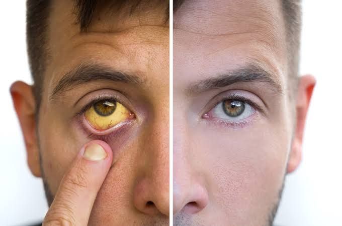
Jaundice, characterized by the yellowing of the skin, mucous membranes, and the whites of the eyes (sclera), is a clinical sign that reflects an underlying medical condition. It is not a disease in itself but rather a visible manifestation of elevated levels of bilirubin in the bloodstream. Understanding jaundice requires a structured approach to its physiology, classification, and potential implications.
The Physiology of Bilirubin
Before defining jaundice, it is crucial to understand the normal metabolic pathway of bilirubin, the substance responsible for the yellow discoloration.
Bilirubin is a yellow waste product that is primarily formed from the breakdown of heme, a component found mainly in hemoglobin within red blood cells. Approximately 70-80% of daily bilirubin production comes from the breakdown of senescent red blood cells by macrophages in the reticuloendothelial system (spleen, liver, bone marrow). The remaining 20-30% is derived from ineffective erythropoiesis and the breakdown of other heme-containing proteins.
The process involves several key stages:
- Production: Heme is converted into biliverdin (a green pigment), which is then reduced to bilirubin. This initial form is called unconjugated bilirubin.
- Transport: Unconjugated bilirubin is lipid-soluble and toxic in high concentrations. It binds to albumin in the bloodstream for transport to the liver.
- Hepatic Uptake and Conjugation: In the liver, hepatocytes (liver cells) take up the unconjugated bilirubin from the blood. Inside the hepatocytes, unconjugated bilirubin is conjugated with glucuronic acid by the enzyme UDP-glucuronosyltransferase (UGT). This process converts unconjugated bilirubin into conjugated bilirubin. Conjugated bilirubin is water-soluble and non-toxic.
- Biliary Excretion: Conjugated bilirubin is actively secreted by hepatocytes into the bile canaliculi, which merge to form larger bile ducts. Bile containing conjugated bilirubin flows from the liver into the gallbladder (for storage) or directly into the small intestine via the common bile duct.
- Intestinal Metabolism: In the small intestine, conjugated bilirubin is deconjugated by bacterial enzymes and further metabolized into urobilinogen.
- Excretion: Most urobilinogen (80-90%) is excreted in the feces, primarily as stercobilin (which gives stool its brown color). A small amount (10-20%) is reabsorbed into the bloodstream. Of this reabsorbed portion, most is taken back up by the liver, while a small fraction is filtered by the kidneys and excreted in urine as urobilin (which contributes to the yellow color of urine).
Jaundice occurs when there is an disruption in this complex pathway, leading to the accumulation of either unconjugated or conjugated bilirubin in the blood.
Defining Jaundice (Icterus Diagnosis)
Jaundice is the clinical manifestation of hyperbilirubinemia, defined as an elevated serum bilirubin level. While the exact threshold can vary slightly, jaundice typically becomes clinically apparent (visible yellowing) when the total serum bilirubin level exceeds approximately 2.5 to 3 mg/dL (40-50 µmol/L). Normal total bilirubin levels are usually below 1 mg/dL.
The yellow discoloration results from the deposition of excess bilirubin pigment in tissues, particularly those rich in elastin, such as the skin and sclera.
Classifying the Causes of Jaundice by Etiology
Categorizing the causes of jaundice based on where they disrupt the bilirubin pathway is fundamental for diagnosis and management. The traditional classification divides causes into three main categories: pre-hepatic, intra-hepatic, and post-hepatic.
1. Pre-hepatic Jaundice:
- Problem: Occurs before the liver’s main processing of bilirubin. The issue lies in an exaggerated load of unconjugated bilirubin being delivered to the liver, exceeding its capacity to conjugate and excrete it. The liver itself is generally healthy and functioning optimally, but it is simply overwhelmed.
- Mechanism: Primarily caused by excessive production of bilirubin, most commonly due to increased breakdown of red blood cells (hemolysis).
- Characterized by:
- Elevated unconjugated bilirubin in the serum.
- Normal or near-normal levels of conjugated bilirubin.
- Normal liver enzymes (AST, ALT, ALP) unless underlying liver damage is also present.
- Increased urobilinogen in urine (due to increased bilirubin load processed by the liver and gut).
- Normal stool color.
- Examples of Causes:
- Hemolytic Anemias: Genetic (e.g., Sickle cell anemia, Thalassemia, Hereditary spherocytosis, G6PD deficiency), Acquired (e.g., Autoimmune hemolytic anemia, Transfusion reactions, Drug-induced hemolysis, Malaria).
- Resorption of Large Hematomas: Extensive bruising or internal bleeding leads to the breakdown of a large volume of red blood cells.
- Ineffective Erythropoiesis: Conditions where red blood cells are produced but destroyed before entering the circulation (e.g., pernicious anemia, thalassemias).
- Gilbert’s Syndrome: A common, benign genetic disorder causing reduced activity of the UGT enzyme, leading to mildly elevated unconjugated bilirubin, often noticeable during stress, fasting, or illness.
2. Intra-hepatic Jaundice:
- Problem: Occurs within the liver itself. The issue is impaired hepatic function, affecting the liver’s ability to efficiently take up, conjugate, or excrete bilirubin.
- Mechanism: Damage to hepatocytes or disruption of the liver’s internal structures interferes with the entire bilirubin processing pathway.
- Characterized by:
- Usually elevated levels of both unconjugated and conjugated bilirubin, although the ratio can vary depending on the specific cause and stage of liver dysfunction.
- Elevated liver enzymes (AST, ALT, ALP) often indicate hepatocellular damage or cholestasis (impaired bile flow).
- Changes in other liver function tests (e.g., albumin, prothrombin time) in severe cases.
- Urine may contain bilirubin (conjugated bilirubin is water-soluble and filtered by kidneys).
- Stool color may be normal or pale depending on the degree of impaired bile flow into the gut.
- Examples of Causes:
- Acute Hepatitis: Viral (Hepatitis A, B, C, D, E), Alcoholic hepatitis, Drug-induced liver injury (e.g., acetaminophen overdose, certain antibiotics, NSAIDs), Autoimmune hepatitis.
- Chronic Liver Diseases leading to Cirrhosis: Chronic viral hepatitis (B, C), Alcoholic liver disease, Non-alcoholic fatty liver disease (NAFLD/NASH), Autoimmune liver diseases, Hemochromatosis, Wilson’s disease.
- Liver Infiltrative Diseases: Metastatic cancer, Lymphoma, Amyloidosis.
- Genetic Disorders affecting Liver Bilirubin Conjugation/Excretion: Crigler-Najjar syndrome (severe UGT deficiency), Dubin-Johnson syndrome, Rotor syndrome (defective hepatic excretion of conjugated bilirubin).
- Primary Biliary Cholangitis (PBC) / Primary Sclerosing Cholangitis (PSC): Autoimmune diseases causing damage to the small intrahepatic bile ducts.
3. Post-hepatic Jaundice (Obstructive Jaundice):
- Problem: Occurs after the liver. The liver is typically functioning normally, producing conjugated bilirubin. However, there is an obstruction of the bile flow in the extrahepatic bile ducts, preventing conjugated bilirubin from reaching the intestine.
- Mechanism: Blockage anywhere along the common bile duct (from the point where the cystic duct joins to the duodenum) causes bile to back up into the liver and then into the bloodstream.
- Characterized by:
- Significantly elevated conjugated bilirubin in the serum.
- Elevated Alkaline Phosphatase (ALP) and Gamma-Glutamyl Transferase (GGT) levels, which are enzymes typically elevated in biliary obstruction.
- Normal or mildly elevated AST/ALT unless the obstruction is prolonged or causes secondary liver injury.
- Urine is dark (contains conjugated bilirubin).
- Stool is pale or clay-colored (lack of stercobilin).
- Often associated with pruritus (itching) due to the accumulation of bile salts under the skin.
- Examples of Causes:
- Gallstones: Most common cause, particularly when stones migrate from the gallbladder into the common bile duct (choledocholithiasis).
- Strictures: Narrowing of the bile ducts due to inflammation, surgery, or chronic pancreatitis.
- Tumors: Malignancies arising from the bile ducts (cholangiocarcinoma), pancreas (pancreatic cancer compressing the duct), ampulla of Vater, or metastatic disease compressing the ducts.
- Pancreatitis: Inflammation of the pancreas can compress the common bile duct.
- Parasites: Certain liver flukes can obstruct bile ducts.
Risk Factors for Jaundice
Several factors can increase an individual’s risk of developing jaundice:
- Newborns: Physiologic jaundice is common in the first few days of life due to an immature liver enzyme system (UGT), increased red blood cell turnover, and enterohepatic circulation of bilirubin. Premature babies are at higher risk.
- Genetic Disorders: Family history of conditions like Gilbert’s syndrome, Crigler-Najjar syndrome, Dubin-Johnson syndrome, or hereditary hemolytic anemias.
- Chronic Liver Disease: Existing conditions such as chronic hepatitis (B, C), alcoholic liver disease, cirrhosis, or non-alcoholic fatty liver disease.
- Excessive Alcohol Consumption: A major risk factor for alcoholic hepatitis and cirrhosis.
- Medications: Use of drugs known to be hepatotoxic (damaging to the liver) or those that interfere with bilirubin metabolism or bile flow.
- Exposure to Hepatitis Viruses: Engaging in behaviors that increase the risk of viral hepatitis transmission (e.g., intravenous drug use, unprotected sex, unscreened transfusions).
- Gallbladder/Bile Duct Issues: History of gallstones or inflammation in the biliary system.
- Certain Autoimmune Diseases: Conditions like autoimmune hepatitis or primary biliary cholangitis.
- Conditions causing Increased RBC Breakdown: Certain blood disorders or exposure to agents causing hemolysis.
Potential Complications of Jaundice
While jaundice itself is a symptom, its underlying cause and the consequences of high bilirubin levels can lead to significant complications:
- Pruritus (Itching): Especially common in obstructive (post-hepatic) jaundice due to the accumulation of bile salts in the skin. Can be debilitating.
- Malabsorption: Poor flow of bile into the intestine, particularly in obstructive jaundice, impairs the digestion and absorption of fats and fat-soluble vitamins (A, D, E, K).
- Coagulopathy: Deficiency of Vitamin K (a fat-soluble vitamin) due to malabsorption can impair the liver’s production of clotting factors, increasing the risk of bleeding.
- Hepatic Encephalopathy: In severe intra-hepatic jaundice due to extensive liver failure, the liver cannot adequately detoxify the blood, leading to the accumulation of toxins (like ammonia) that affect brain function, causing confusion, disorientation, and coma.
- Renal Dysfunction (Hepatorenal Syndrome): A serious complication in advanced liver failure where there is functional kidney failure without intrinsic kidney disease.
- Osteoporosis: Chronic malabsorption of Vitamin D and metabolic bone disease can occur in prolonged cholestasis (impaired bile flow).
- Kernicterus (Bilirubin Encephalopathy): A severe and potentially devastating complication in newborns where very high levels of unconjugated bilirubin cross the immature blood-brain barrier and deposit in the brain, causing permanent neurological damage, deafness, cerebral palsy, and developmental delay. Less common in adults due to a more developed blood-brain barrier.
- Complications of the Underlying Cause: These are often the most serious. Examples include liver failure progression (cirrhosis), spread of malignancy, or complications related to severe hemolysis.
Management Principles for Certain Causes
The management of jaundice is directed at treating the underlying condition responsible for the elevated bilirubin levels. Symptomatic treatment may also be employed.
- Diagnosis is Key: The first step is accurate diagnosis through a detailed medical history, physical examination, blood tests (total and fractionated bilirubin, liver enzymes, and specific tests for suspected causes like viral markers, autoimmune antibodies, or genetic tests), and imaging studies (ultrasound, CT scan, MRI/MRCP, ERCP) to visualize the liver and biliary system. Liver biopsy may be necessary in some cases.
- General Supportive Care: May include adequate hydration, nutritional support (especially if malabsorption is present; potentially requiring supplementation of fat-soluble vitamins), and symptom management (e.g., medications for pruritus like cholestyramine or rifampicin).
- Management Examples Based on Etiology:
- Pre-hepatic Jaundice (e.g., Hemolytic Anemia): Management focuses on treating the cause of the excessive red blood cell breakdown. This may involve blood transfusions, corticosteroids or other immunosuppressants for autoimmune causes, splenectomy in certain conditions (e.g., hereditary spherocytosis), or specific treatments for underlying genetic disorders or infections (e.g., antimalarial drugs). Gilbert’s syndrome typically requires no treatment, only reassurance.
- Intra-hepatic Jaundice (e.g., Hepatitis, Cirrhosis): Treatment is aimed at the specific liver disease. This can include:
- Antiviral medications for chronic viral hepatitis (B, C).
- Abstinence from alcohol for alcoholic liver disease.
- Immunosuppressants for autoimmune hepatitis or primary biliary cholangitis.
- Withdrawal of offending drugs in cases of drug-induced liver injury.
- Supportive care for acute liver failure.
- Management of complications of cirrhosis (e.g., ascites, encephalopathy).
- Liver transplantation may be considered for selected patients with end-stage liver disease.
- Post-hepatic Jaundice (e.g., Obstructive Jaundice): Management requires relieving the obstruction, often urgently.
- Endoscopic Retrograde Cholangiopancreatography (ERCP): This minimally invasive procedure can visualize the bile ducts, remove gallstones, or place stents to bypass strictures or tumors.
- Percutaneous Transhepatic Biliary Drainage (PTBD): If ERCP is unsuccessful, a drain can be placed through the skin into the bile duct to relieve pressure.
- Surgery: May be necessary to remove tumors (e.g., Whipple procedure for pancreatic cancer), excise strictures, or perform procedures like a cholecystectomy (gallbladder removal) if gallstones are the root cause.
- Neonatal Jaundice: Often managed with phototherapy, which uses light to change unconjugated bilirubin into a water-soluble form that can be excreted. In severe cases, exchange transfusion (replacing the baby’s blood with donor blood) may be needed to rapidly lower bilirubin levels and prevent kernicterus.
Conclusion
Jaundice is a significant clinical sign that signals excess bilirubin accumulation in the body. Its presence warrants thorough medical investigation to determine the underlying cause. The systematic classification into pre-hepatic, intra-hepatic, and post-hepatic etiologies based on the disruption in bilirubin metabolism is critical for accurate diagnosis. Identifying risk factors and recognizing potential complications underscore the importance of timely evaluation and management. As a signpost for diverse conditions ranging from benign genetic variations to severe liver failure or malignancy, the management of jaundice is invariably focused on effectively addressing the specific root cause to prevent complications and restore health.


