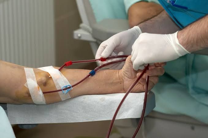
Access to the bloodstream or peritoneal cavity is a cornerstone of care for patients with chronic organ failure, most notably end-stage renal disease (ESRD) requiring dialysis. These access types are essential for delivering life-sustaining therapies. However, they are also potential sources of significant morbidity if not meticulously managed.
The management of patients with vascular and peritoneal access can be broken down into several critical areas:
Understanding the Types of Access and Their Purpose:
-
- Vascular Access: Primarily used for hemodialysis (HD), allowing rapid blood flow rates needed for effective waste removal. Types include arteriovenous fistulas (AVF – created surgically by connecting an artery and a vein), arteriovenous grafts (AVG – using a synthetic tube to connect an artery and vein), and central venous catheters (CVCs – temporary or tunneled lines inserted into a large vein, typically in the neck, chest, or groin). AVFs are generally preferred due to lower complication rates and longer patency.
- Peritoneal Access: Used for peritoneal dialysis (PD), which utilizes the patient’s peritoneal membrane as a filter. A soft, flexible catheter is surgically placed into the abdominal cavity, allowing dialysis fluid (dialysate) to be instilled, dwell, and drained.
Establishing Baseline Assessment Procedures:
-
- Vascular Access (AVF/AVG):
- Inspection: Look for redness, swelling, warmth, drainage, skin breakdown, aneurysm formation, or changes in the access site’s appearance. Note the location and presence of surgical incisional healing status initially.
- Palpation: Feel for the thrill (vibration) – it should be continuous and strong proximally, decreasing distally. Palpate for pulses distal to the access (checking for steal syndrome), tenderness, swelling, induration, or warmth. Note the turgor and integrity of the skin over the access.
- Auscultation: Listen with a stethoscope for the bruit (whooshing sound) – it should be continuous and low-pitched. Changes in pitch (higher suggests stenosis), absence, or broken quality are significant findings.
- Range of Motion: Assess if the access location impedes joint movement (e.g., elbow AVF).
- Peritoneal Access (PD Catheter):
- Inspection: Examine the exit site (where the catheter exits the skin) for redness, swelling, tenderness, drainage (note colour, odour, consistency), pain, or skin breakdown. Assess the tunnel tract (path of the catheter under the skin) for redness or tenderness which can indicate a tunnel infection.
- Palpation: Gently palpate around the exit site and along the tunnel for tenderness, induration, or swelling.
- Catheter Integrity: Check the catheter itself for cracks, kinks, or damage. Ensure caps and connections are secure.
- Dialysate Drainage: Assess the clarity, colour, and volume of drained dialysate. It should typically be clear or slightly straw-coloured. Cloudiness is a hallmark sign of peritonitis.
- Vascular Access (AVF/AVG):
Implementing General Access Care Principles:
-
- Patient Education: Crucial for empowering patients in their own care. Teach them how to inspect their access daily, recognize warning signs (changes in thrill/bruit, redness, swelling, pain, cloudy PD fluid), protect the access (no tight clothing, no blood pressure cuffs, no venipuncture on the access arm), and report concerns promptly.
- Aseptic Technique: Paramount for preventing infection. Strict hand hygiene, proper skin preparation before access use (HD) or connection/disconnection (PD), and sterile technique for dressing changes are essential.
- Regular Surveillance: Scheduled evaluations of access function (e.g., flow rates, venous pressures during HD; ultrafiltration volumes, drain times in PD) can identify potential problems before they become critical.
- Documentation: Maintain accurate records of access assessment findings, complications, interventions, and specialist consultations.
Recognizing and Managing Complications of Vascular Access (Hemodialysis):
-
- Early Complications (Typically Occur within Days to Weeks Post-Creation/Insertion):
- Recognition:
- Thrombosis: Absence of thrill and bruit; cold, pale, painful limb (rare but possible severe compromise).
- Infection: Redness, warmth, swelling, tenderness at the incision or cannulation site; fever; purulent drainage. May also be systemic with CVCs (fevers, chills).
- Bleeding/Hematoma: Oozing or frank bleeding from the site; swelling and bruising around the access.
- Pseudoaneurysm: Pulsatile swelling near a cannulation site, often with overlying skin changes.
- Early Steal Syndrome: Pain or coolness in the fingers/hand distal to the access, especially during dialysis; potentially numbness/tingling.
- Management:
- Thrombosis: Do not attempt to use. Apply gentle pressure if swelling is present. Involve Vascular Surgery / Interventional Radiology immediately for evaluation and potential intervention (thrombectomy, thrombolysis).
- Infection: Assess severity. Obtain cultures (blood, wound swab). Administer empiric antibiotics as per local protocol, adjusted based on culture results. Local site care (cleaning, dressing). Involve Nephrology, potentially Infectious Disease and Vascular Surgery/Interventional Radiology for severe infections or if access patency is threatened. CVC infections often require catheter removal.
- Bleeding/Hematoma: Apply direct pressure. Assess for uncontrolled bleeding. If significant or expanding hematoma, involve Vascular Surgery.
- Pseudoaneurysm: Avoid cannulating the area. Involve Vascular Surgery/Interventional Radiology urgently for assessment and repair to prevent rupture.
- Early Steal Syndrome: Reduce blood flow rate during dialysis if symptoms occur. Keep limb warm. Involve Vascular Surgery for evaluation; surgical revision may be required.
- Recognition:
- Late Complications (Typically Occur Weeks to Months/Years Post-Creation/Insertion):
- Recognition:
- Stenosis: Change in bruit (high-pitched); change in thrill (weaker, intermittent); prolonged bleeding post-dialysis; difficulty cannulating; elevated venous pressures during HD; reduced blood flow rates during HD.
- Thrombosis: Absence of thrill and bruit (often preceded by signs of stenosis).
- Infection: Chronic exit site drainage; redness, warmth, swelling, pain along the access; aneurysm infection; systemic signs (fever, chills) potentially from a CVC.
- Steal Syndrome (Chronic): Persistent pain, coolness, pallor, numbness, or non-healing ulcers in the distal limb.
- Aneurysm/Pseudoaneurysm: Localized or diffuse bulging/swelling of the access vessel; skin thinning or breakdown over the bulge.
- Central Venous Stenosis: Swelling of the access limb, shoulder, neck, or face; prominent collateral veins; difficulty with HD outflow.
- Management:
- Stenosis: Involve Nephrology and Interventional Radiology/Vascular Surgery for imaging (angiography) and intervention (angioplasty, stenting).
- Thrombosis: As with early thrombosis, involve Vascular Surgery/Interventional Radiology immediately.
- Infection: Similar to early infection, but may require long-term antibiotics, surgical debridement, or access ligation/excision depending on severity and access type. Involve Nephrology, Infectious Disease, and Vascular Surgery/Interventional Radiology.
- Steal Syndrome (Chronic): Involve Vascular Surgery for detailed assessment and potential surgical revision (e.g., DRIL procedure – Distal Revascularization and Interval Ligation).
- Aneurysm/Pseudoaneurysm: Avoid cannulating the affected area. Monitor size and skin integrity. Involve Vascular Surgery for evaluation; repair or ligation may be needed if rapidly expanding, symptomatic, or skin is compromised.
- Central Venous Stenosis: Often requires intervention. Involve Nephrology and Interventional Radiology for venography and angioplasty/stenting.
- Recognition:
- Early Complications (Typically Occur within Days to Weeks Post-Creation/Insertion):
Recognizing and Managing Complications of Peritoneal Access (Peritoneal Dialysis):
-
- Early Complications (Typically Occur within Days to Weeks Post-Insertion):
- Recognition:
- Exit Site/Tunnel Infection: Redness, swelling, tenderness, or drainage at the exit site or along the tunnel tract; pain.
- Peritonitis: Cloudy dialysate (most common sign); abdominal pain (diffuse or localized); fever; nausea, vomiting; rebound tenderness on abdominal exam.
- Catheter Malfunction: Poor inflow or drainage (e.g., slow fills, incomplete drains), often positional.
- Peritoneal Leakage: Fluid leaking from the exit site or inguinal/scrotal area; abdominal wall swelling.
- Bleeding: Pink, red, or brown tinged dialysate return.
- Management:
- Exit Site/Tunnel Infection: Obtain exit site swab and possibly catheter tip culture if drainage is present. Initiate topical or oral antibiotics based on protocol/culture results. Meticulous exit site care. Involve Nephrology/PD Team. If severe or progressing to tunnel infection, IV antibiotics or catheter removal may be necessary. Involve Nephrology and potentially Surgery.
- Peritonitis: Obtain dialysate sample for cell count, differential, Gram stain, and culture. Initiate empiric intraperitoneal (IP) antibiotics immediately as per protocol. Adjust antibiotics based on culture and sensitivity results. Pain management. Involve Nephrology/PD Team urgently. Non-responders or fungal peritonitis may require catheter removal. Involve Nephrology, Infectious Disease, and potentially Surgery.
- Catheter Malfunction: Rule out kinked tubing or constipation. Reposition patient. Gentle flushing attempts as per protocol. Involve Nephrology/PD Team. Imaging (e.g., X-ray) may be needed to check catheter position. May require surgical revision or replacement. Involve Surgery.
- Peritoneal Leakage: Hold PD or reduce fill volume. Convert to temporary HD access if significant leak or insufficient dialysis. Monitor site. Involve Nephrology and Surgery. Surgical repair may be needed if persistent.
- Bleeding: Usually transient due to minor vessel injury. Monitor drainages. If persistent or heavy, check blood pressure and coagulation status. May require temporary cessation of PD. Involve Nephrology.
- Recognition:
- Late Complications (Typically Occur Weeks to Months/Years Post-Insertion):
- Recognition:
- Peritonitis & Exit Site/Tunnel Infection: Recurrence of symptoms as described above.
- Catheter Malfunction: Ongoing difficulty with fills/drains; persistent pain during exchanges.
- Hernias: Bulging in inguinal, umbilical, or incisional areas, especially during fill.
- Encapsulating Peritoneal Sclerosis (EPS): A rare but serious complication characterized by thickened peritoneal membrane leading to small bowel obstruction. Patients present with abdominal pain, nausea, vomiting, weight loss, and malnutrition.
- Fluid/Electrolyte Imbalances: Related to inadequate ultrafiltration or excessive glucose absorption.
- Management:
- Recurrent Peritonitis/Infection: Investigate underlying causes (e.g., poor technique, nasal carriage of Staph Aureus). May require catheter removal and replacement later. Involve Nephrology, Infectious Disease, and Surgery.
- Catheter Malfunction: As with early malfunction, rule out kinks/constipation. May indicate catheter migration or fibrin sheath formation. Involve Nephrology/PD Team and Surgery.
- Hernias: Involve Surgery for evaluation and repair, usually requiring a temporary switch to HD.
- EPS: Very complex management. Often requires switching to HD. Involve Nephrology, Gastroenterology, and Surgery with expertise in this rare condition.
- Fluid/Electrolyte Imbalances: Adjust PD prescription (dwell times, fill volumes, glucose concentration). Monitor labs. Involve Nephrology/PD Team.
- Recognition:
- Early Complications (Typically Occur within Days to Weeks Post-Insertion):
The Crucial Role of Interdisciplinary Collaboration:
-
- Effective management relies heavily on teamwork. Promptly involve specialist colleagues:
- Nephrology Team (Nephrologists, PD Nurses, HD Nurses, Access Coordinators): Central to overall management, prescription adjustments, initial complication assessment, and deciding the need for specialist intervention or temporary change in dialysis modality.
- Vascular Surgery: Essential for creating and revising AVF/AVG, managing complex surgical complications (thrombosis, aneurysm, steal), and surgical repair of PD catheter leaks or hernias.
- Interventional Radiology: Key in diagnosing and treating vascular access stenosis/thrombosis (venography, angioplasty, stenting, thrombolysis) and sometimes placing/revising CVCs.
- Infectious Disease: Consulted for complex or recurrent access infections, atypical pathogens, or guidance on difficult-to-treat organisms or long-term antibiotic plans.
- General Surgery: Involved in PD catheter placement/removal and repair of PD-related hernias.
- Wound Care Specialists: Helpful in managing complex exit site issues or skin breakdown over vascular access.
- Effective management relies heavily on teamwork. Promptly involve specialist colleagues:
Conclusion:
Managing patients with vascular and peritoneal access requires continuous vigilance, thorough assessment, and a proactive approach to complication recognition. Healthcare professionals caring for these patients must have a solid understanding of normal access function, be adept at identifying subtle and overt signs of complications, and know the appropriate initial management steps. Crucially, recognizing the limits of initial management and promptly engaging specialist colleagues – including Nephrology, Surgery, Interventional Radiology, and Infectious Disease – is paramount to preserving access function, preventing patient harm, and ensuring the ongoing provision of life-sustaining dialysis therapy. An interdisciplinary approach, coupled with robust patient education, forms the foundation of optimal access management.


