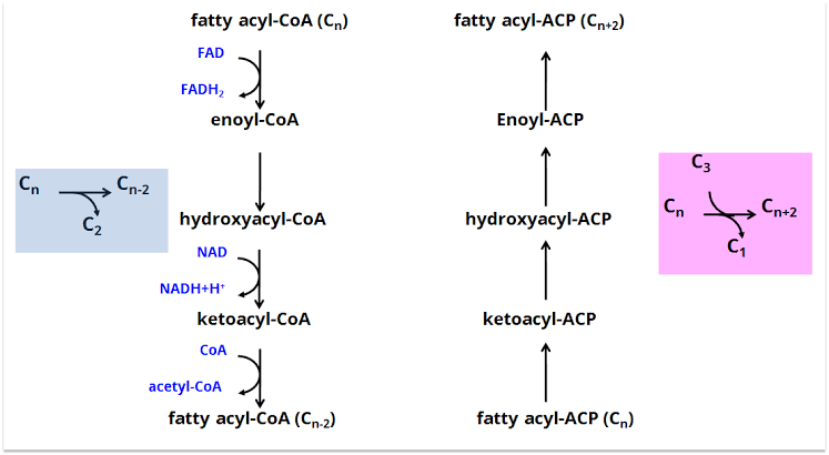
Fatty acid synthesis, also known as lipogenesis, is a crucial metabolic pathway responsible for converting excess metabolic fuel (primarily carbohydrates) into fatty acids for storage as triglycerides. This process primarily occurs in the cytosol of various tissues, most notably the liver, adipose tissue, and lactating mammary glands. It is an energy-requiring process, utilizing ATP and NADPH. The primary substrate is acetyl-CoA, derived from the breakdown of glucose and other sources.
Sources of NADPH Required for Fatty Acid Synthesis
Fatty acid synthesis is a reductive process, requiring a significant amount of reducing power in the form of NADPH. NADPH is essential for the two reduction steps that occur during each cycle of fatty acid elongation catalyzed by the Fatty Acid Synthase complex. The primary sources of cytosolic NADPH in lipogenic tissues are:
- The Pentose Phosphate Pathway (PPP) or Hexose Monophosphate (HMP) Shunt:
- This is the major source of the NADPH needed for fatty acid synthesis and other reductive biosynthetic reactions (like cholesterol synthesis) or detoxification processes (e.g., maintaining reduced glutathione).
- The oxidative phase of the PPP, which occurs in the cytosol, produces two molecules of NADPH for every molecule of glucose-6-phosphate that enters the pathway.
- The key enzymes generating NADPH are:
- Glucose-6-phosphate Dehydrogenase (G6PD): Catalyzes the first committed step, converting glucose-6-phosphate to 6-phosphoglucono-δ-lactone. This reaction reduces NADP+ to NADPH.
- 6-Phosphogluconate Dehydrogenase: Catalyzes the decarboxylation of 6-phosphogluconate (derived from the hydrolysis of 6-phosphoglucono-δ-lactone), producing ribulose-5-phosphate and another molecule of NADPH.
- Tissues highly active in fatty acid synthesis (like the liver and adipose tissue) have high activity of PPP enzymes, ensuring sufficient NADPH supply when carbohydrates are abundant.
- Cytosolic NADP+-Dependent Malic Enzyme:
- This enzyme, also known as cytosolic malate dehydrogenase (NADP+), provides a secondary source of cytosolic NADPH.
- It catalyzes the oxidative decarboxylation of malate to pyruvate, carbon dioxide, and NADPH:
- Malate + NADP+ → Pyruvate + CO2 + NADPH + H+
- This reaction is particularly linked to the citrate shuttle mechanism (discussed below). Cytosolic oxaloacetate, generated from the cleavage of citrate, is reduced to malate (by cytosolic malate dehydrogenase, which uses NADH), and this malate can then be converted to pyruvate by the malic enzyme, thereby generating NADPH.
- While not as prolific as the PPP, the malic enzyme can contribute significantly to the NADPH pool, especially when the citrate shuttle is highly active during active fatty acid synthesis.
These two pathways work in concert in lipogenic tissues to provide the substantial amount of NADPH required to reduce the growing fatty acyl chain.
Key Enzymes of Fatty Acid Synthesis
Fatty acid synthesis involves a complex series of enzymatic reactions. The core machinery consists of two principal enzymatic components:
- Acetyl-CoA Carboxylase (ACC):
- This is the rate-limiting enzyme of fatty acid synthesis.
- It catalyzes the irreversible carboxylation of acetyl-CoA to form malonyl-CoA, utilizing bicarbonate as the source of the carboxyl group and consuming ATP.
- Reaction: Acetyl-CoA + HCO3⁻ + ATP → Malonyl-CoA + ADP + Pi
- ACC is a complex enzyme that requires the vitamin biotin as a prosthetic group. Biotin is covalently attached to the enzyme and acts as a carrier of CO2.
- ACC exists as two major isoforms: ACC1 (or ACCα), primarily found in lipogenic tissues (like liver and adipose tissue) and responsible for supplying malonyl-CoA for fatty acid synthesis, and ACC2 (or ACCβ), found in muscle and heart, largely regulating fatty acid oxidation.
- Due to its rate-limiting nature, ACC is a major site of metabolic regulation, both allosterically (activated by citrate, inhibited by long-chain fatty acyl-CoA) and through covalent modification (phosphorylation, primarily inhibited by glucagon and epinephrine, activated by insulin) and genetic regulation (as discussed below). Malonyl-CoA is the critical two-carbon donor in the fatty acid synthesis pathway.
- Fatty Acid Synthase (FAS):
- In eukaryotes, FAS is a large, multi-enzyme complex found in the cytosol. In mammals, it is a large homodimeric protein, where each monomer contains multiple catalytic domains required for the sequential addition of two-carbon units (derived from malonyl-CoA) to a growing fatty acyl chain.
- The FAS complex carries out a cyclic series of reactions, typically repeating seven times to synthesize palmitate (a 16-carbon saturated fatty acid) from one molecule of acetyl-CoA (used as the ‘primer’) and seven molecules of malonyl-CoA.
- The key functional domains/activities within each monomer of the mammalian FAS complex include:
- Acyl Carrier Protein (ACP): Contains a phosphopantetheine prosthetic group (derived from pantothenic acid, Vitamin B5) that serves as a mobile carrier of the acyl intermediates during synthesis.
- Acetyl-CoA-Malonyl-CoA Transferase (MAT): Transfers acetyl and malonyl groups from their CoA carriers to the sulfhydryl groups of the ACP and the β-ketoacyl synthase (KS) domain.
- β-Ketoacyl Synthase (KS): Catalyzes the condensation reaction between the acetyl group (or growing acyl chain) attached to its cysteine-SH group and the malonyl group attached to the ACP. This releases CO2 and forms a β-ketoacyl group.
- β-Ketoacyl Reductase (KR): Reduces the β-keto group to a β-hydroxyl group, using NADPH.
- β-Hydroxyacyl Dehydratase (DH): Removes water from the β-hydroxyl group to form a double bond.
- Enoyl Reductase (ER): Reduces the double bond to a saturated carbon chain, using NADPH.
- Thioesterase (TE): Hydrolyzes the completed fatty acid chain (usually palmitate) from the ACP, releasing it from the enzyme complex.
- The process starts with acetyl-CoA and seven malonyl-CoA molecules. For each cycle of elongation (7 cycles in total for palmitate), one malonyl-CoA is added, undergoing condensation, reduction, dehydration, and reduction, consuming 2 molecules of NADPH and releasing CO2.
- The overall stoichiometry for palmitate synthesis is:
- Acetyl-CoA + 7 Malonyl-CoA + 14 NADPH + 14 H⁺ → Palmitate + 7 CO2 + 8 CoA + 14 NADP⁺ + 6 H2O
- (Note: 7 malonyl-CoA are formed from 7 acetyl-CoA + 7 ATP + 7 HCO3⁻, so 8 acetyl-CoA are effectively required in total for palmitate synthesis: 1 primer + 7 via malonyl-CoA).
Role of Citrate in Bringing Acetyl-CoA to Cytosol for FA Synthesis
Acetyl-CoA, the primary carbon source for fatty acid synthesis, is mainly produced within the mitochondrial matrix through the oxidative decarboxylation of pyruvate (by pyruvate dehydrogenase), fatty acid β-oxidation, and amino acid catabolism. However, fatty acid synthesis takes place in the cytosol. The inner mitochondrial membrane is impermeable to acetyl-CoA. Therefore, a mechanism is required to transport acetyl-CoA equivalents from the mitochondria to the cytosol. This is primarily achieved through the citrate shuttle.
The steps involved are:
- Formation of Citrate in Mitochondria: When cellular energy levels are high (high ATP) and substrates like glucose are abundant, isocitrate dehydrogenase of the TCA cycle is inhibited by ATP and NADH. This causes citrate to accumulate in the mitochondrial matrix. Citrate is formed from the condensation of acetyl-CoA and oxaloacetate catalyzed by citrate synthase:
- Acetyl-CoA + Oxaloacetate + H2O → Citrate + CoA-SH
- Transport of Citrate to Cytosol: The mitochondrial inner membrane possesses a specific transporter, the tricarboxylate transporter, that facilitates the export of citrate from the mitochondrial matrix into the cytosol in exchange for malate or phosphate.
- Cleavage of Citrate in Cytosol: In the cytosol, citrate is cleaved back into acetyl-CoA and oxaloacetate by the enzyme ATP-Citrate Lyase (ACL). This reaction requires ATP hydrolysis:
- Citrate + ATP + CoA-SH → Acetyl-CoA + Oxaloacetate + ADP + Pi
- Fate of Cytosolic Oxaloacetate: The oxaloacetate produced in the cytosol needs to be returned to the mitochondria to continue the cycle. It is typically reduced to malate by cytosolic malate dehydrogenase (using NADH), and then malate can either be transported back into the mitochondria via the tricarboxylate transporter or converted to pyruvate by the cytosolic NADP+-dependent malic enzyme, generating NADPH (linking back to Section 1):
- Oxaloacetate + NADH + H⁺ → Malate + NAD⁺ (catalyzed by Cytosolic Malate Dehydrogenase)
- Malate + NADP⁺ → Pyruvate + CO2 + NADPH + H⁺ (catalyzed by Cytosolic Malic Enzyme)
- Pyruvate can then re-enter the mitochondria via a specific transporter.
Thus, the citrate shuttle effectively moves acetyl-CoA carbons from the mitochondrial matrix to the cytosol, providing the necessary substrate for ACC and subsequent fatty acid synthesis. The activity of ATP-citrate lyase is upregulated in conditions favoring lipogenesis (e.g., high carbohydrate intake), providing more substrate for fatty acid synthesis.
Genetic Regulation of Acetyl-CoA Carboxylase (ACC)
As the rate-limiting enzyme, ACC is subject to stringent regulation at multiple levels, including allosteric control, covalent modification, and importantly, genetic (transcriptional) regulation. Genetic regulation controls the amount of ACC protein present in the cell, offering a longer-term adaptive response to changes in metabolic state or diet.
The gene expression of ACC (specifically the ACC1 isoform in lipogenic tissues) is primarily regulated by hormones and dietary factors:
- Hormonal Regulation:
- Insulin: Insulin, signaling a state of energy abundance and high blood glucose, is a potent inducer of ACC gene expression. It promotes the transcription of the ACC gene, leading to increased synthesis of the ACC protein. Insulin’s effects are mediated via intracellular signaling pathways (e.g., the PI3K/Akt pathway) that influence the activity of transcription factors. Key transcription factors involved include Sterol Regulatory Element-Binding Protein 1c (SREBP-1c) and Carbohydrate Response Element-Binding Protein (ChREBP). Insulin increases the processing and nuclear translocation of SREBP-1c and promotes the dephosphorylation and nuclear entry of ChREBP, both of which bind to regulatory regions in the ACC promoter, enhancing transcription.
- Glucagon and Epinephrine: These hormones, signaling low energy states and the need for fuel mobilization, generally suppress ACC gene expression. They activate cAMP-dependent signaling pathways (via PKA). While their primary effect on ACC is often acute (phosphorylation and inactivation), sustained high levels can also lead to reduced transcription of the ACC gene, contributing to the long-term suppression of fatty acid synthesis during fasting or stress.
- Dietary Regulation:
- High Carbohydrate, Low-Fat Diet: This dietary pattern strongly upregulates ACC gene expression, particularly in the liver. The abundance of glucose leads to increased insulin secretion and provides substrate for glycolysis and the PPP (generating acetyl-CoA, ATP, and NADPH), creating an environment favorable for fatty acid synthesis. The high glucose levels also activate ChREBP, which works synergistically with SREBP-1c (induced by insulin) to promote ACC transcription.
- Fasting or High-Fat Diet: These conditions generally downregulate ACC gene expression. In fasting, low insulin and high glucagon reduce the signals for ACC synthesis. High-fat diets, even in the presence of sufficient calories, tend to suppress the expression of ACC and other lipogenic enzymes. This is partly due to the presence of polyunsaturated fatty acids which can suppress SREBP-1c activity or other mechanisms.
In summary, the genetic regulation of ACC ensures that the cellular capacity for fatty acid synthesis is appropriately adjusted over the long term in response to hormonal signals reflecting the organism’s nutritional status and energy balance.
Importance of Glycerol Kinase in the Liver
Glycerol kinase is an enzyme that catalyzes the phosphorylation of glycerol to glycerol-3-phosphate (G3P), consuming ATP:
- Glycerol + ATP → Glycerol-3-phosphate + ADP
Glycerol-3-phosphate serves as the backbone molecule upon which fatty acids are esterified to form triglycerides (triacylglycerols). This step is essential for the storage of fatty acids.
The importance of glycerol kinase in the liver stems primarily from its uneven tissue distribution. While triglyceride synthesis occurs in several tissues (including adipose tissue and liver), glycerol kinase activity is high in the liver, kidney, and intestine, but very low or negligible in adipose tissue.
Here’s why this differential distribution is important:
- Glycerol Reclamation: Triglycerides stored in adipose tissue are continually undergoing limited lipolysis, releasing free fatty acids and glycerol into the bloodstream. Unlike fatty acids, glycerol cannot be metabolized by adipose tissue itself due to the lack of glycerol kinase. This released glycerol travels through the circulation and is primarily taken up by the liver (and kidney).
- Liver’s Role as a Metabolic Hub: The high activity of glycerol kinase in the liver allows it to efficiently take up and phosphorylate this circulating glycerol, producing G3P. This reclaimed G3P can then be used for several purposes:
- Triglyceride Synthesis: A significant portion is used as the backbone for de novo triglyceride synthesis in the liver. These newly synthesized triglycerides, along with those assembled from fatty acids taken up from the circulation, are packaged into Very Low-Density Lipoproteins (VLDL) and secreted into the bloodstream for transport to peripheral tissues, including adipose tissue for storage. This represents the liver’s central role in lipid transport.
- Gluconeogenesis and Glycolysis: G3P can be readily converted to dihydroxyacetone phosphate (DHAP), an intermediate of both glycolysis and gluconeogenesis. This allows the liver to utilize glycerol carbons for energy production (via glycolysis) or, particularly during fasting, convert them into glucose (via gluconeogenesis), contributing to the maintenance of blood glucose levels.
- Adipose Tissue Dependence on Glucose: Because adipose tissue lacks significant glycerol kinase, the G3P required for esterifying fatty acids during triglyceride synthesis in adipocytes must be derived from glucose metabolism via glycolysis (glucose → DHAP → G3P, catalyzed by glycerol-3-phosphate dehydrogenase). This explains why glucose uptake and metabolism are crucial for efficient triglyceride synthesis and storage in adipose tissue and how insulin, by promoting glucose uptake, is also important for fat storage in adipocytes.
In essence, hepatic glycerol kinase activity is critical for the liver’s ability to handle circulating glycerol released during lipolysis elsewhere in the body. It facilitates the liver’s role in disposing of this glycerol, either by incorporating it into triglycerides for export or by converting it into carbohydrates via gluconeogenesis, showcasing the liver’s metabolic flexibility and central coordination role.


