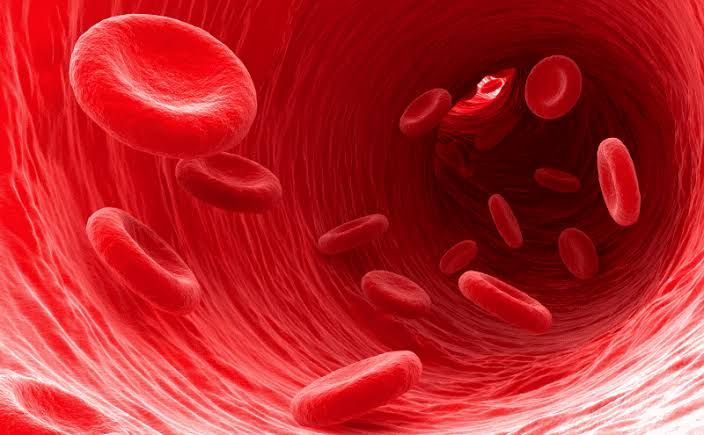
Iron, Vitamin B12 (cobalamin), and Folic Acid (folate) are fundamental micronutrients critical for numerous physiological processes in the human body. They are particularly vital for the formation and maturation of blood cells, a process known as hematopoiesis. Deficiencies in these nutrients can lead to various health issues, most notably specific types of anemia. Understanding their metabolism and function is key to maintaining optimal health and preventing deficiency-related disorders. This document outlines key aspects of these three essential nutrients.
1. Iron: An Essential Mineral
Iron is an indispensable mineral required for oxygen transport, DNA synthesis, energy metabolism, and numerous enzyme functions. Its role in the formation of hemoglobin, the protein in red blood cells that carries oxygen, is perhaps its most well-known function.
- 1.1. Food Sources:
- Heme Iron: Found exclusively in animal tissues (meat, poultry, fish). Heme iron is part of the hemoglobin and myoglobin molecules and is highly bioavailable (easily absorbed). Examples include red meat, liver, and seafood.
- Non-Heme Iron: Found in plant foods and fortified products. Examples include legumes (beans, lentils), tofu, spinach and other leafy greens, nuts, seeds, dried fruits, and iron-fortified cereals and breads. Non-heme iron absorption is lower and significantly influenced by dietary factors. Absorption is enhanced by vitamin C and inhibited by substances like phytates (found in grains, legumes, nuts), tannins (in tea, coffee), and calcium.
- 1.2. Requirements:
- Iron requirements vary significantly based on age, sex, and physiological state.
- Adult Men: Approximately 8 mg per day.
- Adult Women (pre-menopausal): Approximately 18 mg per day (higher due to menstrual blood loss).
- Pregnant Women: Approximately 27 mg per day (due to increased blood volume, fetal development, and placental needs).
- Lactating Women: Approximately 9 mg per day.
- Infants and Adolescents: Have relatively high requirements due to rapid growth.
- Vegetarians and Vegans: May require up to 1.8 times the typical requirement due to lower bioavailability of non-heme iron.
- 1.3. Absorption:
- Iron absorption primarily occurs in the duodenum and upper jejunum of the small intestine.
- Heme iron is absorbed as an intact porphyrin ring and is more efficiently absorbed (about 15-35%).
- Non-heme iron (mostly in the ferric Fe³⁺ state) is reduced to the ferrous Fe²⁺ state (by duodenal reductase enzymes like Dcytb) before transport across the apical membrane of enterocytes via the divalent metal transporter 1 (DMT1).
- Inside the enterocyte, iron can be stored bound to ferritin or transported across the basolateral membrane into the bloodstream via the iron exporter ferroportin.
- Absorption is tightly regulated by the body’s iron status and a key hormone called hepcidin, produced by the liver. High iron stores or inflammation lead to increased hepcidin, which binds to and degrades ferroportin, reducing iron absorption and release from stores. Low iron stores or increased erythropoietic activity suppress hepcidin, increasing iron absorption.
- 1.4. Distribution:
- Circulating iron is bound to the transport protein transferrin, which delivers it to tissues throughout the body.
- The largest proportion of body iron is found in hemoglobin within red blood cells (~65%).
- Significant amounts are stored, primarily as ferritin and hemosiderin, in the liver, spleen, and bone marrow (~25%). Ferritin is the primary storage protein, while hemosiderin is an aggregate formed at higher iron concentrations.
- Smaller amounts are found in myoglobin in muscle tissue, and as components of various enzymes involved in energy metabolism (e.g., cytochromes), DNA synthesis, and antioxidant defense.
- 1.5. Excretion:
- The human body has no specific physiological pathway for active iron excretion.
- Iron is lost passively through obligatory losses such as shedding of mucosal cells from the gastrointestinal and urinary tracts, skin cells, and small amounts in sweat and bile.
- Significant losses occur through bleeding, most notably menstruation in women, which is the primary reason for their higher requirement. Pregnancy and lactation also represent significant iron losses from the mother’s body.
- 1.6. Role in Hematopoiesis:
- Iron is absolutely essential for erythropoiesis, the formation of red blood cells in the bone marrow.
- It is a critical component of the heme group in hemoglobin. Four heme groups combine with a globin protein chain to form a hemoglobin molecule. Without sufficient iron, inadequate amounts of hemoglobin are produced, leading to defective red blood cell formation.
- Iron is delivered to developing red blood cells (erythroblasts) in the bone marrow via transferrin receptors.
- 1.7. Clinical Consequences of Deficiency:
- Iron Deficiency: The most common single nutrient deficiency worldwide. It progresses through stages: depleted iron stores, iron-deficient erythropoiesis, and finally, Iron Deficiency Anemia (IDA).
- Iron Deficiency Anemia (IDA): Characterized by the production of small (microcytic), pale (hypochromic) red blood cells.
- Symptoms: Fatigue, weakness, pallor (pale skin), shortness of breath (dyspnea) on exertion, dizziness, headache, cold hands and feet, brittle nails, hair loss.
- Severe Deficiency: Can lead to pica (craving non-food items like ice or clay), restless legs syndrome, glossitis (sore tongue), angular stomatitis (cracks at the corners of the mouth), and impaired cognitive function, immune response, and temperature regulation.
- Impact on Children: Can impair cognitive development and growth irreversibly if severe and prolonged.
- Impact on Pregnancy: Increased risk of premature birth, low birth weight, and maternal mortality.
2. Vitamin B12 (Cobalamin): A Complex Vitamin
Vitamin B12 is a complex water-soluble vitamin containing a cobalt atom. It is unique among vitamins due to its large size, complex absorption mechanism, and the fact that it is synthesized solely by microorganisms (mainly bacteria).
- 2.1. Food Sources:
- Vitamin B12 is naturally found almost exclusively in animal products. This includes meat, poultry, fish, eggs, and dairy products (milk, cheese, yogurt).
- Some plant-based foods are fortified with B12, such as certain breakfast cereals, nutritional yeasts, plant-based milk alternatives, and meat substitutes. These are important sources for vegetarians and vegans.
- 2.2. Requirements:
- Daily requirements are relatively small, measured in micrograms (µg).
- Adults: Approximately 2.4 µg per day.
- Pregnant Women: Approximately 2.6 µg per day.
- Lactating Women: Approximately 2.8 µg per day.
- Requirements are higher for older adults and individuals with certain medical conditions affecting absorption.
- 2.3. Absorption:
- B12 absorption is a complex process involving multiple steps and factors.
- In the stomach, gastric acid and pepsin release B12 from proteins in food.
- Released B12 binds to R-protein (haptocorrin), a binding protein secreted by salivary glands and gastric parietal cells.
- In the duodenum, pancreatic proteases degrade R-protein, releasing B12.
- B12 then binds to Intrinsic Factor (IF), a glycoprotein secreted by gastric parietal cells. The B12-IF complex is resistant to digestion.
- The B12-IF complex travels to the terminal ileum, where it is absorbed via specific receptors (cubilin). This is the primary mechanism for B12 absorption (‘active’ absorption).
- A small amount of B12 can be absorbed by passive diffusion throughout the small intestine, but this is clinically significant only at very high doses (pharmacological levels).
- Disruption at any step (e.g., insufficient stomach acid, lack of IF due to pernicious anemia or gastric surgery, damage to the terminal ileum due to Crohn’s disease or surgery) can impair B12 absorption.
- 2.4. Distribution:
- After absorption, B12 is released from IF and binds to transport proteins called transcobalamins (TCs).
- Transcobalamin II (TCII) is the primary delivery protein, transporting B12 to tissues, particularly the liver, bone marrow, and other rapidly dividing cells.
- Most of the body’s B12 is stored in the liver (50-90%) in the form of methylcobalamin and adenosylcobalamin. Liver stores are substantial and can last for several years (3-5 years or even more) even in the absence of dietary intake.
- Some B12 circulates bound to Transcobalamin I (TCI), which is a storage form.
- 2.5. Excretion:
- B12 excretion is minimal compared to most water-soluble vitamins.
- Small amounts are lost in the urine, but significant amounts are secreted in the bile into the intestine.
- Most of the biliary B12 is reabsorbed in the ileum via the enterohepatic circulation, conserving body stores. Only a small fraction is lost in the feces.
- 2.6. Role in Hematopoiesis:
- Vitamin B12 is a critical coenzyme required for DNA synthesis. It works closely with folic acid in the methylation cycle.
- Specifically, B12 is needed for the enzyme methionine synthase, which converts homocysteine to methionine. This reaction is coupled with the regeneration of tetrahydrofolate (THF) from 5-methyltetrahydrofolate. THF is necessary for the synthesis of purine and pyrimidine bases, which are building blocks of DNA.
- Without adequate B12 (or folate), DNA synthesis is impaired. This particularly affects rapidly dividing cells like red blood cell precursors in the bone marrow, leading to impaired maturation.
- 2.7. Clinical Consequences of Deficiency:
- Megaloblastic Anemia: Impaired DNA synthesis leads to the production of large (macrocytic), immature red blood cells in the bone marrow that fail to divide properly. These cells (megaloblasts) are eventually destroyed, resulting in anemia. This anemia is morphologically identical to that caused by folate deficiency.
- Neurological Damage: A hallmark of B12 deficiency, which does not occur in folate deficiency. B12 is needed for the synthesis of myelin (the protective sheath around nerves) and certain neurotransmitters. Deficiency can cause peripheral neuropathy (tingling, numbness, weakness), gait abnormalities, cognitive impairment (memory loss, confusion), psychiatric changes (depression, irritability), and in severe cases, spinal cord degeneration. These neurological effects can occur without anemia or precede it.
- Other Symptoms: Fatigue, weakness, glossitis (smooth, sore tongue), digestive issues, and increased homocysteine levels (a risk factor for cardiovascular disease).
- Causes: Common causes include pernicious anemia (autoimmune destruction of parietal cells leading to IF deficiency), malabsorption syndromes (e.g., Celiac disease, Crohn’s disease affecting the ileum), gastric surgery, chronic use of certain medications (e.g., proton pump inhibitors, metformin), inadequate dietary intake (especially in vegans/vegetarians lacking fortified foods or supplements), and bacterial overgrowth in the small intestine.
3. Folic Acid (Folate): Essential for Cell Division
Folic acid is the synthetic form of the vitamin used in fortified foods and supplements, while folate refers to the various forms found naturally in foods. Folate is a water-soluble vitamin crucial for DNA synthesis, cell division, and metabolism of amino acids.
- 3.1. Food Sources:
- Rich sources of natural folate include dark leafy green vegetables (spinach, kale, broccoli), legumes (beans, lentils, chickpeas), citrus fruits and juices, avocados, asparagus, and nuts.
- Folic acid is widely used to fortify grains, cereals, bread, pasta, and rice in many countries, significantly contributing to dietary intake. Food folate can be easily destroyed by heat and prolonged cooking.
- 3.2. Requirements:
- Folate requirements are expressed as Dietary Folate Equivalents (DFE) to account for the higher bioavailability of folic acid compared to food folate. 1 µg DFE = 1 µg food folate = 0.6 µg folic acid from fortified foods or supplements consumed with food = 0.5 µg folic acid from supplements consumed on an empty stomach.
- Adults: Approximately 400 µg DFE per day.
- Pregnant Women: Approximately 600 µg DFE per day (crucial for preventing neural tube defects).
- Lactating Women: Approximately 500 µg DFE per day.
- 3.3. Absorption:
- Dietary folates are typically conjugated to multiple glutamate residues (polyglutamates). These must be hydrolyzed (deconjugated) to the monoglutamate form by conjugase enzymes in the lumen of the small intestine before absorption.
- Folic acid, the synthetic form, is already in the monoglutamate form and is therefore more readily absorbed than food folate.
- Absorption primarily occurs in the upper small intestine (jejunum) via carrier-mediated transport.
- 3.4. Distribution:
- Folate circulates in the bloodstream bound to albumin and folate-binding proteins.
- It is transported to various tissues, with high concentrations in the bone marrow, liver, and gastrointestinal tract (tissues with high cell turnover).
- The liver is the main storage site, containing about half of the body’s total folate. However, liver stores are relatively limited compared to B12 (lasting only a few months).
- 3.5. Excretion:
- Excess folate is primarily excreted in the urine.
- Some folate is also secreted into bile and undergoes enterohepatic circulation, but this process is less efficient than that for B12, contributing to shorter body stores.
- 3.6. Role in Hematopoiesis:
- Folate is essential for the synthesis of DNA and RNA. It functions as a coenzyme carrying single-carbon units in many metabolic reactions.
- Crucially, folate (in its active form, THF) is required, alongside vitamin B12, for the synthesis of nucleotides (adenine, guanine, thymine, cytosine) needed for DNA replication and cell division.
- Like B12, folate is necessary for the proper maturation of red blood cell precursors in the bone marrow. A deficiency impairs DNA synthesis, leading to defective cell division and the production of megaloblastic cells.
- 3.7. Clinical Consequences of Deficiency:
- Megaloblastic Anemia: Morphologically indistinguishable from B12 deficiency anemia, characterized by large, immature red blood cells (macrocytosis).
- Neural Tube Defects (NTDs): Folate is critical during the early stages of pregnancy (often before a woman knows she is pregnant) for the proper closure of the neural tube, which develops into the brain and spinal cord. Folate deficiency significantly increases the risk of NTDs such as spina bifida and anencephaly. This is why folic acid fortification and supplementation are strongly recommended for women of childbearing age and during pregnancy.
- Other Symptoms: Fatigue, weakness, pallor, shortness of breath, glossitis, gastrointestinal disturbances.
- Interactions with B12: High doses of folic acid can mask the megaloblastic anemia of B12 deficiency, correcting the blood picture while allowing the potentially irreversible neurological damage from B12 deficiency to progress. It is therefore important to determine the cause of megaloblastic anemia before initiating treatment.
- Potential Links: Some research suggests that folate deficiency may be associated with increased homocysteine levels (similar to B12 deficiency), potentially contributing to cardiovascular disease risk, and may play a role in certain cancers and cognitive decline, although more research is needed in these areas.
- Causes: Inadequate dietary intake (common, especially without fortification), malabsorption disorders (e.g., Celiac disease, tropical sprue), increased requirements (pregnancy, lactation, rapid growth, certain diseases like hemolytic anemia), chronic alcohol abuse, and interactions with certain medications (e.g., methotrexate, anticonvulsants).
In conclusion, iron, vitamin B12, and folic acid are fundamental to human health, with critical interconnected roles in DNA metabolism and blood cell formation. While their food sources, specific absorption mechanisms, and storage capacities differ, their shared imperative for healthy hematopoiesis means that deficiency in any one can lead to significant clinical consequences. Understanding these metabolic pathways and the signs of deficiency is vital for effective diagnosis, prevention, and management of related health conditions.


