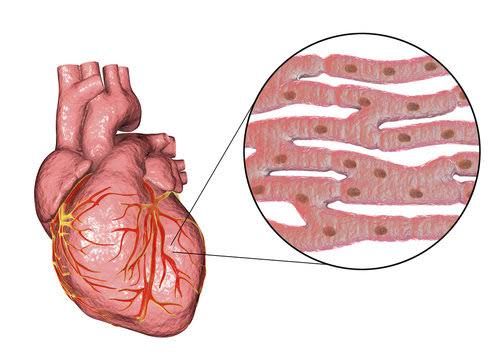
The cardiac muscle, or myocardium, is one of the most metabolically active tissues in the body. Functioning continuously throughout life, it requires a constant and substantial supply of energy in the form of adenosine triphosphate (ATP) to power contraction, relaxation, ion pumps, and cellular maintenance. Unlike skeletal muscle, which can tolerate periods of anaerobic metabolism during intense exertion, the heart is primarily an aerobic organ, highly reliant on oxidative phosphorylation within its abundant mitochondria for ATP production. The heart exhibits remarkable metabolic flexibility, capable of utilizing various substrates for energy depending on their availability and the physiological state of the organism. Understanding the heart’s energy metabolism is crucial for comprehending its function in health and disease.
Here is an explanation of key aspects of cardiac muscle energy metabolism:
Major Sources of Energy for the Cardiac Muscle
The sheer energy demand of the heart necessitates a continuous and robust ATP supply. The primary fuels the heart utilizes are molecules that can be efficiently oxidized through the Krebs cycle and oxidative phosphorylation to yield large amounts of ATP.
- Fatty Acids: Free fatty acids (FFAs) are the dominant fuel source for the heart under normal, resting conditions, accounting for approximately 60-90% of its energy requirements. Circulating FFAs, primarily bound to albumin, are taken up by cardiomyocytes through specific transporters. Once inside the cell, they undergo beta-oxidation within the mitochondria, breaking down the fatty acid chain into acetyl-CoA units. These acetyl-CoA molecules then enter the Krebs cycle, generating reduced coenzymes (NADH and FADH₂) that drive oxidative phosphorylation, the process producing the vast majority of ATP. Fatty acids are energy-dense substrates, making them an efficient fuel for continuous work.
- Glucose: Glucose is another vital energy source, contributing approximately 10-40% of the heart’s ATP production under normal conditions. Glucose enters cardiomyocytes primarily via GLUT4 transporters, which are partly regulated by insulin in the heart (though contraction is a more significant stimulus for GLUT4 translocation). Glucose undergoes glycolysis in the cytoplasm, producing pyruvate. Under aerobic conditions, pyruvate is transported into the mitochondria and converted to acetyl-CoA, which then enters the Krebs cycle, similar to the process for fatty acids. Glycolysis also produces a small amount of ATP directly. Glucose is particularly important during periods of increased workload or changes in circulating substrate levels.
- Lactate: Lactate, often considered a metabolic waste product in other tissues (like intensely working skeletal muscle), is a valuable fuel source for the aerobic heart. Lactate is taken up by cardiomyocytes via monocarboxylate transporters (MCTs) and can be converted back to pyruvate by lactate dehydrogenase (LDH) within the cytoplasm. This pyruvate then enters the mitochondria and is oxidized via the Krebs cycle and oxidative phosphorylation, providing ATP. Lactate can contribute significantly to cardiac energy production, especially during moderate exercise when circulating lactate levels rise.
- Ketone Bodies: While typically minor contributors under normal fed conditions, ketone bodies (primarily acetoacetate and beta-hydroxybutyrate) can serve as alternative fuels for the heart, particularly during prolonged fasting or diabetic ketoacidosis. The heart has the necessary enzymes (thiolase, HMG-CoA synthase, HMG-CoA lyase, etc., collectively known as ketolytic enzymes) to convert ketone bodies back to acetyl-CoA, which enters the Krebs cycle for oxidation. Their role becomes more prominent when other primary fuels are scarce.
- Amino Acids: Amino acids contribute a small percentage to cardiac energy metabolism under specific conditions but are generally not considered primary fuel sources.
The heart’s ability to switch between these substrates based on availability is a critical aspect of its metabolic flexibility. For example, during prolonged fasting or low carbohydrate intake, fatty acid oxidation increases significantly, and ketone bodies become a more important fuel. After a carbohydrate-rich meal, glucose utilization may increase. This adaptability ensures a stable energy supply despite fluctuations in substrate availability.
Ketone Bodies Synthesis and Utilization During Starvation
Ketone bodies are produced primarily in the liver and serve as an alternative energy source for peripheral tissues, including the heart and brain, during periods of glucose scarcity, such as prolonged fasting or uncontrolled diabetes (diabetic ketoacidosis).
- Synthesis (Ketogenesis): Ketogenesis occurs exclusively in the mitochondria of liver cells. During starvation, low insulin levels and high glucagon levels promote the breakdown of stored triglycerides in adipose tissue, releasing large amounts of free fatty acids into the bloodstream. These fatty acids are taken up by the liver and undergo extensive beta-oxidation, producing abundant acetyl-CoA. In the starved state, the capacity of the liver’s Krebs cycle to process all this acetyl-CoA is exceeded, and oxaloacetate (a key Krebs cycle intermediate derived from glucose metabolism) is reduced (as gluconeogenesis uses it to produce glucose). This surplus of mitochondrial acetyl-CoA, coupled with low oxaloacetate levels, diverts acetyl-CoA towards ketone body synthesis. The main steps involve:
- Condensation of two acetyl-CoA molecules to form acetoacetyl-CoA.
- Addition of a third acetyl-CoA to form HMG-CoA (beta-hydroxy-beta-methylglutaryl-CoA).
- Cleavage of HMG-CoA to form acetoacetate and acetyl-CoA.
- Acetoacetate can be reduced to beta-hydroxybutyrate (the most abundant ketone body in circulation) or spontaneously decarboxylated to acetone (a volatile compound exhaled). Ketone bodies (acetoacetate and beta-hydroxybutyrate) are then released from the liver into the bloodstream. The liver itself lacks the enzymes necessary to utilize ketone bodies as fuel, so it exports them to other tissues.
- Utilization (Ketolysis): Peripheral tissues, including the heart and skeletal muscle, and notably the brain after prolonged starvation, can efficiently utilize ketone bodies for energy. The process, called ketolysis, occurs in the mitochondria of these cells.
- Beta-hydroxybutyrate is first oxidized back to acetoacetate by beta-hydroxybutyrate dehydrogenase.
- Acetoacetate is then converted to acetoacetyl-CoA by the enzyme beta-ketoacyl-CoA transferase (or succinyl-CoA:3-ketoacid CoA transferase), which transfers a CoA group from succinyl-CoA. This enzyme is absent in the liver, preventing hepatic ketone body utilization.
- Acetoacetyl-CoA is finally cleaved by thiolase into two molecules of acetyl-CoA. These acetyl-CoA molecules then enter the Krebs cycle and are oxidized via oxidative phosphorylation, producing ATP.
During prolonged starvation, ketone bodies become a major circulating fuel source. For the heart, ketone body oxidation increases significantly, often accounting for a substantial portion of its energy needs. This provides an alternative fuel when glucose availability is low and helps spare the limited glucose supply for the brain, which relies heavily on glucose (though it adapts to use ketones during prolonged starvation). This metabolic shift is a crucial adaptive mechanism for survival during periods of nutrient deprivation.
Specificity of Lactate Metabolism in Hypoxic Heart Muscle
Lactate metabolism in the heart is particularly interesting because the heart is unusual among tissues in its ability to readily consume lactate as a fuel source under normal aerobic conditions. However, this dynamic is drastically altered under hypoxic or ischemic conditions.
- Normal Aerobic State: As discussed earlier, under normal oxygen availability, the heart is a net consumer of lactate. External lactate is taken up via MCTs and converted to pyruvate, which enters the mitochondria for complete oxidation. This contributes significantly to ATP production, especially when circulating lactate levels are elevated (e.g., during exercise or stress).
- Hypoxic/Ischemic State: When the heart muscle experiences hypoxia (low oxygen) or ischemia (restricted blood flow, leading to low oxygen and nutrient supply and metabolite accumulation), the primary pathway for ATP production – oxidative phosphorylation – is severely compromised because oxygen is the final electron acceptor. To try and maintain some ATP production, the heart shifts towards anaerobic glycolysis.
- Glucose uptake increases, and glycolysis accelerates.
- However, without sufficient oxygen, pyruvate cannot efficiently enter the Krebs cycle or be converted to acetyl-CoA. Instead, pyruvate is converted to lactate by the enzyme lactate dehydrogenase (LDH) to regenerate NAD⁺ from NADH. NAD⁺ is essential for the continuation of glycolysis.
- This leads to a rapid production and accumulation of lactate within the cardiomyocyte and its release into the extracellular space and circulation, primarily because uptake and utilization pathways (which are oxygen-dependent for subsequent oxidation) are impaired.
- The normal balance where the heart is a net lactate consumer is reversed; it becomes a net lactate producer.
- Consequences of Lactate Accumulation: While serving a temporary purpose of regenerating NAD⁺ for glycolysis, the increasing concentration of lactate, coupled with the co-produced protons (H⁺), leads to severe intracellular acidosis. This acidosis has detrimental effects:
- Inhibition of glycolytic enzymes, limiting further ATP production via this route.
- Impairment of contractile proteins (actin and myosin), reducing force generation.
- Inhibition of sarcoplasmic reticulum calcium pumps, disrupting calcium handling crucial for contraction and relaxation.
- Disruption of ion gradients across the sarcolemma via inhibition of pumps like the Na⁺/K⁺-ATPase.
- Potential contribution to cell damage and death.
Therefore, the specificity of lactate metabolism in the hypoxic heart lies in the reversal of its role. From being a preferred fuel source under aerobic conditions, it becomes a marker and contributor to metabolic dysfunction and injury under anaerobic (ischemic/hypoxic) conditions due to the necessary but ultimately detrimental switch to unsustainable anaerobic glycolysis and the resultant acidosis.
Specificity of Metabolism of the Cardiac Muscle Under Pathological Conditions (Diabetes)
Diabetes Mellitus, particularly type 2 diabetes characterized by chronic hyperglycemia and insulin resistance, significantly impacts cardiac metabolism, contributing to the development of diabetic cardiomyopathy and increasing vulnerability to ischemic injury. While the heart’s ability to utilize fatty acids and glucose is normally flexible, chronic exposure to the metabolic environment of diabetes causes detrimental adaptations.
- Altered Substrate Preference: In the diabetic heart, there is a pronounced shift towards increased reliance on fatty acid oxidation, often at the expense of glucose utilization, despite high circulating glucose levels. This is partly due to:
- Insulin Resistance: Insulin signaling is impaired, affecting the translocation of GLUT4 glucose transporters to the cell surface, thus reducing glucose uptake.
- Increased Fatty Acid Availability: Insulin resistance in adipose tissue leads to increased lipolysis and higher circulating free fatty acid levels, which are readily taken up and oxidized by the heart.
- Transcriptional Changes: Chronic exposure to high glucose and fatty acids alters gene expression, promoting the uptake and oxidation of fatty acids while suppressing glucose transport and utilization pathways.
- Consequences of Excessive Fatty Acid Oxidation: While fatty acids are a primary fuel, excessive reliance on them in the diabetic heart leads to several problems:
- Mitochondrial Overload: The continuous high flux of fatty acids can exceed the capacity of the Krebs cycle and oxidative phosphorylation, leading to incomplete fatty acid oxidation.
- Accumulation of Toxic Lipid Metabolites: Incomplete oxidation results in the buildup of potentially toxic intermediates, such as acylcarnitines, ceramides, and diacylglycerols. These metabolites can interfere with insulin signaling (exacerbating insulin resistance), impair calcium handling, induce oxidative stress, and activate pro-apoptotic pathways. This phenomenon is often referred to as “lipotoxicity.”
- Reduced Metabolic Efficiency: Fatty acid oxidation requires more oxygen per molecule of ATP produced compared to glucose oxidation. This reduced oxygen efficiency can strain the heart’s oxygen balance, particularly under stress.
- Increased Reactive Oxygen Species (ROS) Production: Excessive substrate flux through the mitochondria, especially fatty acids, increases the production of ROS as a byproduct of oxidative phosphorylation. This oxidative stress damages cellular components (lipids, proteins, DNA), contributing to inflammation, fibrosis, and cell death.
- Impaired Glucose Metabolism: Despite high ambient glucose, reduced glucose uptake and utilization limit the beneficial aspects of glucose metabolism, such as the pentose phosphate pathway (important for antioxidant defense) and replenishment of Krebs cycle intermediates.
- Mitochondrial Dysfunction: Chronic metabolic stress in diabetes leads to mitochondrial abnormalities, including decreased number, altered morphology, impaired respiration, and reduced ATP production efficiency. This compromises the very engine room of cardiac energy supply.
The sum of these metabolic derangements in the diabetic heart creates a state of metabolic inflexibility and inefficiency. The heart becomes overly dependent on fatty acids, prone to lipotoxicity and oxidative stress, and metabolically less capable of adapting to increased workload or ischemic challenges. These changes underpin the functional decline characteristic of diabetic cardiomyopathy, manifesting as impaired contractility and relaxation, and contributing to increased cardiovascular risk in diabetic patients.
In conclusion, the cardiac muscle’s energy metabolism is complex, highly regulated, and adaptable. While relying primarily on fatty acid oxidation under normal conditions and exhibiting flexibility in utilizing other substrates like glucose and lactate, this delicate balance can be severely disrupted by conditions such as hypoxia and chronic diseases like diabetes, leading to metabolic dysfunction and ultimately compromising cardiac function. Understanding these specific metabolic pathways and their perturbations is essential for developing therapeutic strategies aimed at preserving cardiac health.


